Indonesian Journal of Biotechnology
VOLUME 22(1), 2017, 1–5 | RESEARCH ARTICLEExpression of haloacid dehalogenase gene and its molecular protein
characteriza on from
Klebsiella pneumoniae
ITB1
Ridani Rino Anggoro1and Enny Ratnaningsih1,∗
1Biochemistry Research Division, Department of Chemistry, Ins tut Teknologi Bandung, Jalan Ganesha 10, Bandung 40132, West Java,
Indonesia
∗Corresponding author:enny@chem.itb.ac.id
ABSTRACTOrganohalogen compounds are widely used industrially and agriculturally, as well as in households as flame retar-dants and refrigerants. However, these compounds can become significant pollutants through their accidental or deliberate release into the environment in large quan es. Dehalogenase is an enzyme with the poten al to be used in the removal of organohalogen contaminants. A previous study successfully subcloned a 690 bp of haloacid dehalogenase gene (hakp1) fromKlebsiella pneumoniaeITB1 into a pET-30a(+) expression system to achieve high enzyme produc vity. IPTG was used as an inducer to express a pET-hakp1recombinant clone inEscherichia coliBL21 (DE3). The molecular mass of the haloacid dehalogenase Hakp1 protein was 30 kDa as determined by SDS-PAGE. Zymogram analysis showed that this recombinant protein has dehalogenase ac vity as shown by the forma on of AgCl white precipitate. A quan ta ve assay of haloacid dehalogenase Hakp1 gave a specific ac vity of 84.29 U/mg with the op mum temperature of 40°C at pH 9. Predicted three-dimensional structure of Hakp1 showed α/β mo f folding which comprised of cap and core domain. The predicted ac ve sites of Hakp1 were Asp8, Glu10, Leu22, Phe23, Trp90, Ser125, Ser126, Lys159, and Asp184 with Asp8, Glu10, Ser126, and Lys159 act as binding residue. This recombinant haloacid dehalogenase clone provides an alterna ve agent for effec ve bioremedia on of organohalogen pollutants.
KEYWORDShaloacid dehalogenase;Klebsiella pneumoniaeITB1; organohalogen
1. Introduc on
Organohalogen compounds are compounds that contain carbon-halogen bond. Large-scale synthesis and extensive uses of these organic chemicals in many areas of agricul-ture and industry have led to widespread distribution of harmful compounds in the environment and created pollu-tion problems. These chemicals, which are produced in-dustrially and introduced into the environment as novel compounds, or at a concentration that exceeds the normal amount, are called xenobiotics (Top and Springael 2003). Organohalogen compounds, which constitute of more than 75% compounds listed as “priority pollutants”, have been widely used as herbicides, pesticides, fungicides, solvents, plasticizers, paints, printing ink, adhesives, hydraulic and heat transfer fluids, flame retardants, refrigerants, addi-tives for cutting oils, textile auxiliaries, and intermediates for other fine chemical synthesis (Fetzner and Lingens 1994;Janssen et al. 1994).
These chemically synthesized organic compounds are not readily degraded in the environment, and many are accumulate in soil water, groundwater, lake, and river (Esteve-Núñez et al. 2001;Pervova et al. 2002). Besides
local and regional contamination, organohalogen contin-ues to be a global issue pollutants, partly because its transport through water and air helps these compounds to spread across the Earth (Iwata et al. 1993). The persis-tence of these compounds in the environment causes con-siderable human health problems because of their toxicity and bioaccumulation in the food chain and ground water (Brokamp et al. 1997;Dórea 2008). It is well-known that some organohalogen compounds degrade slowly and form toxic intermediates which may affect cellular metabolic processes (Slater et al. 1995;Janssen et al. 2001).
bromoacetic acid (BAA), and dibromoacetic acid (DBA). HAA, which is expressed as the sum of the concentrations of these acids, is currently limited there to 60 ppb (Xie and Zhou 2002). The current concentration of HAA in the Sidoarjo (Rosyidi 2010) and Sukabumi (Indraningsih et al. 2006) water supply sampled was approximately 50 ppb. Because of their widespread occurrence, toxicity to plants and aquatic organisms, and most importantly their suspected human carcinogenicity, there is a great need to find treatment methods for haloacetic acids.
In nature, some microorganisms are known as able to degrade organohalogen compounds and play a major role in biodegradation and decontamination (Weightman and Slater 1980; de Lorenzo 2008). Microbes degrade organohalogen compounds in order either to exploit them for growth as a carbon source and/or as a means of protec-tion against its toxicity (Müller and Lingens 1986). There are several key requirements for a microorganism to de-grade xenobiotics: (a) the ability to transport the com-pound into the cell where enzyme action can occur; (b) degradative catabolic genes must be expressed producing functional enzymes; (c) and the product of enzyme must be able to enter metabolism pathways (i.e. to be a growth substrate) (Weightman and Slater 1980;Weightman et al. 1985). Dehalogenases are key enzymes in the degradation of organohalogen compounds (Fetzner and Lingens 1994;
Janssen et al. 1994).
A problem in using a wild-type bacterial dehaloge-nase for industrial biotransformation is rare because of their basal level availability and its low efficiency in de-grading organohalogen pollutants. The advance of molec-ular biotechnology provides a solution to increase de-halogenases production by microbes through cloning and subcloning of the gene encoding dehalogenase into ex-pression system for high enzyme productivity. Previous studies had been successfully cloned the haloacid dehalo-genase gene fromKlebsiella pneumoniae ITB1 (hakp1) into pET-30a(+) expression vector, named as pET-hakp1
(Anggoro and Ratnaningsih 2017). This system would make good expression because it contains a strong regu-latable promoter for gene expression. In order to analyze the function ofhakp1gene, the pET-hakp1should be trans-formed intoEscherichia coliBL21 (DE3) and expressed by IPTG induction, which is performed in this research.
2. Materials and methods
2.1. Bacterial strains and chemicals
All bacterial strains were obtained from the Biochem-istry Laboratory of ITB. E. coli BL21 (DE3) was used as bacterial hosts. Monochloroacetic acid (MCA), mercury(II)thiocyanate (Hg(SCN)2), iron(II)ammonium
sulfate (Fe(NH4)2(SO4)2) were obtained from Merck.
Isopropyl-B-D-Thiogalactopyranoside (IPTG) from Am-resco was used as inducer for expression. Kanamycin from Bioline were used as antibiotic to screen positive
re-combinant clones. Natrium chloride, yeast extract, and tryptone from Liochemfil were used as ingredients in Luria-Bertani (LB) medium.
2.2. Transforma on and clone selec on
The pET-hakp1 recombinant clone isolated from E. coli TOP10 was transformed into competent E. coli
BL21(DE3) cells using heat shock method (Sambrook and Russell 2001). The obtained transformants were plated on LB medium supplemented with 50 µg kanamycin for an-tibiotic screening. TheE. coliBL21 (DE3) colonies har-boring pET-hakp1recombinant clone would survived on this medium due to the presence of kanamycin resistance gene in pET-30a(+).
2.3. Expression and extrac on of haloacid dehaloge-nase
Single colony ofE. coliBL21(DE3) harboring pET-hakp1
was grown at 37°C on 100 mL liquid LB medium supple-mented with 50 µg kanamycin. Induction was performed by adding 1 mM IPTG when the 450 nm optical density of this culture reached around 0.6. These induced cells were then harvested by centrifugation at 5,000 rpm for 10 min after 4-h induction, washed and resuspended in 10 mL of 50 mM Tris-acetate buffer (pH 7.5). Cells were disrupted by sonication for 30 min at 4°C. A crude extract was ob-tained after the cell debris had been removed by centrifu-gation.
2.4. Es ma on of haloacid dehalogenase molecular mass
The molecular mass of denatured protein from the crude extract was estimated by sodium dodecyl sulfate-polyacrylamide gel electrophoresis (SDS-PAGE) utilizing protein ladder standard. Gel was visualized using staining Commassie Brilliant Blue solution.
2.5. Zymogram assay
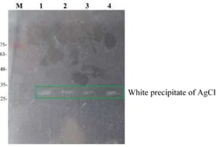
Qualitative assay of haloacid dehalogenase activity in the crude extract was determined by zymogram. Crude ex-tracts were electrophorized using Native-PAGE, and the resulted gel was incubated with 50 mM monochloroacetic acid (MCA) at 40°C for 10 min, and then soaked and in-cubated at room temperature for 1 h in 0.1 M AgNO3
so-lution. Haloacid dehalogenase activity was showed by the formation of AgCl white precipitate.
2.6. Enzyma c assay
forma-(a) (b)
FIGURE 1SDS-PAGE electropherogram of the pET-hakp1gene expression inE. coliBL21(DE3). (a) Overexpression analysis; M: Promega Broad Range Protein Ladder; 0: Total cell protein before IPTG induc on; 1-4: Total cell protein a er IPTG induc on from 1 to 4 h; Deb: Total protein in cell debris; Eks: Protein content in the medium (represen ng extracellular protein); Lis: Total protein in the cell lysate. (b) Molecular mass analysis of Hakp1. M: Promega Broad Range Protein Ladder; 1-2: Cell lysate protein from recombinantE. coliBL21 (DE3); C : Cell lysate protein from non-recombinantE. coliBL21 (DE3).
tion of 1 µmol of chloride ion per min. Protein was deter-mined using Bradford reagent with bovine serum albumin as standard (Bradford 1976).
2.7. Bioinforma c analysis
The three-dimensional structure of deduced Hakp1 tein was predicted using Swiss Model and I-TASSER pro-gram. The active site of this protein was analyzed us-ing Sequences Annotated by Structure (SAS). Molecular docking was done using Autodock Vina to determine the binding site, ligand conformation, and affinity energy.
3. Results and discussion
3.1. Expression of recombinant hakp1 gene on E. coli BL21(DE3) biofilm qualita ve analysis by SEM Expression of hakp1 gene in pET-hakp1 recombinant clone was studied by induction of IPTG. The SDS-PAGE analysis on the cell pellet protein showed that the over-expression was occurred after 4-h induction (Figure1a). On this gel, haloacid dehalogenase was seen more con-centrated in the cell lysate compare to that in the cell de-bris. This fact indicated that the haloacid dehalogenase was present as soluble protein. Molecular mass estimation of this haloacid dehalogenase recombinant was obtained at around 30 kDa (Figure1b). This size was larger than 25.5
kDain silicoanalysis of Hakp1 protein, which was
possi-bility due to the addition of fusion 6x His-Tag, S-Tag, and enterokinase site on its N-terminus.
3.2. Dehalogenase ac vity of recombinant haloacid dehalogenase
The zymogram assay showed that the haloacid dehaloge-nase produced by pET-hakp1has detectable activity on MCA. This result was proved by the formation of AgCl white precipitate observable on the gel (Figure2). The size of this active halaocid dehalogenase was approximately 30 kDa.
Further assay of recombinant haloacid dehalogenase activity were determined by identified chloride ion
re-FIGURE 2Zymogram assay of recombinant haloacid dehalogenase; M: 1stBase prestained protein ladder; 1–4: Cell lysate protein.
leased upon incubation with 5 mM MCA, by varying pH (Figure3a) and temperature (Figure3b) to obtain optimum condition of enzymatic reaction. The result suggests that the maximum activity of the recombinant haloacid dehalo-genase was 84.29 U/mg at 40°C and pH 9. At this con-dition, the total chloride ion detected was 684 µM repre-senting 13.68% of MCA degradation. This activity was three times higher compare to haloacid dehalogenase ac-tivity from the wild-typeK. pneumoniaeITB1 (Tahya and Ratnaningsih 2015). The specific activity was not as high as expected because the haloacid dehalogenase crude used was still impure.
3.3. Bioinforma cs of protein Hakp1
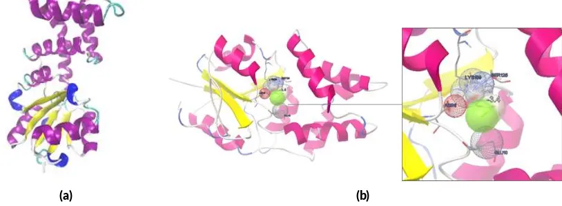
The predicted three-dimensional structure of Hakp1 was done by Swiss-Model and I-TASSER programs (Figure
(a) (b)
FIGURE 3Ac vity assay of recombinant haloacid dehalogenase (a) Effect of pH on enzyme ac vity. (b) Effect of temperature on enzyme ac vity
(a) (b)
FIGURE 4Bioinforma cs analysis of Hakp1. Predicted three-dimensional structure (a) and molecular docking (b) of Hakp1 protein with MCA ligand.
pneumoniaeITB1 has Asp8, Glu10, Leu22, Phe23, Trp90,
Ser125, Ser126, Lys159, and Asp184 residues, which are similar to Asp9, Ser154, Lys159, and Asp188 of haloacid dehalogenase superfamilies Enolase-phospatase E1 pro-tein fromHomo sapiens(Wang et al. 2005). Molecular docking result suggested that Asp8, Glu10, Ser126, and Lys159 are the binding residues in Hakp1 (Figure 4b). The identified lowest affinity energy of Hakp1 was -3.4 kcal/mol.
4. Conclusions
The pET-hakp1system have been successfully express a 30 kDa recombinant haloacid dehalogenase. This recom-binant dehalogenase has 84.29 U/mg optimum activity at 40°C and pH 9. Dehalogenase activity assay showed that the enzymes can degrade 13.68% chloride ion from 5 mM monochloroacetic acid substrate. Predicted three-dimensional Hakp1 structure showed α/β folding motif with Asp8, Glu10, Ser126, and Lys159 as binding residues. These result suggested that the recombinant haloacid de-halogenase clone has potentially used as bioremediation
agent for organohalogen pollutants in the environment. Further studies with enzyme purification and gene muta-tion needed to be done to improve the purity and activity of the recombinant haloacid dehalogenase.
Acknowledgments
We would like to acknowledgeLembaga Pengelola Dana
Pendidikan(LPDP) Indonesia for full financial support on
this research.
Authors’ contribu ons
ER, RRA designed the study. RRA carried out the labora-tory work. ER, RRA analyzed the data. ER, RRA wrote the manuscript. All authors read and approved the final version of manuscript.
Compe ng interests
References
Anggoro RR, Ratnaningsih E. 2017. Subcloning of haloacid dehalogenase gene fromKlebsiella pneumo-niaestrain ITB1 into pET-30a expression vector. vol-ume 3 ofProceedings of the 7th Annual Basic Science
International Conference. Malang: Brawijaya
Univer-sity. p. 99–104.
Bergmann JG, Sanik J. 1957. Determination of trace amounts of chlorine in naphtha. Anal Chem. 29(2):241–243. doi:10.1021/ac60122a018.
Bradford MM. 1976. A rapid and sensitive method for the quantitation of microgram quantities of pro-tein utilizing the principle of propro-tein-dye binding. Anal Biochem. 72(1):248–254. doi: 10.1016/0003-2697(76)90527-3.
Brokamp A, Schwarze R, Schmidt FRJ. 1997. Ho-mologous plasmids from soil bacteria encoding d,l-halidohydrolases. Curr Microbiol. 34(2):97–102. doi:10.1007/s002849900151.
de Jong RM, Dijkstra BW. 2003. Structure and mech-anism of bacterial dehalogenases: different ways to cleave a carbon-halogen bond. Curr Opin Struct Biol. 13(6):722–730. doi:10.1016/j.sbi.2003.10.009. de Lorenzo V. 2008. Systems biology approaches to
bioremediation. Curr Opin Biotechnol. 19(6):579– 589. doi:10.1016/j.copbio.2008.10.004.
Dórea JG. 2008. Persistent, bioaccumulative and toxic substances in fish: human health consid-erations. Sci Total Environ. 400(1-3):93–114. doi:10.1016/j.scitotenv.2008.06.017.
Esteve-Núñez A, Caballero A, Ramos JL. 2001. Bi-ological degradation of 2,4,6-trinitrotoluene. Microbiol Mol Biol Rev. 65(3):335–352. doi:10.1128/MMBR.65.3.335-352.2001.
Fetzner S, Lingens F. 1994. Bacterial dehalogenases: biochemistry, genetics, and biotechnological applica-tions. Microbiol Rev. 58(4):641–685.
Indraningsih, Bahri S, Sani Y. 2006. Beberapa faktor yang mempengaruhi keamanan pangan asal ternak di Indonesia. Wartazoa 16:1–13.
Iwata H, Tanabe S, Sakai N, Tatsukawa R. 1993. Dis-tribution of persistent organochlorines in the oceanic air and surface seawater and the role of ocean on their global transport and fate. Environ Sci Technol. 27(6):1080–1098. doi:10.1021/es00043a007. Janssen DB, Oppentocht JE, Poelarends GJ. 2001.
Mi-crobial dehalogenation. Curr Opin Biotechnol. 12(3):254–258. doi: 10.1016/s0958-1669(00)00208-1.
Janssen DB, Pries F, Van der Ploeg JR. 1994. Ge-netics and biochemistry of dehalogenating en-zymes. Annu Rev Microbiol. 48(1):163–191. doi:10.1146/annurev.mi.48.100194.001115.
Müller R, Lingens F. 1986. Microbial degradation of halo-genated hydrocarbons: A biological solution to
pollu-tion problems? Angew Chem Int Ed Engl. 25(9):779– 789. doi:10.1002/anie.198607791.
Pervova MG, Kirichenko VE, Pashkevich KI. 2002. Deter-mination of chloroacetic acids in drinking water by re-action gas chromatography. J Anal Chem. 57(4):326– 330. doi:10.1023/A:1014902431520.
Rebhun M, Heller-Grossman L, Manka J. 1997. For-mation of disinfection byproducts during chlo-rination of secondary effluent and renovated water. Water Environ Res. 69(6):1154–1162. doi:10.2175/106143097x125902.
Rosyidi M. 2010. An effect of a breakpoint chlorination BPC coliform total number from hospital wastewater in Sidoarjo. Undergraduate thesis. [Surabaya]: Institut Teknologi Sepuluh Nopember.
Sambrook J, Russell DW. 2001. Molecular cloning: a laboratory manual. Cold Spring Harbor, NY: CSHL Press.
Slater JH, Bull AT, Hardman DJ. 1995. Micro-bial dehalogenation. Biodegradation 6(3):181–189. doi:10.1007/BF00700456.
Tahya CY, Ratnaningsih E. 2015. Cloning and sequenc-ing of haloacid dehalogenase gene from Klebsiella
pneumoniae ITB1. Procedia Chem. 16(Supplement
C):121–128. doi:10.1016/j.proche.2015.12.039. Top EM, Springael D. 2003. The role of mobile genetic
elements in bacterial adaptation to xenobiotic organic compounds. Curr Opin Biotechnol. 14(3):262–269. doi:10.1016/s0958-1669(03)00066-1.
Wang H, Pang H, Bartlam M, Rao Z. 2005. Crystal struc-ture of human E1 enzyme and its complex with a substrate analog reveals the mechanism of its phos-phatase/enolase activity. J Mol Biol. 348(4):917–926. doi:10.1016/j.jmb.2005.01.072.
Weightman AJ, Slater JH. 1980. Selection of
Pseu-domonas putida strains with elevated dehalogenase
activities by continuous culture growth on chlori-nated alkanoic acids. Microbiology 121(1):187–193. doi:10.1099/00221287-121-1-187.
Weightman AJ, Weightman AL, Slater JH. 1985. Toxic ef-fects of chlorinated and brominated alkanoic acids on
Pseudomonas putidaPP3: selection at high
frequen-cies of mutations in genes encoding dehalogenases. Appl Environ Microbiol. 49(6):1494–1501.