Modeling neuronal dynamic coding in primary visual cortex
Qian Yang
a, Xianglin Qi
a,*, Wang Yunjiu
a,baLaboratory of Visual Information Processing,Institute of Biophysics,Academy of Sciences,
Beijing100101,People’s Republic of China
bCenter for Brain-Mind Science,Academy of Sciences,Beijing100101,People’s Republic of China
Abstract
The visual system can be considered as a multi-layered and dynamic image processing system. According to experimental evidence, the receptive field (RF) organization is characterized by spatio-temporal properties. The modified extended Gabor (MEG) function model was proposed to describe the main spatio-temporal properties of RF at different levels of visual pathway. Based on the MEG model, a three-layered dynamic coding model was constructed for a complex cell. The responses of the complex cell depend on synaptic events from a simple cell assembly within a time window. The membrane potential evolution equation was applied to the analysis of the length of a time window. The simulation results demonstrated that a complex cell plays as a coincidence detector in encoding synaptic events within the time window. © 2000 Elsevier Science Ireland Ltd. All rights reserved.
Keywords:Receptive field; Dynamic coding; Coincidence detector; Neural representation
www.elsevier.com/locate/biosystems
1. Introduction
The visual system can be considered a multi-layered and dynamic image processing system. Its task is to extract some key information from the environment, then encode, transmit and process them through different levels on the visual path-way. It is very important to understand how the information from the eyes is coded and repre-sented in the brain. Many researchers have pro-posed various viewpoints on encoding and representing visual information by theoretical and experimental approaches in terms of the experi-mental results from physiology, anatomy and
psy-chophysics. The traditional viewpoint is firing rate coding where the firing spike frequency is re-garded as the neuronal information carrier, such as in the mean firing rate hypothesis based on some unicellular electro-physiological recording (Barlow, 1972) and synchronization oscillation hypothesis of neural spike (Gray and Singer, 1989). Another is the temporal pattern of spike train which was considered as a representation of neuronal information, e.g. timing of spikes based on active potential (Hopfield, 1995). There are other hypotheses on neural coding, for example, the neuronal population hypothesis (Hebb, 1949), the icon alphabet hypothesis (Tanaka et al., 1991; Fujita et al., 1992) or the sparse and coarse cod-ing (Rolls and Treves, 1990; Olshausen and Field, 1997). The emergent dynamics of dynamical cell * Corresponding author.
E-mail address:[email protected] (Xianglin Qi).
assemblies has been explored based on the hy-pothesis that a cortical neuron functions effec-tively as a coincidence detector. Moreover, the principle of spatio-temporal coding was proposed in terms of the hypothetical emergent dynamics (Softky, 1995b; Fujii et al., 1996). This functional dynamic neuronal assembly provides a dynamical foundation of bi-directional interactions for the linkage of distant modules to create integrated information.
In the primary visual cortex, a great number of experiments have been carried out based on the above standpoint of mean firing rate, such as receptive field (RF), orientation selectivity and so on. Particularly, neuronal RF is regarded as a basic structural and functional unit for informa-tion coding. During the recent years, experimental evidence accumulated shows that RF organization is characterized by spatio-temporal properties. The dynamic RF has been discovered in various neurons of retina, lateral geniculate nucleus (LGN) and visual cortex of cat or monkey (DeAngelis et al., 1995). In fact, these neurons in the visual pathway act like encoders of spatial and temporal visual stimuli. They encode patterns of modulation of light, in space and time, into trains of nerve impulses.
In this paper one first proposed the MEG model to describe the dynamic behavior of neu-ronal RFs in the primary visual system. Then a three-layer dynamic coding model was con-structed for a complex cell based on the MEG model. The responses of the complex cell depend on synaptic events from simple cell assembly within a time window. The membrane potential evolution equation was applied to the analysis of the length of a time window. The simulation results demonstrated that a complex cell plays the role of coincidence detector in encoding synaptic events within the time window.
2. Mathematical model of RF
Based on the physiological results in mam-malians, many mathematical models of visual in-formation processing have been proposed. Some models can only describe the spatial structures of
neuronal RFs of retina and LGN (Rodieck, 1965). Some models related to Gabor functions can describe the static properties of RF in space domain and spatial frequency domain (Marcelja, 1980Daugman, 1985; Daugman, 1988). The math-ematical models for spatio-temporal RF have been proposed since 1985 (Adelson and Bergen, 1985; Pan et al., 1985; Wang et al., 1985; Watson and Ahumada, 1985; Heeger, 1987; Shapley et al., 1991; Kawakami and Okamoto, 1996; Wang et al., 1996).
2.1. Mathematical description of MEG model
From the viewpoint of system analysis, a family of extended Gabor functions (EGF) was con-structed as the model of spatio-temporal RFs (Wang et al., 1985). In this paper a modified EG model (MEG) was proposed. In the MEG mod-els, Gabor function is still adopted as a term related to spatial variables and the temporal term is substituted by two order integral kernel func-tion of G function tz−1e−t. When
z=2,G func-tion indicates te−t. Then the modified Gabor
function model (MGM) is expressed as follows, (1) Isotropic MG model in polar coordinates (first type of MGM)
where r and are spatial polar coordinates, vr is
spatial frequency and vt temporal frequency, sr
spatial variation,ur spatial phase andut temporal
phase, T1, T2, tand K are constants.
(2) Anisotropic MG model in Cartesian coordinates
(ii) Spatio-temporal inseparable MG model
where x, y are spatial coordinates vx, vy spatial
frequencies, vt is temporal frequency sx and sy
are spatial variations uxy is spatial phase ut
tem-poral phase, uxyt spatio-temporal phase and the
other symbols are the same as in Eq. (1).
2.2. Simulation of dynamic RF by MEG model
The above MEG models include a set of parameters with physiological significance. When the parameters were taken properly, the proper-ties of RF on space domain, time domain or space-time joint domains were simulated by means of the MEG model. In this paper the simulation results of spatial and spatio-temporal structures of RF will be demonstrated in the visual cortex.
2.2.1. Spatial properties of RF
If Eqs. (2) and (3) merely include spatial and spatial frequency variables, the static RFs in spa-tial domain are specified. They can be expressed as follows:
Fig. 1 illustrates the spatial RF structure of a simple cell from striate cortex. Panel (A) is the measured RF profile for a simple cell from cat striate cortex observed by DeAngelis (DeAngelis et al., 1995). Panels (B) and (C) are the simulation results by Eq. (4). Fig. 1(B) is density plot of 3D spatial profile of RF. Fig. 1(C) is 3D spatial responsive profiles of a simple cell.
2.2.2. Dynamic properties of RF in space-time domain
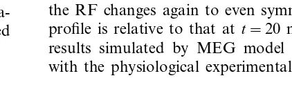
Recently, the development of powerful map-ping techniques has facilitated the spatio-temporal properties of RFs for neurons in the visual path-way. If the RF is a spatio-temporal entity, the dynamic RFs can be divided into space-time sepa-rable and insepasepa-rable. The dynamics of space-time inseparable RF structure of a simple cell from the striate cortex is shown in Fig. 2. Fig. 2(A) indicates the experimental data recorded from a simple cell of a cat as reported in DeAnge-lis et al. (1995), TINS Vol. 18, No. 10. Fig. 2(B) demonstrates the simulation results by the MEG model using Eq. (3). The parameters are taken as vx=p/deg, vy=0/deg, sx=1 deg, sy=1.2 deg,
vt=p/(120 ms), t=60 ms, uxyt=0.6p. For each
panel of (A) and (B), 2D spatial RF profiles are shown as isoamplitude contour maps for different times t. The excitatory sub-region is indicated by solid contour lines and the inhibitory by broken contour lines. Below each contour plot is a 1D spatial profile that is obtained by integrating the 2D profile along the y-axis, which is parallel to the cell’s preferred orientation. The temporal se-quences of RF profile in Fig. 2 are continuous. The sub-regions of the RF appear to move right-ward along the x-axis over time within a spatial window. At t=20 ms, the 1D profile is approxi-mately even symmetric, while at t=100 ms, the RF profile is odd symmetric. Then att=180 ms, the RF changes again to even symmetric and the profile is relative to that at t=20 ms. The above results simulated by MEG model are consistent with the physiological experimental data.
Fig. 2. Dynamics of the spatio-temporal inseparable receptive field (RF) structure of simple cells from striate cortex. (A) The experimental results as reported in TINS, vol. 18, no. 10, 1995, DeAngelis et al. (B) Simulation results by the modified extended Gabor (MEG) model. The parameters are taken asvx=p/deg,vy=0/deg,sx=1 deg,sy=1.2 deg,vt= −p/(120 ms),t=60 ms, uxyt=0.6p.
3. Spatio-temporal coding model of a complex cell
3.1. Structure of model
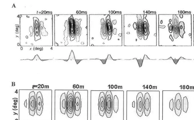
A complex cell receives the synaptic events from the simple cell assembly. The RF of a com-plex cell is composed of a number of parallel similar simple cells. Generally, excitatory and in-hibitory areas of RFs for complex cells overlap each other and vary with time. The maximal response was obtained when the pulses of stimuli reach to complex cells simultaneously. A three-layer network model was constructed based on simple cell assembly with dynamic RF in order to explain this kind of coding pattern. The structure of the model is demonstrated in Fig. 3. There are three parts shown as (A), (B) and (C) in Fig. 3. The first layer is an array N×N of input units denoted by (A). The second layer denoted by (B) consists of simple cells whose possesses characters of spatio-temporal filter and the sizes of grid are the same as the first layer. Part B is the spatio-temporal filter of simple cells denoted by SCi,
i=1, 2, . . .,N. The panel, T, stands for the tem-poral properties of simple cells, and panel S, the
spatial properties of simple cells. Spatio-temporal integrated layer of complex cell belongs to the part C.uij(t) indicates the response of the complex
cell in this layer which was obtained by the coinci-dent detector from the output of simple cells SCi
at t.
3.2. Mathematical description of the model
In the second layer of the model, the evolution of each individual simple cell is indicated by Eq. (3) of spatio-temporal inseparable modified ex-tended Gabor function. An assembly consisting of this kind of simple cell acts as a filter to a complex cell. The mutual relation among local regions of synaptic connection within this assembly leads to asymmetric distribution of the filtering layer. The asymmetry means local in spatial and having refined temporal structure in time.
analysis. The strongest response of a complex cell appears in its RF while the two stimuli simulta-neously happened. The timing of spike trains of simple cells is considered as a temporal integrated variable. The response of a complex cell can be expressed by the membrane potential evolution equation as follows: input layer and the second layer p×q represents the size of discrete spatial window. Wij(k,l,T)
indicates the weight of connecting the input unit on (i,j) node with the simple cell on (k,l) node at
T. Ikl(T) external input at T andUij(t) threshold
of firing at t.
3.3. Simulation results
Three types of input stimuli were designed: Gaussian function, random function and sinu-soidal function. The length of the time windows were set to 2, 20 or 50 ms individually.
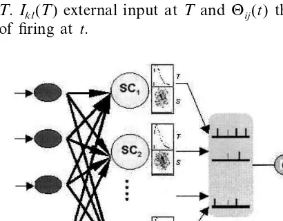
The number of simple cells in the filtering layer of the model is 5776 arranged as a 76×76 lattice. There are eight gray levels which represent the amplitude of neuronal response at a spatial loca-tion and a given time. The simulated results show the different dynamic patterns of cell-assembly in the second layer under the three kinds of stimuli (see Fig. 4). For example, when the Gaussian function stimulus is presented, the distribution of the codes is divided into three sub-regions which emerge as a light – dark – light pattern along the diagonal direction at 10 ms and dark – light – dark pattern at 40 ms. The polarity is completely re-versed at 60 ms. In the same way, there exists five sub-regions and polarity reversals except the re-sponse amplitudes have some changes, when ran-dom function and sinusoidal function are provided. For the different inputs, the behaviors of these coding trains demonstrated finer struc-tures under more complicated stimulus when the parameters of filter window and the initial weights of connections were kept consistently. The cell-as-sembly has self-adaptive and self-organized func-tion by this type of synapses connecfunc-tion.
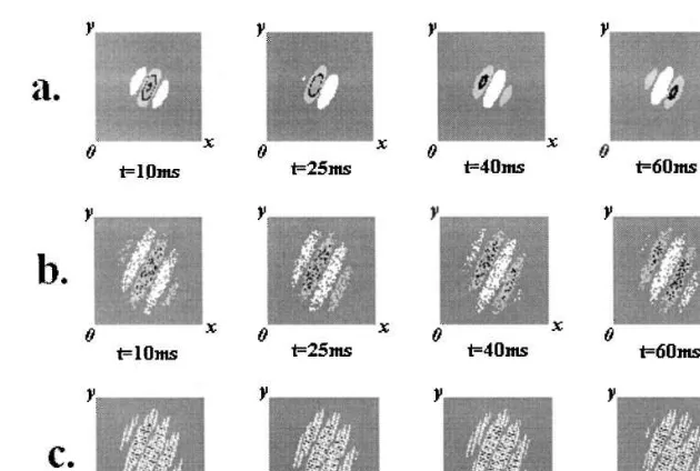
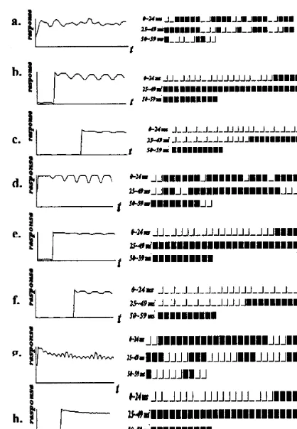
In Fig. 5, the interval in the coincidence win-dow, such as 2, 20, 50 ms, represents simultaneous synaptic events coding and the traditional firing rate coding. However, for three different input patterns, the variability of a complex cell’s spike train in 2 ms-interval time window (see Fig. 5a, d and g) is stronger than that of in 20 ms (Fig. 5b, e and h) or 50 ms interval time window (see Fig. 5c, f and i). This result shows that the time window with an action of coincidence detection should be several milliseconds. In contrast to the traditional coding strategy, coding space of 2 ms interval is bigger and leads to a wide range of the spatio-temporal patterns to uniquely represent one of the properties of an input. Moreover, the periodicity of codes for random function input is stronger within a shorter period than longer ones, but their coding efficiencies are reversed.
Fig. 4. The spatio-temporal codes of simple cells assembly. The abscissa is the spatial coordinatex, and the ordinate is the spatial coordinatey. Each row represents the evolution process from 10 to 60 ms with an interval of 15 ms or so. All of the neuronal coding amplitudes in each patch are normalized by gray scales with eight different levels represented from zero to 0.86, respectively. The results in a, b and c correspond to the different inputs. (a, Gaussian function (top); b, random function (middle); c, sinusoidal grating function (bottom)).
4. Discussion
The EG and MEG models with a variety of parameters can describe the main spatial, tempo-ral and spatio-tempotempo-ral properties of RF at dif-ferent levels of the visual system. Specifically, these models can simulate the spatio-temporal separability and inseparability of the RFs. The results of computer simulation were found to agree with the physiological data. But there are many parameters in the EG and MEG models. This is a key problem for simulating the proper-ties of the RFs. The estimated methods have been explored for the spatial frequencies, preferred ori-entation angle of RF, the size of RF, temporal frequencies, spatial phase and temporal phase. The concept of neural RFs defined in a parameter space was studied. In this paper the purpose is to apply the MEG model to the construction of a neural coding model of complex cell.
The time window within which the complex cell processes many synaptic events from the simple cell assembly have been estimated by simulation. The result implies that the window should be a
few milliseconds in length like the conclusions from another model (Sakumura and Aihara, 1998). The biophysical mechanism for the model is based on the nonlinear summation of excitatory inputs in each of the dendrites as a current sink for inputs. It shows that the coincident activity of the membrane may narrow the time window (also see Egger et al., 1999). In the time window of a complex cell, the cell morphology and the spatio-temporal distribution of the pre-synaptic spikes enriches its coding power, such as in the auditory coincidence detection (Hagai Agmon-Snir et al., 1998). Although it seems to be more difficult to analyze the complex cell information on the spike train, the synchronized neuronal activity over a short period may be sufficient to bring about exquisite diversiform neural codes of complex cell in the visual cortex.
Acknowledgements
Fig. 5. Temporal coding plots of complex cells in visual cortex. The panels include two parts, the response curve (left) and the spike code (right). The coincident integrated windows last 2 ms (from a to c), 20 ms(from d to f) or 50 ms (from g to i), respectively. The elapsed time is 60 ms. In the right column, there are three rows in each patch represented by three contin-uous time regions: 0 – 24, 25 – 49 and 50 – 59 ms (black is firing, white is silent).
Egger, V., Feldmeyer, D., Sakmann, B., 1999. Coincidence detection and changes of synaptic efficacy in spiny stellate neurons in rat barrel cortex. Nat. Neurosci. 2, 1098 – 1105. Fujii, H., Ito, H., Aihara, K., Tsukada, M., 1996. Dynamical cell assembly hypothesis — theoretical possibility of spa-tiotemporal coding in the cortex. Neural Network 9, 1303 – 1350.
Fujita, I., Tanaka, K., Ito, M., Cheng, K., 1992. Columns for visual features of objects in monkey inferotemporal cortex. Nature 360, 343 – 346.
Gray, C.M., Singer, W., 1989. Stimulus-specific neuronal oscil-lations in orientation columns of cat visual cortex. Proc. Natl. Acad. Sci. USA 86, 1698 – 1702.
Hagai Agmon-Snir, Carr, C.E., Rinzel, J., 1998. The role of dendrites in auditory coincidence detection. Nature 393, 268 – 272.
Hebb, D.O., 1949. The Organization of Behavior — A Neuro-physiological Theory. Wiley, New York.
Heeger, D.J., 1987. Model for the extraction of image flow. JOSA A87 (4), 1455 – 1471.
Hopfield, J.J., 1995. Pattern recognition computation using action potential timing for stimulus representation. Nature 376, 33 – 36.
Kawakami, S., Okamoto, H., 1996. A cell model for the detection of local image motion on the magnocellular pathway of the visual cortex. Vision Res. 36, 117 – 148. Marcelja, S., 1980. Mathematical description of the responses
of simple cortical cells. JOSA 70, 1297 – 1300.
Olshausen, B.A., Field, D., 1997. Sparse coding with an overcomplete basis set: a strategy employed by V1? Vision Res. 37, 3311 – 3325.
Pan, Z.H., Qi, X.L., Wang, Y.J., 1985. A mathematical model of primary information processing in vertebrate visual system (II). Acta Biophys. Sinica 1, 189 – 198.
Rolls, E.T., Treves, A., 1990. The relative advantage of sparse versus distributed encoding for associative neuronal net-works in the brain. Network 1, 407 – 421.
Sakumura, Y., Aihara, K., 1998. A neornal time window for coincidence detection. IEICE E81-A (9), 1818 – 1823. Shapley, R., Reid, R.C., Soodak, R., 1991. Spatiotemporal
receptive fields and direction selectivity. In: Landy, M.S., Movshon, J.A. (Eds.), Computational Models of Visual Processing. MIT Press, Cambridge, MA, pp. 109 – 118. Softky, W.R., 1995b. Simple codes versus efficient codes. Curr.
Opin. Neurobiol. 5, 239 – 247.
Tanaka, K., Saito, H., Fukuda, Y., Moriya, M., 1991. Coding visual images of objects in the inferotemporal cortex of the macaque monkeys. J. Neurophysiol. 66, 170 – 189. Wang, Y.J., Qi, X.L., Pan, Z.H., 1985. A mathematical model
of primary information processing in vertebrate visual system (I). Acta Biophys. Sinica 1, 123 – 133.
Wang, Y.J., Qi, X.L., Chen, Y.Z., 1996. Simulations of recep-tive-field dynamics. TINS 19, 385 – 386.
Watson, A.B., Ahumada, A.J., 1985. Model of human visual-motion sensing. JOSA A85 (2), 322 – 341.
.
References
Adelson, E.H., Bergen, J.R., 1985. Spatio-temporal energy models for the perception of motion. JOSA A85 (2), 284 – 299.
Barlow, H.B., 1972. Single units and sensation: a neuron doctrine for perceptual psychology? Perception 1, 371 – 394. DeAngelis, G.C., Ohzawa, I., Freeman, R.D., 1995. Receptive-field dynamics in the central visual pathways. TINS 18, 451 – 458.