www.elsevier.nlrlocateraqua-online
Experimental induction of jaw, gill and pectoral fin
malformations in Japanese flounder, Paralichthys
oli
Õ
aceus, larvae
Tohru Suzuki
), Anand Shanker Srivastava, Tadahide Kurokawa
Metabolism Section, National Research Institute of Aquaculture, Fishery Agency, Nansei, Mie 516-0193, Japan
Accepted 20 October 1999
Abstract
The phenotypes of skeletal malformations induced in pharyngeal arches and pectoral fins of
Ž . X
Japanese flounder larvae by retinoic acid RA , disulfiram, 2,2 -dipyridyl and azetidine-2-carbo-xylic acid were characterized, RA controls gene expression essential for pharyngeal and pectoral fin development; disulfiram is an inhibitor of RA synthase, 2,2X-dipyridyl and azetidine-2-carbo-xylic acid are inhibitors of collagen synthesis. In larvae exposed to RA at shield stage for 1 h, the Meckel’s cartilage did not form in mandible arch. Exposure to RA from hatching period shifted the growth direction of the pharyngeal cartilages posteriorly. Disulfiram did not affect the cartilage formation when given before hatching, even though it shortened the trunk. However, disulfiram exerted teratogenic effects when given after hatching time, inducing bending pharyngeal cartilages and S-shaped pectoral fin plate. 2,2X-Dipyridyl given from hatching also caused bending of pharyngeal cartilages and S-shaped pectoral fin plates. Azetidine-2-carboxylic acid reduced the size of cartilages, without causing remarkable malformation. Thus, it was demonstrated that both RA and inhibitor of its synthesis, and inhibitors of collagen synthesis exert specific teratogenic effects on both pharyngeal and pectoral skeletons of flounder larvae.q2000 Elsevier Science
B.V. All rights reserved.
Keywords: Skeletal malformations; Paralichthys oliÕaceus; Retinoic acid
)Corresponding author. Tel.:q81-5996-6-1830; fax:q81-5996-6-1920.
Ž .
E-mail address: [email protected] T. Suzuki .
0044-8486r00r$ - see front matterq2000 Elsevier Science B.V. All rights reserved.
Ž .
1. Introduction
Skeletal malformation often occurs in fingerlings cultured in tanks. Information on the mechanisms to induce skeletal malformation will be helpful to develop strategies for preventing the malformation. In most teleosts including flounder, cartilage components
Ž .
in the pharyngeal arches mandible, hyoid and five sets of gill arches , neurocranium
Ž
and pectoral fin develop from embryonic to early larval stage before first feeding Bisbal
.
and Bengston, 1995; Kimmel et al., 1995 . So, the development of these skeletal components seems to be susceptible to the effects of maternal factors stored in eggs.
The skeletal components in the pharyngeal arches develop from the cranial neural
Ž .
crest through various processes Noden, 1983 . At the early somite stage, cranial neural crest cells, which give rise to cartilage precursor cells and some of connective tissue cells, migrate from the dorsal part of hindbrain into pharyngeal arches composed of gut endoderm and surface ectoderm. The cartilage precursor cells migrate from certain limited rhombomeric segments of the hindbrain into their specific pharyngeal arch, into
Ž
the mandible, hyoid and gill arches present on both sides of the foregut Sadaghiani and
.
Vielkind, 1990; Schilling and Kimmel, 1994; Kimmel et al., 1995 . Since the precursor cells soon begin to divide rapidly, the paired pharyngeal arches grow antero-medially and fuse at the medial line. Then, the precursor cells mature to chondrocytes, which
Ž .
produce cartilage matrix Suzuki and Kurokawa, 1996 . Lines of studies using tetrapod embryos demonstrated that segmentation of pharyngeal arches and their regional
identi-Ž .
ties are controlled by Hox genes Hunt et al., 1991; Kontges and Lumsden, 1996 . In
¨
addition, growth and differentiation of cartilage precursor cells require signal molecules,
Ž . Ž
such as sonic hedgehog shh , secreted from the gut endoderm Epperlein and Lehmann,
.
1975; Wedden, 1987; Helms et al., 1997; Suzuki et al., 1999 .
Pectoral fin cartilages, which originated from the lateral plate mesoderm, develop simultaneously with the pharyngeal cartilages, unlike other trunk skeletons appearing in the late larval stage in flounder. During tetrapod limb formation, shh is secreted from the
Ž .
zone of polarizing activity ZPA and functions as a signal to determine the anterior–
Ž .
posterior axis and growth speed of cartilaginous skeletons Laufer et al., 1994 . Shh
Ž
mRNA is demonstrated to be expressed at ZPA of pectoral fin buds Akimenko and
.
Ekker, 1995 . Thus, the molecular system controlling pattern formation of the pectoral
Ž .
fin is supposed to be homologous to that of fore limb of tetrapods Sordino et al., 1995 . By revealing the phenotypes of malformations induced by chemicals of which inhibitory effect towards skeletal development is understood, it comes possible to predict which developmental process is affected to cause malformations when similar abnormal-ity happens in the fingerlings. It is known that the pharyngeal skeletons are malformed
Ž .
by retinoic acid RA , which is essential to initiate the expression of Hox and shh genes
Ž
but affect their normal expression when exogenously given Alexandre et al., 1996;
. Ž .
Suzuki et al., 1998 . Disulfiram inhibits RA synthase retinalaldehyde dehydrogenase ,
Ž
which converts retinal to RA, and affects notochord development Marsh-Armstrong et
.
malforma-Ž
tions in head and trunk Vikkula et al., 1994; Johnston and Bronsky, 1995; Rintala et al.,
. Ž . X Ž 2q
1997 . Since azetidine-2-carboxylic acid proline-analogue , and 2,2 -dipyridyl Fe
. Ž
chelating agent depress collagen synthesis Hurych and Chvapil, 1965; Alescio, 1973;
.
Blanck and Peterkofsky, 1975 , we expected that they have potential to deform cartilaginous skeletons. From these reasons, we selected RA, disulfiram, azetidine-2-carboxylic acid and 2,2X-dipyridyl as chemicals to test teratogenic effects towards skeletal formation of flounder larvae. This paper is aimed at characterizing the pheno-types of malformations induced by these chemicals.
2. Materials and methods
2.1. Embryos
Ž .
Japanese flounder Paralichthys oliÕaceus embryos were collected from a hatchery tank at the National Research Institute of Aquaculture, Mie, as previously described
ŽSuzuki and Kurokawa, 1996 . Experiments were carried out in April and May, 1997,.
when spawning occurred at around 6 AM. Collected embryos were developed at 178C in a 50-l plastic tank.
2.2. Treatment of embryos and larÕae with chemicals
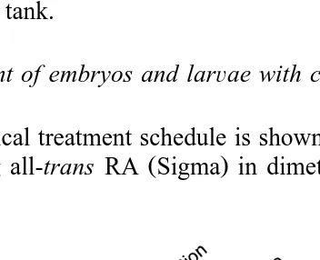
The chemical treatment schedule is shown in Fig. 1. RA stock solution was prepared
Ž . Ž . y3
by dissolving all-trans RA Sigma in dimethyl sulfoxide DMSO, Wako at 10 M.
Fig. 1. Experimental time diagram showing chemical treatment schedule. The upper diagram indicates the time
Ž .
Ž .
RA stock solution was dissolved in filtered seawater FSW . In the assay of RA effects
Ž . Ž .
at early embryonic stage RA-1 in Fig. 1 , shield embryos 26 h after fertilization were incubated with RA for 1 h, washed three times and further developed in FSW. In the
Ž . Ž
assay of RA effects at early larval stage RA-2 in Fig. 1 , newly hatched larvae 2.5 days
. Ž
after fertilization , corresponding the prim-5 stage of zebrafish development Kimmel et
.
al., 1995 , were cultured in the presence of RA for 40 h, and then kept in FSW.
y3 Ž .
Stock solution of 5=10 M disulfiram Wako was prepared in DMSO prior to
Ž
use, and dissolved in FSW. To test the effects of disulfiram at embryonic stage DF-1 in
. Ž
Fig. 1 , embryos were incubated with disulfiram from the 5-somite stage 30 h after
.
fertilization to the hatching period, and then washed and further developed in FSW. To
Ž .
assess the effects at early larval stage DF-2 in Fig. 1 , newly hatched larvae were incubated with disulfiram to open-mouth stage. For the control of RA and disulfiram treatments, embryos were treated with 0.1% DMSO.
X Ž . Ž .
2-2 -Dipyridyl Wako and azetidine-2-carboxylic acid Aldrich were directly dis-solved in FSW. Newly hatched larvae were developed in the presence of these chemicals
ŽDP and AC in Fig. 1 ..
In all experiments, embryos or larvae were cultured in 40 ml FSW containing the
Ž .
chemicals using plastic dishes 10 cm diameter at 178C. Two hundred embryos were put into a dish at the beginning of each incubation period, and each experiment was done in triplicate. At 6.5 days after fertilization when normal larvae developed to the open-mouth stage and start feeding, larvae were fixed in 4% paraformaldehyde in 10 mM phosphate buffered saline, pH 7.5. They were stained with Alcian-blue 8GX, dehydrated through a series of graded methanol and finally immersed in 80% glycerol,
Ž .
as previously described Suzuki and Kurokawa, 1996 .
3. Results
The pharyngeal and pectoral fin cartilages of untreated flounder larvae were
visual-Ž .
ized with Alcian-blue at the open-mouth stage 6.5 days after fertilization , and
Ž .
schematically reconstructed Fig. 2 . The treated larvae were fixed at day 6.5 and stained
Ž .
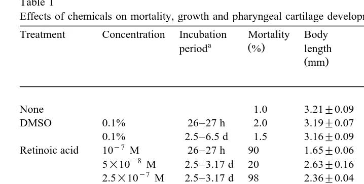
with Alcian-blue Fig. 3 . The mortality during this study and morphometric character-istics are indicated in Table 1.
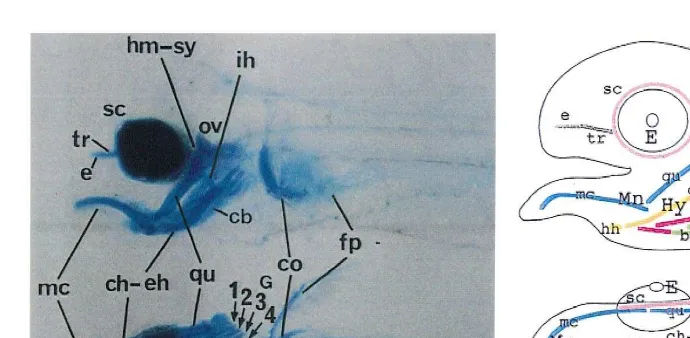
3.1. Cartilages in normal flounder larÕae
Cartilaginous skeletons formed in the mandible, hyoid and 1st–4th gill arches,
Ž .
Ž .
Fig. 2. Cartilaginous skeletons in normal flounder larvae at 6.5 days after fertilization the open-mouth stage .
Ž .A Alcian-blue staining to visualize cartilages. Upper is lateral view and lower is ventral view. Bar; 200mm.
Ž .B Schematic arrangement of cartilages. Cartilages of the same segment share the same color. Abbreviations: bb, basibranchial cartilage; cb, ceratobranchial cartilage; ch–eh, ceratohyal–epihyal cartilage; co, coracoid-scapula cartilage; e, ethmoid plate; E, eye; fp, fin plate cartilage; G, gill arch; hb, hypobranchial cartilage; hh, hypohyal cartilage; hm-sy, hyomandibular-sympletic cartilage; Hy, hyoid arch; ih, interhyal cartilage; mc, the Meckel’s cartilage; Mn, mandible arch; nt, notochord; Ov, otic vesicle; qu, quadrate cartilage; sc, sclerotic cartilage; tr, trabecula cartilage.
cartilages run anteroventrally from the ventral side of otic vesicles. The interhyal cartilage dorsally contacted with the hyomandibular-sympletic cartilages. The anterior ends of ceratohyal–epihyal cartilages were situated at the level of the rostral ends of the eyes. The ceratobranchial, hypobranchial and basibranchial cartilages formed in the gill arches. Trabeculae and ethmoid cartilages, which compose neurocranium, were visible in the lateral view of larvae. The trabecula cartilages extended anteriorly between the eyes from the anterior part of the otic vesicles. Coracoid-scapula and fin plate cartilages formed at the pectoral fins. Sclerotic cartilages can be seen surrounding the eyes.
3.2. Effects of RA
The phenotypes of malformations induced by RA in larvae exposed either at early embryonic or early larval stages were examined. The structure of pharyngeal and pectoral fin cartilages were not affected by 0.1% DMSO, vehicle of RA, at either stage. Gastrulating embryos were first incubated with 10y6, 10y7 and 10y8 M RA for 1 h at
the shield stage. At 10y6 M, all embryos died during the somite stage. At 10y7 M, 10%
of larvae were alive at 6.5 days post fertilization and two obvious malformations were observed in surviving larvae. The first malformation observed was that the Meckel’s
Ž .
visible so that their morphology could not be compared with that of normal embryos. At 10y8 M, no pharyngeal cartilage malformation and mortality was seen.
Ž .
When larvae were exposed to RA for 40 h from hatching period prim-5 stage , strong malformations occurred in mandible, hyoid and gill arches at a concentration of 5=10y8 M RA. The Meckel’s cartilages were reduced in size, to 37% the length of
Ž .
Table 1
Effects of chemicals on mortality, growth and pharyngeal cartilage development
Treatment Concentration Incubation Mortality Body Length of Length of
a Ž . b
period % length pharynx of the Meckel’s
Žmm. Žmm. cartilage
Retinoic acid 10 M 26–27 h 90 1.65"0.06
y8
Distance between the rostral end of the Meckel’s cartilage and the posterior end of the 4th gill ceratobranchial cartilage was measured.
more severe malformations could be observed in the survived larvae. The cartilages of all pharyngeal arches were reduced in size, and in particular the Meckel’s cartilage was
Fig. 3. Skeletal malformations induced in flounder larvae. Larvae were stained with Alcian-blue at 6.5 days
Ž . y7
after fertilization, when untreated embryos develop to the open-mouth stage. A Larva incubated with 10
Ž . Ž .
M RA for 1 h at shield stage. Note the absence of the Meckel’s cartilage, and forked cartilage arrow . B Larvae incubated with 5=10y8 M RA for 40 h from hatching. Note the truncated Meckel’s cartilage. The
Ž .
pharyngeal cartilages grew downwards, and the trabecula cartilage curved downwards, anteriorly. C Embryos incubated with 2.5=10y7 M RA for 40 h from hatching. Note truncated pharyngeal cartilages extending
Ž .
posteroventrally and absence of trabecula cartilage. The fin plate cartilage was bent. D Alignment of control
Ž . y6 Ž .
upper and treated larvae with 1=10 M disulfiram from the 5-somite to hatching middle and from
Ž .
hatching lower . Note truncated trunk and swollen notochord, but normal skeletons in the middle larvae, and
Ž . Ž .
abnormal cartilages in lower embryo. E, F Higher magnification of middle and lower embryos in D ,
Ž . Ž .
respectively. In E , the skeletons are normal. In F , the Meckel’s, ceratohyal–epihyal and ceratobranchial
Ž . Ž .
cartilages were bent arrowheads . The pectoral fin plate cartilages were deeply bent, in S-shape. G, H
y3 X Ž .
Embryos incubated with 2=10 M 2,2 -dipyridyl from hatching. In G , the Meckel’s cartilage is elongated,
Ž . Ž .
ceratohyal–epihyal and ceratobranchial cartilages are bent arrowheads . In H , the Meckel’s and trabecula
Ž . Ž .
cartilages were also bent arrowhead , and pectoral fin plate cartilages were infolded. I Embryos incubated with 5=10y3 M 2,2X
-dipyridyl from hatching. Note the irregular-shaped Meckel’s, ceratohyal–epihyal and
Ž . y3
sclerotic cartilages with bents, and infolded fin plate cartilages. J Embryos incubated with 1.7=10 M
Ž . Ž . Ž .
azetidine-2-carboxylic acid from hatching. Note truncated fin plate cartilages. In B , C and E–J , upper is
Ž . Ž . Ž . Ž . Ž .
Ž .
extremely truncated to about 6.5% of control embryos incubated with DMSO Fig. 3C . The hyoid and gill arches extended posteroventrally, indicating that the growth direction of pharyngeal arches was severely affected by RA. It was also remarkable that the trabecula cartilage did not form. The pectoral fins were bent. The survived larvae in all experimental groups treated with RA showed identical malformations.
3.3. Effects of disulfiram
The effects of disulfiram on cartilage formation were assessed by exposing flounder to this chemical at embryonic and early larvae stages. In the larvae treated with 1=10y6 M disulfiram from the 5-somite to hatching period, no malformation of
pharyngeal cartilages was seen in terms of morphological or morphometric
character-Ž .
istics Fig. 3D,E . Contrast to the normal development of pharyngeal area, the trunk
Ž .
became severely shortened, making the body length 67% of control embryos Fig. 3D . Remarkably, the notochord was irregularly shaped. Thus, disulfiram treatment at embry-onic stage did not affect the pharyngeal skeletons, though it induced strong abnormality in the trunk region.
In the larvae incubated with 1=10y6 M disulfiram from the hatching time, pharyngeal cartilages were reduced in size and the cartilages of each arch showed
Ž .
malformations Fig. 3D,F . The Meckel’s cartilages were 63% the length of control embryos, and were irregularly bent. The ceratohyal–epihyal and epibranchial cartilages were sharply bent. The fin plate cartilages were also deeply bent, appearing S-shape as seen from the ventral side, when treated after the hatching time, whereas they developed
Ž .
normally when treated at embryonic stage Fig. 3E,F . Such malformation could be seen in all survived larvae. Thus, it appears that disulfiram exerts teratogenic effects towards pharyngeal and pectoral fin skeletons and notochord in a stage-specific manner.
3.4. Effects of 2,2X-dipyridyl
In the larvae incubated with 2=10y3 M 2,2X
-dipyridyl from hatching, strong malformations occurred in the pharyngeal arches. In the mandible arches, the Meckel’s
Ž
cartilages elongated 1.2-fold in length, compared with those of control embryos Fig.
.
3G . In the hyoid and gill arches of these embryos, ceratohyal–epihyal and cerato-branchial cartilages were bent into two pieces. In 10% of the embryos, the Meckel’s
Ž .
cartilages was also bent, showing more severe malformations Fig. 3H . In these embryos, fin plate cartilages were also bent, showing S-shape in ventral view.
X Ž y3 .
In the embryos incubated with higher concentration of 2,2 -dipyridyl 5=10 M ,
Ž .
pharyngeal cartilages were reduced in size Fig. 3I ; e.g., the length of Meckel’s cartilage was approximately half of the control embryos. Furthermore, irregular forma-tion of the pharyngeal and sclerotic cartilages was apparent, due to repeated bending. At this concentration, the fin plate cartilages of all survived embryos were bent, appearing S-shaped.
3.5. Effects of azetidine-2-carboxylic acid
above 3.4=10y3 M. In the embryos incubated with 1.7=10y3 M
azetidine-2-carbo-Ž .
xylic acid, the pharyngeal cartilages were reduced in size Fig. 3J ; e.g., the Meckel’s cartilages were 78% the length of those of untreated embryos. The pectoral fin plate cartilages were also reduced in size. Except for truncation, remarkable malformation, such as bending, did not occur.
4. Discussion
We selected several chemicals as candidates to affect skeletal development, and analyzed their teratogenic effects on the cartilaginous skeletons of pharyngeal arch and pectoral fin of flounder larvae. Several phenotypes could be characterized on the malformations induced by RA, disulfiram and 2,2X-dipyridyl.
4.1. Teratogenic effects of RA
Pulse RA-treatment of gastrulating embryos at shield stage caused strong malforma-tion in pharyngeal skeletons with two noticeable phenotypes; absence of the Meckel’s cartilage in the mandibular arches; fusion of cartilage components in mandibular and hyoid arches. Zebrafish incubated with RA at the same stage also shows identical
Ž
malformations with those observed in flounder Alexandre et al., 1996; Ellies et al.,
.
1997 . On the other hand, RA given after the hatching period disorganized the growth direction of pharyngeal cartilages. Thus, it was indicated that RA induces different phenotypes of malformations in pharyngeal skeleton, depending on developmental stages.
In vertebrates, a series of Hox genes lining on four clusters are expressed in domains, of which anterior borders form a hierarchy of 3X™5X restricted pattern at the hindbrain
Ž .
and pharyngeal area, determining regional identity of their segments Hunt et al., 1991 . Experimentally given RA shifts anteriorly the anterior borders of Hox expression domains in the central nervous system, causing homeotic transformations in brain
ŽMorriss-Kay et al., 1991 . We observed that, in flounder, pulse-treatment with RA at.
the shield stage anteriorly expands the Hoxd-4 expression domain at pharyngeal area as
Ž .
well as central nervous system Suzuki et al., 1998 . Therefore, it is speculated that the absence of the Meckel’s cartilage is the phenotype of a homeotic transformation of pharyngeal arches, in which the identity of mandible arch was transformed to that of hyoid arch. RA also affects the migration passway of cranial neural crest cells, in such a way that two populations of cartilage precursor cells that normally enter the mandible
Ž .
and hyoid arches are mixed to form fused arches Lee et al., 1995 . Such mixture of cartilage precursor cells during migration probably causes the fusion of cartilage components of mandibular and hyoid arches.
In flounder, the cartilage precursor cells originating from the cranial neural crest form cell aggregates in the pharyngeal arches at the hatching period, and then develop to cartilaginous skeletons by the first feeding. The trabeculae cartilage, which also
origi-Ž
nates from the cranial neural crest and forms a compartment of neurocranium Langille
.
Ž .
of cartilage precursor cells is extremely active Suzuki and Kurokawa, 1996 . Our data indicate that RA given at this stage causes directional shift and suppression of the cell growth. It was recently demonstrated that shh, a polarizing signal, is expressed at the pharyngeal endoderm in flounder larvae at hatching and exogenous RA suppresses the
Ž .
shh expression Suzuki et al., 1999 . These results suggest that RA given after hatching induces down-regulation of shh expression at pharyngeal area, disordering the pattern of pharyngeal skeletons.
Exogenous RA exerts a strong teratogenic effects toward pharyngeal skeleton, as described. However, Hox and shh genomic genes possess RA response element at their
X
Ž
5 upstream region, and require RA for initiation of transcription Ogura and Evans,
.
1995; Chang et al., 1997 . So, a certain level of endogenous RA is essential for normal skeletal development. In Xenopus eggs, precursors of RA are stocked in the yolk, in the
Ž .
form of a complex with vitellogenin by Schiff-base linkage Irie et al., 1991 . In zebrafish, a low level of RA and an excess of retinal, the precursor of RA, exist even in
Ž .
8-cell embryos Costaridis et al., 1996 . We exposed flounder embryos to disulfiram, which inhibits the conversion of retinal to RA, expecting that disulfiram affects the pharyngeal arch development if RA de novo converted from retinal functions in the initiation of Hox and shh expressions. Contrary to our expectation, disulfiram given at embryonic stage exerted no effect on the skeletal formation. This result suggests that RA stored in egg directly functions in pharyngeal arch development. Since 10y8 M RA did not affect the pharyngeal arch development, adequate concentration of RA stocked in eggs for normal flounder development is supposed to be 10y8 M or less.
4.2. Teratogenic effects of inhibitors of cartilage synthesis
Type II collagen and chondroitin sulfate, a kind of proteoglycans, are the main components of the cartilage matrix. Type II collagen fibrils contribute to the structural
Ž .
strength of cartilage Sandell et al., 1992 . Collagen molecules includes ;20% proline, about half of which are enzymatically hydroxylated to stabilize the molecular structure. The prolyl hydroxylase which catalyzes the hydroxylation requires Fe2q and ascorbic
Ž .
acid for enzymatic activity Tuderman et al., 1977; Kleinman et al., 1981 . Due to such properties of collagen synthesis, there are two types of inhibitors against the synthesis;
Ž .
proline analogues such as azetidine-2-carboxylic acid in mouse Alescio, 1973 ; and
2q X Ž .
Fe chelating agents such as 2,2 -dipyridyl Hurych and Chvapil, 1965 . Among these two chemicals, 2,2X-dipyridyl exerted a powerful teratogenic effect towards cartilaginous skeletons of pharyngeal arches and pectoral fin of flounder. It is demonstrated that azetidine-2-carboxylic acid affects in vitro organogenesis by suppressing collagen
Ž .
synthesis Alescio, 1973 . That this chemical did not severely affect the skeletal formation probably means that endogenous proline compensates for the effect of azetidine-2-carboxylic acid in embryos.
The most noticeable phenotypes of malformation induced by 2,2X-dipyridyl was the
Ž .
bending distortion of the cartilaginous skeletons both in pharyngeal arches and pectoral
Ž
fin. In the list of zebrafish mutants with malformed pharyngeal skeletons Neuhauss et
. m 452
deficit in mechanical stability of the cartilage, caused by mutation. Referring this data, it is supposed that deposition of unstable under-hydroxylated collagen by 2,2X-dipyridyl reduces the mechanical stability of the cartilage, causing bent of cartilaginous skeletons. That high concentration of 2,2X-dipyridyl severely truncated the cartilages can be explained by strong inhibition of collagen synthesis.
Bending cartilaginous skeletons also formed in larvae incubated with disulfiram after hatching period. RA also functions in a regulatory network that controls resorption and
Ž .
growth of cartilage by regulating the proteoglycan synthesis Morales 1994 . It is speculated that RA converted from retinal works in the proteoglycan synthesis in larvae and that disulfiram affects the proteoglycan synthesis through inhibition of RA conver-sion, making cartilaginous skeleton mechanically unstable. Alternatively, disulfiram might exert side effect on the enzymatic activity of prolyl hydroxylase.
Our data indicate that abnormality of cartilage synthesis results in the bending of cartilaginous skeletons in pharyngeal arches and pectoral fin. It is almost impossible that larvae produced for aquaculture are exposed to 2,2X-dipyridyl or disulfiram. Fragile collagen is formed by ascorbic acid deficiency, which also inhibits the hydroxylation of
Ž .
collagen as disulfiram Peterkofsky, 1976 . If bending of cartilaginous skeleton occurs in fingerlings, one possible cause is deficiency in vitamin C.
Not much attention has been paid to the abnormality of cartilaginous skeletons of cultured larvae. This is probably, in part, because larvae with strongly malformed pharyngeal skeletons cannot survive due to incapability of feeding. Furthermore, since development of pharyngeal skeletons is closely related with brain development, their strong malformation probably occurs with high mortality, as observed here in RA tests. We think that skeletal structure should be diagnosed when high mortality occurs in cultured larvae and that our data on malformation help such diagnose.
Acknowledgements
Ž .
This study was supported by a Bio-Media Project BMP-97-II-2-6 sponsored by the Ministry of Agriculture, Forestry and Fisheries of Japan.
References
Akimenko, M.-A., Ekker, M., 1995. Anterior duplication of the Sonic hedgehog expression pattern in the pectoral fin buds of zebrafish treated with retinoic acid. Dev. Biol. 170, 243–247.
Alescio, T., 1973. Effects of a proline analogue, azetidine-2-carboxylic acid, on the morphogenesis in vitro of mouse embryonic lung. J. Embryol. Exp. Morphol. 29, 439–451.
Alexandre, D., Clarke, J.D.W., Oxtoby, E., Yan, Y.-L., Jowett, T., Holder, N., 1996. Ectopic expression of
Hoxa-1 in the zebrafish alters the fate of the mandibular arch neural crest and phenocopies a retinoic
acid-induced phenotype. Development 122, 735–746.
Bisbal, G.A., Bengston, D.A., 1995. Development of the digestive tract in larval summer flounder. J. Fish Biol. 47, 277–291.
Ž .
Chang, E.-E., Blader, P., Fischer, N., Ingham, P.W., Strahle, U., 1997. Axial HNF3¨ b and retinoic acid receptors are regulators of the zebrafish Sonic hedgehog promoter. EMBO J. 16, 3955–3964.
Costaridis, P., Horton, C., Zeitlinger, J., Holder, N., Maden, M., 1996. Endogenous retinoids in the zebrafish embryo and adult. Dev. Dyn. 205, 41–51.
Ellies, D.L., Langille, R.M., Martin, C.C., Akimenko, M.-A., Ekker, M., 1997. Specific craniofacial cartilage dysmorphogenesis coincides with a loss of dlx gene expression in retinoic acid-treated zebrafish embryos. Mech. Dev. 61, 23–36.
Ž .
Epperlein, H.H., Lehmann, R., 1975. The ectomesenchymal–endodermal interaction system EEIS of
Triturus alpestris in tissue culture: 2. Observations on the differentiation of visceral cartilage.
Differentia-tion 4, 159–174.
Helms, J.A., Kim, C.H., Hu, D., Minkoff, R., Thaller, C., Eichele, G., 1997. Sonic hedgehog participates in craniofacial morphogenesis and is down-regulated by teratogenic doses of retinoic acid. Dev. Biol. 187, 25–35.
Hunt, P., Gulisano, M., Cook, M., Sham, M.-H., Faiella, A., Wilkinson, D., Boncinelli, E., Krumlauf, R., 1991. A distinct Hox code for the branchial region of the vertebrate head. Nature 353, 861–864. Hurych, J., Chvapil, M., 1965. Influence of chelating agents on the biosynthesis of collagen. Biochim.
Biophys. Acta 97, 361–363.
Irie, T., Azuma, M., Seki, T., 1991. The retinal and 3-dehydroretinal in Xenopus larÕis eggs are bound to lipovitellin 1 by a Schiff base linkage. Zool. Sci. 8, 855–863.
Johnston, M.C., Bronsky, P.T., 1995. Prenatal craniofacial development: new insights on normal and abnormal mechanisms. Crit. Rev. Oral Biol. Med. 6, 25–79.
Kimmel, C.B., Ballard, W.W., Kimmel, S.R., Ullmann, B., Schilling, T.F., 1995. Stages of embryonic development of the zebrafish. Dev. Dyn. 203, 253–310.
Kleinman, H.K., Klebe, R.J., Martin, G.R., 1981. Role of collagenous matrices in the adhesion and growth of cells. J. Cell Biol. 88, 473–485.
Kontges, G., Lumsden, A., 1996. Rhombencephalic neural crest segmentation is preserved throughout¨
craniofacial ontogeny. Development 122, 3229–3242.
Langille, R.M., Hall, B.H., 1988. Role of the neural crest in development of the cartilaginous cranial and
Ž .
visceral skeleton of the medaka, Oryzias latipes Teleostei . Anat. Embryol. 177, 297–305.
Laufer, E., Nelson, C.E., Johnson, R.L., Morgan, B.A., Tabin, C., 1994. Sonic hedgehog and Fgf-4 act through a signaling cascade and feedback loop to integrate growth and patterning of the developing limb bud. Cell 79, 993–1003.
Lee, Y.M., Osumi-Yamashita, N., Ninomiya, Y., Moon, C.K., Eriksson, U., Eto, K., 1995. Retinoic acid stage-dependently alters the migration pattern and identity of hindbrain neural crest cells. Development 121, 825–837.
Morales, T.I., 1994. Transforming growth factor-b and insulin-like growth factor-1 restore proteoglycan metabolism of bovine articular cartilage after depletion by retinoic acid. Arch. Biochem. Biophys. 315, 190–198.
Morriss-Kay, G.M., Murphy, P., Hill, R.E., Davidson, D.R., 1991. Effects of retinoic acid excess on expression of Hox-2.9 and Krox-20 and on morphological segmentation in the hindbrain of mouse embryos. EMBO. J. 10, 2985–2995.
Neuhauss, S.C.F., Solnica-Krezel, L., Schier, A.F., Zwartkruis, F., Stemple, D.L., Malicki, J., Abdelilah, S., Stainier, D.Y.R., Driever, W., 1996. Mutants affecting craniofacial development in zebrafish. Development 123, 357–367.
Noden, D.M., 1983. The role of neural crest in patterning avian cranial skeletal, connective and muscle tissues. Dev. Biol. 96, 144–165.
Ogura, T., Evans, R.M., 1995. A retinoic acid-triggered cascade of HOXB1 gene activation. Proc. Natl. Acad. Sci. U.S.A. 92, 387–391.
Peterkofsky, B., 1976. The effect of ascorbic acid on collagen polypeptide-synthesis and proline hydroxylation during the growth of cultured fibroblasts. Arch. Biochem. Biophys. 152, 318–328.
Rintala, M., Metsaranta, M., Saamanen, A.-M., Vuorio, E., Ronning, O., 1997. Abnormal craniofacial growth¨ ¨¨ ¨ ¨
Sadaghiani, B., Vielkind, J.R., 1990. Distribution and migration pathways of HNK-1-immunoreactive neural crest cells in teleost fish embryos. Development 110, 197–209.
Sandell, L.J., Morris, N., Robbins, J.R., Goldring, M.B., 1992. Alternatively spliced type II procollagen mRNAs define distinct populations of cells during vertebral development; differential expression of amino-propeptide. J. Cell Biol. 114, 1307–1319.
Schilling, T.F., Kimmel, C.B., 1994. Segment and cell type lineage restrictions during pharyngeal arch development in the zebrafish embryo. Development 120, 483–494.
Sordino, P., van der Hoeven, F., Duboule, D., 1995. Hox gene expression in teleost fins and the origin of vertebrate digits. Nature 375, 678–681.
Suzuki, T., Kurokawa, T., 1996. Functional analyses of FGF during pharyngeal cartilage development of
Ž .
flounder Paralichthys oliÕaceus embryo. Zool. Sci. 13, 883–891.
Suzuki, T., Oohara, I., Tadahide, T., 1998. Hoxd-4 expression during pharyngeal arch development in
Ž .
flounder Paralichthys oliÕaceus embryo sand effects of retinoic acid on expression. Zool. Sci. 15, 57–67.
Suzuki, T., Oohara, I., Tadahide, T., 1999. Retinoic acid given at late embryonic stage depresses Sonic
hedgehog and Hoxd-4 expression in the pharyngeal area and induces skeletal malformation in flounder
ŽParalichthys oliÕaceus embryos. Dev. Growth Differ. 41, 143–152..
Tuderman, L., Myllyla, R., Kivirikko, K.I., 1977. Mechanism of the prolyl hydroxylase reaction: 1. Role of¨
co-substance. Eur. J. Biochem. 80, 341–348.
Vikkula, M., Metasranta, M., Ala-Kokko, L., 1994. Type II collagen mutations in rare and common cartilage¨
diseases. Ann. Med. 26, 107–114.