B-1
SCALE MORPHOLOGY OF CUNING FISH (
Caesio cuningBloch,1791)
(CAESIONIDAE) USING DEKSTOP SCANNING ELECTRON MICROSCOPE
Abdul Razak
Departement Biology, Mathematics and Natural Sciences Faculty, State University of Padang, Prof. Hamka Street, Air Tawar, West Sumatera
Abstract
Scale morphology is importance in fish taxonomy today. Scale ultrastructure of Cuning fish (Caesio cuning) from Padang Waters, Pasir Kandang, Padang West Sumatera was studied using desktop scanning electron microscope (D-SEM Phenom Pro-X) since 2 January-27 February 2014. Scale as object research from below dorsal fins and lateral line region were removed. Scales was cleaned by water and dried naturally were prepare for the dekstop scanning electron microscope technique. The type scales of Cuning fish is ctenoid. Variations are found in components of scale morphology of this fish in diffrent body parts (below dorsal fins and lateral line position). Focus observation is located anterior, and central of the scales. Radii are found only on the anterior part of scale. Primary radii is more tertiary radii. These radii is divided of the anterior part of scale into few region and thus the scale have been sectioned. Lobes of anterior is diffrent . Inter circular is without granules and like straight of curve line is observed in some circuli. Lepidont form is diffrent, when we observed in below dorsal ins and lateral line position. Arrangement cteni of the scales in this fish could be used important for taxonomic characters.
Key words : scale morphology, Caesio cuning, taxonomy and dekstop scanning electron microscope
INTRODUCTION
The Cuning Fish or The Yellowtail Fusilier (Caesio cuning, Bloch,1791) can be recognised by its colouration. This species occurs in tropical marine waters of the Indo-West Pacific. This fish is usually seen swimming in midwater where it feeds on zooplankton.
The body of Cuning Fish have color with Upper body if not yellow, grayish blue; lower sides and belly white or pinkish. This fish, we found on coastal areas, usually over rocky and coral reefs. The Cuning Fish swim make up schools in midwater and feeds on zooplankton. The Cuning Fish is oviparous, with numerous, small pelagic eggs.
This fish caught mostly by fish traps in western Thailand and Malaysia. In the Gulf of Thailand, this fish caught by a variety of methods including drive-in nets, fish traps and gill nets in Indonesia, the Philippines and Papua New Guinea (www.fishbase.org, 2014)
Furthermore, for confirmation this species, Cuning Fish have first spine dorsal and spine anal fish is short (Carpenter, 1987). As part of body fish, scale is derivative of dermal. Scales are the overlapping series of hard plates that cover a fish's body. Scales can protect the body
This method was introduce by Agassiz (1833-1834). The fishes is classified into for groups based on scale morpology (Jawad and Al Jufaili, 2007 in Esmaeili, 2009).
During late 19th century until now, scale is observed by SEM, information about scale in systematics increased significantly. Detailed structure of the fish scale can be important and support in identification of fish to several groups and species level (Esmaeili et al, 2007). In Indonesia, research about scale of fishes is seldom.We have problems, especially, tools for observed like scanning electron microscopy (SEM). With SEM, we can detail observe scale morphology of Cuning fish. Today, the Cuning Fishes have been harmless condition by human exploitation.We need some information for conserve this marine fishes.
Correct identification of fish is essential process in study taxonomy. In other side, essential to formulate conservation and management for fish commercial / I think, this study about scale morphology can develop for fish identification combine with information technology on the future. This research first step for that. Today, many technique has been scanning electron microscope (D-SEM Phenom Pro-X). The observation focused on central, anterior and posterior side of scale morphology.
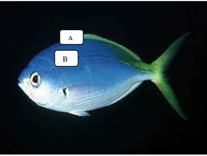
Figure 1. Caesio cuning (Bloch 1791) (A) scale below dorsal fins (B) scale from lateral line position (Photo courtesy of John Randall http://reefkeeping.com/issues/2005-01)
A
B-3
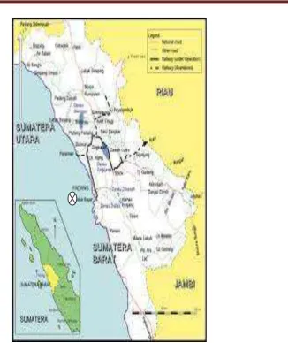
Figure 2. Padang waters, location of sample (http://www.google.com/imgres)
RESULT AND DISCUSSION
We put scales from dorsal below and lateral line position have reason. According Esmaeli (2007), the scales from below dorsal fin and lateral line position is largest better than on the other parts of body fish. For research, the scales is important the same morphological
proportion. The both location have been designated as “key scale”.
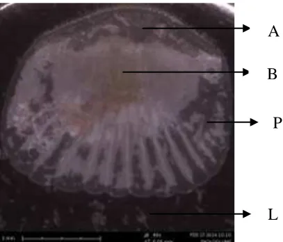
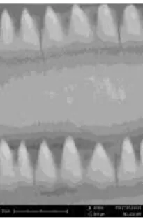
This is element of scale from dorsal fin below (fig 3). The scale element is divided into anterior position (A), posterior position or caudal (P), lateral field (L). Anterior field is embedded in the skin. The types scale of The Cuning Fish is ctenoid. We found ctenii in anterior part. With photos SEM seen element of the scale of the Cuning Fish.
In Anterior part of scale from below dorsal fins, we fund focus. The focus is part of the scale that developed during ontogenesis. The focus is divided two part, first parta we call cephalic of focus and caudal of focus (Esmaeili, et al.,2007).
posterior parts of scale below dorsal fins. Lepidont is important structures for support species specifity in classification process. The taxa have different lepidont Kaur and Dua (2004). The shape lepidonts in this research is interesting. Lepidont of scale below dorsal fins is different which on scale on lateral line position (see fig 11 and fig 17).
.
Figure 3. Anterior position (A), (B) focus, posterior position or caudal (P), lateral field (L).
Figure 4. Cteni of scale of below dorsal fins the Cuning Fish
Figure 5. Posteriot parts of scale below dorsal fins Cuning Fish
A
B
P
B-5
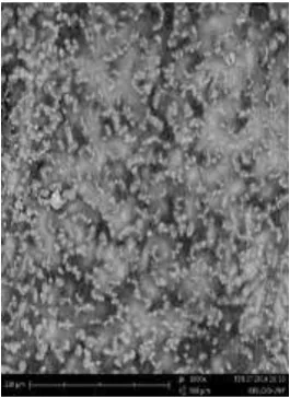
Figure 6. Several Radii and Lepidont
Figure 7. Two Radii of the Cuning Fish
Figure 8. Lepidont interrupted tranversal radii of scale below dorsal fins of the Cuning Fish
Figure 10. Lepidont of of the Cuning Fish (3000 x)
Figure 11. Lepidont of scale on below dorsal fins of the Cuning Fish (5000x)
The next of the result is scale from lateral line position. This part like scale on the dorsal fin below. But in this scale position we found mucous pore. Few mucous found in focus region. The same has been reported by Esmaeili et al. (2007). The scale of the fish is divided into anterior position (A), posterior position or caudal (P), lateral field (L). Anterior field is embedded in the skin.
B-7
Figure 13. Ctenii as part of anterior of the scale in Lateral Line Position.
Figure 14. Several Radii and Circuli
Figure 16. Mucous Pore of Scale on Lateral Line Position (5000 x)
Figure 17. Lepidont of Scale on Lateral Position (5000x)
Figure 18. Lepidont of Scale on Lateral Line Position (10000x)
In Anterior part of scale from below dorsal fins, we fund focus. The focus is part of the scale that developed during ontogenesis. The focus is divided two part, first parta we call cephalic of focus and caudal of focus (Esmaeili, et al.,2007).
B-9 according species.
Next, the circuli are interrupted by tranverse radii. The circuli is line growth start appearing. The circuli found on anterior part and there are not found on posterior part. The circuli is different and overcrowded in anterior parts widely separated in lateral parts. The circuli is formed by secreted calcium salts from skin and their deposition on the scale. The arrangement of circuli is relevant with shape of scale (Esmaeili, et al.,2007). According Kaur and Dua (2004) the arrangement of circuli around focus is essentials for species specifity.
We are going to compare the result and Amor et al., (2010) research about scale of Yellowsstriped Goat Fish (Upeneus vittatus, Fosrkal, 1775), he found variation in scales between sexes. The typical of scales is ctenoid. Amor et al., (2010) did not found lepidont and mucous pore in the scale female and male of Yellowsstriped Goat Fish.
REFERENCES
Carpenter, K.E., 1987. Revision of the Indo-Pacific fish family Caesionidae (Lutjanoidea), with descriptions of five new species. Indo-Pac. Fish. (15):56 p.
http://www.fishbase.org/summary/Caesio-cuning.html. diakses : 10 April 2014 jam 11.00 WIB