P-ISSN.2503-0817, E-ISSN.2503-0825
CrossMark
Abstract
Objective: To describe and compare the procedure of superior labial frenum frenectomies with conventional techniques and incisions below the clamp technique.
Methods: Two female patients came to the Department of Periodonsia, Dental Hospital Hasanuddin University, to go through frenectomies. The first patient was 28 years old, with labialis superior frenulum reaching the attached gingiva, gingival recession 1-2 mm with calculus deposits, and she was referred to undergo frenectomy with incision below the clamp. The second patient was a 15-year-old; she presented with labialis superior frenulum extending up to palatine papilla, central diastema. She was also referred to undergo frenectomy with conventional techniques.
Results: The conventional technique is carried out by engaging the frenum with a haemostat that is inserted into the depth of the vestibule, and incisions are placed on the upper- and the under-surface of haemostat, which is then followed by suturing the wound and covering the wound with periodontal pack. Incision below the clamp technique is done by placing a haemostat in adjacent position and parallel to the lip mucosa; incision carried out below the clamp, then followed by suturing at the mucolabial fold and periodontal pack. Conclusions: Patients were very satisfied with the results that were achieved. The technique of using incision below the clamp is a sound alternative treatment with good aesthetics and involves much lesser bleeding during frenectomies which involves the use of a scalpel.
Keywords: Incision below the clamp, Frenum, Frenectomy, Scalpel, Bleeding
Cite this Article: Thahir H, Djais AI, Wendy S, Achmad MH, Akbar FH. 2018. Management of maxillary labial frenum and comparison between conventional techniques and incision-belowthe-clamp technique (case report). Journal of Dentomaxillofacial Science 3(1): 61-66. DOI:10.15562/ jdmfs.v3i1.634
Management of maxillary labial frenum and
comparison between conventional techniques and
incision-below the-clamp technique
:
case report
Hasanuddin Thahir1, Arni I. Djais1, Shek Wendy1, Muhammad H. Achmad2, Fuad H. Akbar3*
Introduction
Recently, more people have begun to realize the importance of aesthetic appearance of the oral cavity, considering the main reason that better aesthetics would enhance their appearance and personality and self-confidence when smiling. A great smile is attributed to the presence of variety of factors: harmonization of shape, location, and sizes of teeth in relation to alveolar bones and gingiva tissue that is a part of the oral cavity. Frenulum attachment inside the cavity is an important factor that affects the appearance of smile, because this will determine the shape of lips and teeth feasibility.1
Frenulum is a tiny fold consisting mucosal membranes, fibrous tissues, and muscle fibres that attach the inner lips or cheek to the alveolaris process, gingiva, and periosteum. It stabilizes the movement of lips or cheek and tongue. Generally, the oral cavity is made up of the following: labialis, buccalis, and lingualis frenulums. Labialis frenulum is divided into superior labialis frenulum of the upper lip and the inferior labialis frenulum of the lower lip. Superior labialis frenulum is the residual embryological struc-ture that connects upper labial tubercles to the pala-tine papilla and shape of triangle.2
Frenulum attachment inside the oral cavity varies; therefore, it requires special attention during the observation of oral cavity. Normal frenulums are attached apically on the free margin gingiva and ends on the mucogingival junction and in some cases approach gingiva margin (abnormal). This
1Departement of Periodontics,
Faculty of Dentistry, Hasanuddin University, Makassar, Indonesia
2Department of Pediatric, Faculty
of Dentistry, Hasanuddin Univer-sity, Makassar, Indonesia
3Department of Dental Public
Health, Faculty of Dentistry, Hasanuddin University, Makassar, Indonesia
*Corresponding to: Fuad H. Akbar,
Department of Dental Public Health, Faculty of Dentistry, Hasanuddin University, Makassar, Indonesia [email protected]
Received: 24October 2017
Revised: 22 October 2017 Accepted: 15November 2017 Available online: 1 April 2018
abnormal frenulum attachment can occur when development of teeth and jaws not followed by the apical migration of the attachment. Abnormal frenulum attachment can be detected visually by pulling the upper lip to observe the movement of papilla edge or by performing blanch test, in which upper lips are lifted and held until the area becomes
ischaemic and turns pale.3
Placek had classified frenulum attachment into four types.4,5
Attachment of labialis frenulum might induce pathologic issues and create complications involving periodontological tissues from gingivitis, gingiva recess and even central diastema. High superior labialis frenulum attachment may create an upward pull of healthy gingiva and prevent dental cleansing, and thus making it prone for plaque accumulation and gingivitis, and this will develop into sulcus and pocket, and eventually advanced periodontal tissues will develop. In addition, this will also lead to local gingival recess, extreme sepa-ration or gaps in central incisive teeth and will affect patient’s psychological condition. Abnormal frenu-lum attachment can also disturb tooth prostheses condition and hinder teeth movement in ortho-dontic care to manage central diastema and relapse after orthodontic management; furthermore, it will influence tissue healing after periodontal care.6
The successful of periodontal therapy is dependent on proper diagnosis, proper case
selection and patient cooperation.7
Abnor-mal superior labialis frenulum can be treated by radical exicision of frenulum, includes its attachment to the bones belows (frenectomy) and partial excision to correct the abnormal attachment (frenetomi). Frenektomi or frene-tomi procedures generally can be performed
by using scalpel, electrosurgery, or laser.8
Conventional technique frenectomy by using scalpel is the most generic and common technique used, but have high risk of massive hemmorage. Generally, frenectomy procedures
may create w o u n d t h a t expanded in
diamond shape because pull of lips
muscula-tures and triggers bleeding from open
capillaries. This i s s u e i n f l i c t e d e f f o r t t o minimalize bleeding especially in usage of scalpel, b y r e p l a c i n g w i t h electrosurgery and laser, or can be performed with incision m o d i f i c a t i o n o f available surgery tech-niques,9,10 one of them is incision below the
clamp.11,12 This case report aims to describe
the incision below the clamp technique and compare it with conventional technique that have been utilized since long.
Case Report
First PatientA 25 years old woman was presented at Depart-ment of Periodontal dental Hospital Hasanuddin University with complaint of tenderness on region of front teeth, she had difficulties in cleaning those teeth region.
63 Journal of Dentomaxillofacial Science ( ) April 2018; 3(1): 61-66 | doi: 10.15562/jdmfs.v3i1.63
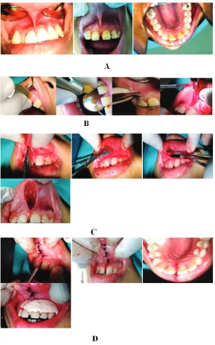
This complaint was felt since 1 year ago and gets more disturbing 1 month ago, she never went to dentist before. Patient stated that she had never had any systemic diseases. Physical examination showed small inflammations and recesses in gingiva in the labial area of two central incisive, wide, and tall frenulum expanded to the gingiva region and was named as gingival classification. Based on patient’s complaints and physical examination, it was decided that the most appropriate management of therapy would be frenectomy using scalpel by making an incision below the clamp. The whole procedure was explained to patient, and the patient had signed her consent to undergo the planned program figure 2A; the figure shows high frenulum attachment with gingival classification, with recess gingiva measuring 1 mm in length on tooth 11, and 2 mm on tooth 12, labial view).
Second Patient
A 15-years-old teenage girl came to the orthodontic outpatient clinic, Hasanuddin University Teaching Hospital, in order to correct teeth formation. Patient complained of gap between her front tooth in the upper jaw. This condition had made her lose her self-confidence. Even though she had this teeth formation since childhood, up until her visit that day to the clinic she had never consulted and sought intervention from a professional dentist. Her parents stated that she had never had any systemic disease or allergies to any substance. The girl was referred to periodontics department to undergo further observation and care to study specifically the periodontal tissue. From the physical examination, it was found that the central diastema with
Figure 3 One week post-frenectomy and suture removal, and one month post-frenectomy
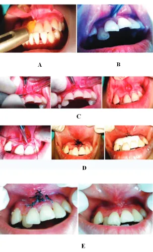
frenu-lum had extended to the gingiva region, causing enlargement of the palatal papillary incisiva. Blanch test was positive. The therapy management for the patient was to use the conventional technique of frenectomy using scalpel. The whole procedure was explained to the patient and her parents; the patient had signed the consent form to undergo the treatment programme figure 2A; clinical features before carrying out frenectomy and high frenulum attachment with papilla penetrating classification and midline diastema).
Conventional Technique
During her first visit, the patient underwent initial therapy with dental hygiene education (DHE) and scaling and root planning. A week later, she came for her second visit and observation, during this visit frenectomy procedure was performed conven-tionally using a scalpel. Surgery-site disinfection carried out by the application of Povidone-iodine solution and local anaesthetics infiltration on the area surrounding the superior frenulum labialis and palatal extension of superior frenulum labialis see figure 2A.
In the next step, conventional frenectomy was performed by using scalpel. The haemostat was placed in the innermost part of vestibulum, and incision was performed over and above the haemo-stat with blade no.15 figure 2B. Blunt dissection was performed by using scissors to release fibrous and epithelial attachment on the excised area figure 2B. Suturing at the base of the vestibulum and lips mucosal was performed using silk no. 5-0, with the expectation that the wound would not spread out beyond the incision area and to curtail excessive bleeding figure 2C. The surgery was performed further to make a wide incision on the extension of superior labial frenulum in the palatal region, and fibrous tissues bony attachment was released by raspatorium figure 2D. This step was followed by irrigation procedure; irrigation was performed by using saline solution followed by periodontal pack placement figure 2D.
After the surgery, patient was given the follow-ing medications: clindamycin, 500 mg, for 10 days; Mefenamic acid, 500 mg, again for 10 days; and Minosep mouthwash for 2 weeks. The patient was instructed to maintain dental hygiene and adhere to post-surgery care measures, such as avoiding hot drinks and solid, rough, and sticky food. She was also advised to avoid use of mouthwash the first day following surgery and was also told to follow a soft food diet for up to 2 days. The patient was asked to visit follow-up one week after the procedure in order to evaluate the wound-healing and removal of sutures, with further requirement that she should visit the one month thereafter; during the second
visit, clinical examination revealed the presence of longitudinal scars and residual fibrous tissues in the labial region figure 3.
Incision Below the Clamp (IBC) Technique
Initial care plan included the following steps: dental hygiene education, scaling, and root planning before carrying out the frenectomy procedure. A week later, the patient underwent frenectomy procedure that involved the use of scalpel, a conven-tional method. Extra- and intra-oral disinfection was performed by using Povidone-iodine solution followed by the infiltration of local anaesthetics on the left and right sides of superior labialis in the frenulum labialis region figure 4A.
The next procedure carried out was frenectomy by using scalpel as per conventional IBC technique. IBC was carried out by placing the haemostat on the innermost region of vestibulum and positioned near and parallel to lips mucosal area; incision was performed beneath the haemostat using blade no.15 figure 4B and figure 4C. This step was followed by irrigation with saline solution. Immediate suturing was performed on the base of the vestibulum and lips mucosal area with silk thread no. 5-0. This step was followed by periodontal pack placement figure 4D.
After the surgery, the patient was given the following medications: clindamycin, 500 mg, for 10 days; Mefenamic acid, 500 mg, for 10 days; and Minosep mouthwash, for 2 weeks. The patient was instructed to maintain dental hygiene and follow the steps prescribed for post-surgery care: avoiding hot drinks and solid, rough, and sticky food; avoiding the use of mouthwash the first day after surgery; and following soft food diet for up to 2 days. The patient was asked to return one week after surgery for evaluation and suture removal. One month after frenectomy, a very apparent shifting of frenulum attachment was observed in the mucogingival junction and healed scars without fibrous tissues figure 4E and figure 4F.
Discussion
65 Journal of Dentomaxillofacial Science ( ) April 2018; 3(1): 61-66 | doi: 10.15562/jdmfs.v3i1.63
labialis frenulum is marked by attachment near the margin of gingiva region or over interdental papilla and even extending into the palatal region.4
This condition causes gingiva margin retraction and is commonly associated with difficulties in optimal teeth cleaning, thus causing gingivitis, and stretches gingiva sulcus, accelerates plaque accumulation, eventually leading to the periodontal conditions as presented in the first patient. In the first patient, we observed the high attachment of frenulum and classified it as gingival type. On the other hand, we believe high frenulum attachment might also cause a wide gap between two the regions of inci-sions, central maxillaris and central diastema, as presented in the second patient. The second patient presented with high attachment of frenulum and also thickening of palatinal papilla, and this condition was classified as papilla penetrating. The gap between the two incisions affects appearance because the presence of fibrous tissues are actually an extension of the frenulum towards the palatinal region and fibrous tissues would pose obstacles in orthodontic treatment.5,6
Frenectomy is an obligatory treatment that must be performed and indicated in abnormal frenulum. Frenectomy is commonly carried out to prevent or correct central diastema, prevent obstacles and relapse in orthodontic treatment, facilitate adequate dental cleaning, and prevent gingiva recess. Frenectomy can be performed by using scalpel, electrosurgery, or laser technique. In our study, frenectomy was carried out using the scalpel. Conventional frenectomy using the scalpel is the most common procedure because it is simple, cheap, and practical.6,8
Nevertheless, while carrying out this procedure, there is a higher probability of complications arising with the use of conventional techniques in frenectomy. A wide incision wound might appear, followed by excessive bleeding during surgery. Excessive bleeding would create discomfort for patient and traumatize them as well as the treating doctor and thus would negatively affect the success of procedure. Dentist would be comfortable and relaxed in cleaning residual fibrous tissue in which the attachment expands up to the palatal region; this feature was present in our second patient.
Hence, for the reasons discussed above, efforts were made to determine the best method to minimize bleeding. Electrosurgery and laser are preferred if bleeding is likely to become an issue based on pre-surgery diagnostics. Electrosurgery and laser in frenectomies were proven to be effective in minimizing bleeding; they are not time-consuming procedures, there is no need to suture and apply periodontal pack, and compli-cations
are minimum, such as swelling after the surgery, and eventually patient would feel more comfort-able.6–9
These procedures involve the use of high-frequency energy through needle-shaped electrodes that produce heat to lacerate the infected tissue and any blood that erupts around the wound is immediately dried out. However, electrosurgery and laser techniques need special apparatus (elec-trosurgery unit and laser: diode, carbon dioxide, Nd:YAG, Er:YAG, and Er,Cr:YSGG) and demand a highly skilled operator, and the laser technique involves high operational and maintenance costs. In addition, there are some drawbacks to electro-surgery and laser, including the following: tissue surrounding the site would necrotize (thermal necrosis) as a result of excessive contact with the tools, the procedure is contraindicated to patients with pacemaker, and moreover it produces smoke that would be inhaled by the patient during the procedure. Modified surgical techniques are available, and many of them have been developed to overcome issues regarding abnor-mal frenulums.8–10
Conclusion
Frenectomy with conventional technique is rela-tively secure, safe, and practical and does not involve the use of sophisticated measures like elec-trosurgery and laser; however, bleeding remains a significant drawback. Incision below the clamp technique is a sound alternative to conventional techniques and poses a lesser risk of excessive bleeding and provides for better aesthetics. This method is simple and provides comfort to both the patient and the operator.
Acknowledgment
The author would like to thank the patient who has been willing to share his case for reported and for his cooperation to come for control treatment.
Con
flict of Interest
The authors report no conflict of interest.
Re
ferences
1. Nowazari H, Rich S K, Moslemi N, Johari M, Farahani R.
Smile Enhancement Through Periodontal Surgery. Thrita 2015;4: 1-6.
2. Priyanka M, Sruthi R, Ramakrishnan T, et al. An Overview
of frenal attachments. J Indian Soc Periodontol 2013;17: 12-15.
3. Devishree, Gujjari SH, Shubhashini PV. Frenectomy: a
review with the reports of surgical techniques. J Clin Diagn Res 2012;6: 1587-1592.
4. Reddy S. Essentials of clinical periodontology and
periodontics. 3rd ed. India: Jaypee; 2011. p. 372-375.
5. Cho N, Jeon H, Ko Y, et al. Maxillary labial frenum and its
relationship to developing dentition in Koarean children. J Korean Acad Pediatr Dent 2014;41: 266-270.
6. Delli K, Sculean A, Katsaros C, et al. Fact and myth
regarding the maxillary midline frenym and its treatment: A systematic review of the literature. Quintessence Int 2013;44: 177-187.
7. Reddy S, Abdelmagyd H, Shetty S, et al. Minimal invasive periodontal surgery: a review. J Dentomaxillofac Sci 2017;2: 81-85.
8. Sharma P, Salaria SK, Gowda RKN, et al. Frenectomy-a
brief review. Int J Contemporary Med Res 2014;1: 37-52.
9. Lawande SA, Lawande GS. Surgical management of
abber-rant labial frenum for controlling gingival tissue damage: a case series. Int J Biomed Res 2013;4: 574-578.
10. Patel RM, Varma S, Suragimath G, et al. Comparison of labial fenectomy procedure with conventional surgical technique and diode laser. J Dent Lasers 2015;9: 94-99.