VOL. 3, NO. 1, pp. 63 – 68, January, 2013
Comparison of Hyclone and Biowest: Effect to hMSC Proliferation,
Morphology, and Osteogenic Differentiation
Tania Saskianti1*, Kanawa Misami2, Yukio Kato2
1
Department of Pediatric Dentistry, Airlangga University, Surabaya, Indonesia
2
Department of Dental and Medical Biochemistry, Graduate School of Biomedical Sciences, Hiroshima University, Hiroshima, Japan
ABSTRACT
Mesenchymal stem cells (MSC) are expanded in a basal culture medium supplemented with fetal bovine serum (FBS) with or without additional growth factors. The serum contains basic components such as hormones and growth factors which provide robust MSC proliferation. However, the effects of different serum in the isolation and proliferation of primary MSC remains unclear. In this study we compared effect of serums on stimulating MSC proliferation and osteogenic differentiation. Therefore we evaluated the impact of Hyc1 and Bw1 containing medium on primary MSC morphology. Primary MSC grown in both serum containing medium retain the ability to differentiate into osteoblast. Bw1 containing serum showed slightly, unsignificanty higher potential as compared to Hyc1. However more detailed analysis on the composition of each serum on overall MSC morphology and proliferation must be further explored. The result of this study should form a basis for further studies examining specific substance needed in MSC proliferation and differentiation in more detail.
Keywords: FBS, MSC, proliferation
INTRODUCTION
Human bone marrow mesenchymal stem cells (MSC) represent an appealing source of adult stem cells for cell therapy and tissue engineering. As it has been estimated that human MSC comprise 0.001 to 0.01% of total bone marrow mononuclear cells [1], this population requires extensive in vitro cell-culture expansion to obtain sufficient numbers for clinical applications without loss of stemness, i.e. preserving their multipotency and proliferative capacity in the undifferentiated state. Usually, MSC are expanded in a basal culture medium (i.e. Dulbecco’s Modified Eagle’s Medium or Minimum Essential Medium alpha) supplemented with fetal bovine serum (FBS) with or without additional growth factors (i.e. FGF-2). Serum contains basic components, such as hormones and growth factors which provide robust MSC proliferation. However, the effect of different serum in the isolation and proliferation of primary MSC remain unclear.
*
Corresponding author: Tania Saskianti
Department of Pediatric Dentistry, Airlangga University, Surabaya, Indonesia
Email: [email protected]
Spesifically, there were two serum lots used in MSC culture in our lab, including fetal bovine serum (FBS) from Whittaker Bioproducts (Walkersville, MD), and Thermo Fisher Scientific (Logan, UT). Both lots were selected among ~10 lots, since MSC seed to these lots showed the maximal levels of proliferation and osteogenic differentiation. These FBS lots have been useful in many studies with secondary or primary MSC. Their concentrations as well as compositions markedly vary among individual lots prepared using different animals and different times.
MATERIALS AND METHODS
Primary MSC Isolation, Proliferation, and Cultures
Bone marrow mononuclear fraction was obtained from the iliac crest of 9 patients according to protocol approved by ethical authorities at Hiroshima University. In this study, we selected patients whose bone marrow sites were opened during autologous bone marrow cell transplantation to obtain marrow aspirates.
Aspiration of mononuclear fraction was mixed immediately with 3 ml of culture medium. We used Dulbecco`s Modified Eagle`s Medium (DMEM) low glucose (Sigma-Aldrich, Germany), supplemented with 10% Fetal Bovine Serum (Hyc1 or Bw1), 1% mixture of penicillin-streptomycin (Sigma-Aldrich, Germany) (Medium A), and 200 units/ml heparin. The cells were centrifuged at 500xg for 5 minutes and resuspended with medium A without heparin. Mixture of mononuclear fraction contains haemopoeitic cells were calculated using automated blood cells counter.
Mononuclear fraction contains haemopoeitic cells were seeded at a density of 1x10⁵ cells/cm2 in 100-mm tissue culture dish (Corning). We used fresh medium containing Hyc1 or Bw1 to expand MSC and incubated at 37°C in an atmosphere of 95% relative humidity and 5% CO2. Three days after seeding, floating cells were removed, and attached cells were fed with fresh medium supplemented with 1 ng/ml of FGF-2, which was added every 2-3 days. After the cells reaching confluent, the medium was discarded, the system was washed with PBS, cells were detached using 0.05% Trypsin 1mM EDTA 4Na (Gibco 25200-56 or equivalent). After trypsin neutralization with DMEM supplemented with 10% FBS, the cell suspension was centrifuged at room temperature for 5 minutes at 500×g, the supernatant was removed and the cells were resuspended, counted (Beckman Coulter) and distributed in culture dishes for the next culture. On day 14th, the ability of primary MSC adherence in the plastic dish were assess by counting all cells number with a Coulter particle counter (Beckman Coulter) after trypsinization.
Secondary MSC Proliferation and Culture lines of MSCs (passage 2) had been cultured until reach passage 3. For cell proliferation analyses, confluent 3rd cultured of MSC were detached with 0.05% Trypsin 1mM EDTA 4Na (Gibco 25200-56 or equivalent) and seeded at 3000 cells/cm2 in 24 well tissue culture plate (Corning) in the presence of two different types of serum containing medium: Hyc1 or Bw1, supplemented or not supplemented with 1 ng/ml of FGF-2. Total cells count was performed after days 7th by counting all cell numbers with a Coulter particle counter (Beckman Coulter) after trypsinization.
Cell Morphology, Alizarin Red Staining, and Cellular Senescence Immunohistochemical Staining
Second passage of primary MSC (MSC9) was used for these experiments. Morphology of attached monolayer cells were observed with phase contrast microscope (Olympus) at 100x magnification. For osteogenic differentiation, MSCs were seeded at 3000-6000 cells/well in 48 well tissue culture plate (Corning). The cells were cultured in both Hyc1 and Bw1 containing medium until reach confluent and transferred to STK3 medium for another 10 days. Medium change was performed every 3 days. To monitor osteogenic differentiation, cultures were stained with Alizarin Red, washed with distilled water, and visualized. For cell senescence, 48 well tissue cultured plated-MSC cultured in both serum conditions were determined using a Senescence Cells Histochemical Staining Kit (Sigma), and follow the protocol given by the company.
RESULTS AND DISCUSSION
Result
Effect of Two Different Serum on
Proliferation of Primary and Secondary MSC
primary MSC cultured with two different serum, Hyc1 seemed to provide better adhesion and proliferation for 6 lines of MSC (Figure 1). On the other hand, Bw1 containing medium provided lesser support of adhesion and proliferation of 4 lines primary MSC (Figure 1). Hyc1 contained medium are important in cell adhesion and proliferation of primary MSC, they maybe showed the similar pattern in secondary cultured MSC. These primary MSC proliferation experiment result were consistent with
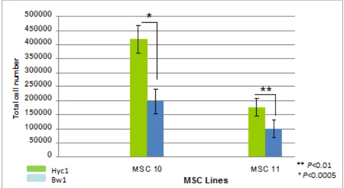
proliferation experiment result for secondary MSC in all two different serum containing medium (Figure 2). There were a slightly proliferation difference between secondary MSC grown in Hyc1 and Bw1 containing medium. In other words, when MSC were grown in Hyc1 containing medium, they showed higher adhesion and proliferation rate. In contrast, other commonly used serum, Bw1, maintained adhesion and proliferation of MSC in a slower rate.
Figure 1. Low expansion of primary MSC using Bw1 contained medium. Proliferation of primary MSC lines. Cell count -based quantification of each primary MSC line (MSC 1-9) grown for 14 days in 10% FBS (Hyc1 or Bw1) in the presence of FGF-2 (1ng/ml). The mononuclear fraction of bone marrow MSC isolated from ilium of nine patients were seeded at 1×10⁵ cells per cm2 in 100 mm culture dish.
Figure 2. Hyc1 contained medium are important in cell proliferation of secondary MSC. Proliferation of MSCs (MSC 10&11) after passage in the presence of FGF-2 (1 ng/ml). The MSCs obtained from the 3rd passage cultures were seeded at 3×10³ cells/cm2 and maintained in the medium containing different serum (Hyc1 or Bw1). Two
independent studies were carried out in triplicates. Each bar represents average ± SD, **P<0.01, +P<0.000
Effect of Two Different Serum on
Morphology of MSC
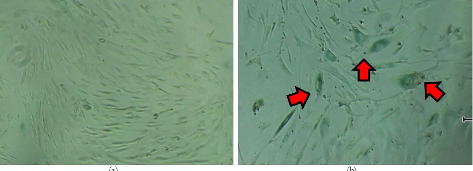
In addition, we also evaluate the impact of Hyc1 and Bw1 containing medium, on primary
medium tended to grow in altered morphology: more flattened, elongated adherent cells, and had a bigger nucleus (Figure 3b) as compared with cells grown in Hyc1, which grew as a small spindle shaped, uniform monolayer of cells (Figure 3a). Enlarged cells which usually present in senescent cells were stained with Senescence cells histochemical staining kit (Sigma-Aldrich,
USA) (Figure 4). The specific cell senescence biomarker, β-galactosidase, was not expressed on cells grown in Hyc1 (Figure 4a). In the case of Bw1 containing medium, β-galactosidase were showed its high activity on primary MSC, it was observed positively stained by cell senencence histochemical staining (Figure 4b).
(a) (b)
Figure 3. A small spindle shaped, uniform monolayer of cells grew in Hyc1. Morphology of primary MSC expanded in vitro in the presence of FGF-2 (1 ng/ml). MSC were isolated from the illium of one patient (MSC 10) grown in Hyc1 (a) or Bw1 (b) containing medium. Mononuclear fraction contain haemopoeitic cells were seeded at 2×10⁶ cells in 6 well culture plate
and maintained until 14 days. (Magnification 100x)
(a) (b)
Figure 4. β-galactosidase, was not expressed on cells grown in Hyc1. Cellular senescence of MSC grown in Hyc (a), and Bw1 (b). The 2nd passage cultures of MSC were transferred into Senescence Cells Histochemical Staining Kit (Sigma-Aldrich, USA). (Magnification 40x)
Effect of FGF-2 on Osteogenic
Differentiation
To examine the osteogenic potential, MSC grown on the both two types of serum containing medium were transferred into the osteogenic different medium (STK3). As shown in Figure 5, primary MSCs grown for ten days in STK3 retained the ability to differentiate into osteocyte as shown by Alizarin red staining. In this experiment, MSC grown in Bw1 containing
medium (Figure 5b) showed a slightly higher differentiation potential than those with Hyc1, but not significantly (Figure 5a). Thus, serum condition did not affect osteogenic differentiation of MSC.
Discussion
(a) (b)
Figure 5. Serum condition did not affect osteogenic differentiation of MSC. Osteogenic potential of MSC expanded in different serum-containing medium (a) Hyc1 or (b) Bw1. hMSC obtained from the 2nd passage cultures were maintained under STK3 medium for 10 days, and stained with Alizarin red
research of human MSC, the ability to expand large numbers of cells at high density quickly by using effective medium may prove very useful. The important achievements in this area will be the importance of the serum choice.
Assays to assess the proliferative activity of cells grown in culture or harvested from tissue samples are a core tool for monitoring the health and growth rate of a cell population. To test the ability of the different types of serum containing medium to support MSC proliferation, we used Cell count assay. Cell count is a direct measure of proliferation which has been shown to be a cost-effective method for describing cell proliferation.
In addition, primary human bone marrow-derived MSC expanded in Hyc1 from 6 donors revealed robust proliferation. The same result goes to secondary MSC culture. Eventhough between Hyc1 and Bw1 containing medium, proliferation rate of secondary MSC from two donors were not significantly different but still there was a difference in their proliferation. It is noteworthy that along with cell proliferation, a drastic change in secondary MSC morphology was observed in the Bw1 containing medium. Whereas primary MSC expanded in Hyc1 containing medium displayed a small, spindle-shaped morphology, cells grown in Bw1 exhibited a bigger cells and nucleus. This morphology allowed cells in Hyc1 containing medium to be grown at much higher density compared with cells grown in Bw1.
In accordance with MSC morphology, similar result shown by cell senescent immunohistochemical staining that expanded primary MSC cultured in Bw1 containing medium displayed positive expression of beta galactosidase in a higher number. Whereas in primary MSC expanded in Hyc1 containing
medium, only small number of positively-stained cells were found.
Primary MSC grown in both serum containing medium retain the ability to differentiate into osteoblast. Bw1 containing serum showed slightly, unsignificanty higher potential comparing with Hyc1. A more detailed analysis on the composition of each serum may on overall MSC morphology and proliferation must be further explored.
CONCLUSION
The establishment of medium for the isolation and expansion of human MSC represent a necessary step in developing the tools required to study human MSC in a consistent and reproducible manner. This study shows the importance of choosing fetal bovine serum to expand MSC. By choosing the highest effective fetal bovine serum, an inherently variable reagent, will provide the ability for researchers from different laboratories to conduct studies with similar reagents. The result from this study should form a basis for further studies examining specific substance needed in MSC proliferation and differentiation in more detail.
REFERENCES
1. Pittenger MF, Mackay AM, Beck SC, Jaiswal RK, Douglas R, Mosca JD, Moorman MA, Simonetti DW, Craig S, Marshak DR (1999) Multilineage potential of adult human mesenchymal stem cells. Science. 284:143-147.
2. Misami K (2010) Isolation of primary human bone marrow MSC. Hiroshima (Unpublished data).