JTLS | J. Trop. Life. Science 96 Volume 4 | Number 2 | May | 2014
Pharmacognostic Investigation of
Clerodendrum phlomidis
Linn. f. Root
Dinesh Kumar1*, Ajay Kumar1, Om Prakash2
1
Institute of Pharmaceutical Sciences, Kurukshetra University, Kurukshetra-136119, India 2
Manav Bharti University, Solan-173229, Himachal Pradesh, India
ABSTRACT
The present study was aimed to perform the pharmacognostic evaluation of Clerodendrum phlomidis
Linn. f. root in terms of organoleptic, fluorescence analysis, macro-microscopy and physicochemical parameters. The characteristic macroscopic study showed that the root consists of 715 cm long, 0.2 -3.0 cm thick pieces which are cylindrical, tough and yellowish-brown externally, with hard fracture and slightly astringent taste. The main microscopic characters of the root show exfoliating cork, having 10-15 rows of tangentially elongated, thick-walled cells. Cortex consists of round to oval parenchymatous cells, a few containing rhomboid shaped calcium oxalate crystals. Endodermis consists of 3-4 layers of non-lignified, thick-walled rounded parenchymatous cells followed by a single pericyclic layer. Phloem consists of isodiametric, thin-walled, parenchymatous cells whereas xylem contains lignified pitted vessels. Medullary rays consisting of biseriate layer of lignified and radially elongated parenchymatous cells is narrower in the xylem region during wider in the phloem region. The physicochemical analysis of the root, i.e., total ash, water-soluble ash, sulphated ash are 7.8, 0.9 and 10.3 (% w/w) respectively. Further successive extraction of the root powder with petroleum ether, chloroform, alcohol, water yielded 2.2, 2.4, 12.4 and 9.6 (% w/w) extracts respectively. Fluorescence study imparted characteristic colours to the root powder when observed under visible, short and long wavelength light. Various pharmacognostic parameters evaluated in this study helps in botanical identification and standardization of Clerodendrum phlomidis L. root part in crude form and provide the authentic data for the researchers and scientists involved in carrying out further research on this plant part.
Keywords:Clerodendrum phlomidis L., Microscopy, Physico-chemical, Fluorescence analysis
INTRODUCTION
Clerodendrum phlomidis Linn. f. (Family: Verbe-naceae), known as Wind-killer or Arni in Hindi, is a common shrub of barren plains, low hills and tropical deserts, distributed more or less throu-ghout India, Myanmar, Pakistan, Sri Lanka and south-east Asia [1,2]. Traditionally, C. phlomidis L. is effectively used in treating bronchitis, headac-he, inflammation, weakness, drowsiness, digestive problems and joint pains [3,4]. C. phlomidis has shown antidiarrhoeal[5], antifungal, antiam-nesic, antidiabetic and antihepatotoxic activities [6-9]. Phytochemical analysis of this plant has reported
*Corresponding author: Dinesh Kumar
Institute of Pharmaceutical Sciences, Kurukshetra University, Kurukshetra-136119, Haryana, India
E-mail: [email protected]
various constituents like β-sitosterol, ceryl alco-hol, scutellarein, pectolinaringenin, γ-sitosterol, clerodin, clerosterol, clerodendrin-A, apige-nin, hispidulin, luteolin, α-L-rhamnopyranosyl-(1→2)
-α-D-glucopyranosyl-7-O-naringin-4ʹ-O-α D-glu-copyranoside-5-methylether, Lup-20(29)-en-3-triacontanoate, tetratriacontanol, 24β -ethyl-cho-lesta-5,22E, 25-triene-3β-ol [10-15]. In the pre-sent study, Pharmacognostical investigations have been carried out on the roots of
Clerodendrum phlomidis L. to describe the parameters useful for the characterization and standardization of this drug part both in scraped and powdered form.
MATERIALS AND METHODS
JTLS | J. Trop. Life. Science 97 Volume 4| Number 2| June| 2014
All the chemicals and reagents used in this study were of analytical grade and procured from Rankem Limited India and Hi-Media laborato-ries, Mumbai, India.
Collection of Plant materials
The plant material (roots) were collected from the campus of Kurukshetra University, Kuru-kshetra during April 2011 and authenticated by Dr. H.B Singh, NISCAIR (NISCAIR/RHMD/ Consult/-2010-11/1471/69).
Macroscopic evaluation
Various organoleptic and macroscopic charac-ters like color, shape, size, taste, odor, fracture and configuration, etc. of C. phlomidis L. root we-re studied and compawe-red with available data [16].
Microscopic evaluation
Microscopic studies were performed qualitati-vely and quantitatiqualitati-vely with the help of a compo-und microscope [Model: SKC-400, Suswox Op-tik, Sudheer Scientific Works, India].
Qualitative microscopy
In this study, transverse sections of root were examined under photographic compound micro-scope. Phloroglucinol and conc. Hydrochloric acid in the ratio 1:1 were used as staining rea-gents. The several identifying features of the drug were studied with or without staining and recor-ded as per already reported methods [17,18].
Root microscopy
Few fine root samples immersed in a test tube containing sufficient water and boiled for few minutes. Thereafter, the softened pieces were transversally cut into fine sections, stained with staining reagent 0.1% w/v phloroglucinol follo-wed by concentrated hydrochloric acid. The stai-ned sections were observed for different layers of cells/ tissues and recorded other identifying fea-tures photomicrographic [19,20].
Powder microscopy
To a small amount of root powder taken over a microscopic slide, 1-2 drops of 0.1% w/v phlo-roglucinol and concentrated hydrochloric acid (1:1) were added, mixed well, covered with a co-ver slip and examined.Various identifying
featu-res of cell components were observed and recor-ded using photomicrograph [21].
Fluorescence analysis
Powdered root was treated with various chemical reagents and observed under ultraviolet light (short and long wavelength) to study its fluorescence behavior as per reported procedure [22]. To small quantity of the powder taken on clean microscopic slide, 1-2 drops of the freshly prepared reagent phloroglucinol and concentra-ted hydrochloric acid (1:1) solution were added, mixed and kept aside for 1-2 minutes. Thereafter, the slide was observed in visible light, short (254 nm) and long (365 nm) ultraviolet radiations where the colors observed by application of dif-ferent reagents in difdif-ferent radiations were recor-ded.
Physicochemical analysis
In this study, root powder was evaluated for the determination of physicochemical conditions as loss on drying, total ash, acid insoluble ash, water-soluble ash, sulphated ash and extractive values as per reported method [23].
RESULTS
Macroscopic study of root
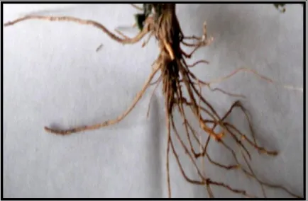
Macroscopic examination of the root (Figure 1) shows that it consists of 7-15 cm long and 0.2 -3.0 cm thick pieces which are occasionally bran-ched, cylindrical, tough, yellowish-brown exter-nally, thin bark, the outer surface rough due to exfoliation, with hard fracture and slightly astri-ngent taste.
JTLS | J. Trop. Life. Science 98 Volume4| Number2| May| 2014
Microscopic study of root
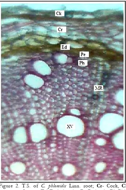
Transverse section of the root (Figure 2) shows exfoliating cork, consisting of about 10-15 layers of tangentially elongated, thick-walled cells. Cortex consists of round to oval parenchymatous
Figure 2. T.S. of C. phlomidis Linn. root; Cr- Cork, Ck- Cortex, Ed-Endodermis, Pe-Pericycle Ph -Phlo-em, XV-Xylem vessel, MR-Medullary rays
cells, a few containing rhomboid shaped calcium oxalate crystals. Endodermis consists of 3-4 layers of non-lignified, thick-walled rounded pa-renchymatous cells. Pericycle consists of a single layer of non-lignified, thin-walled rounded paren-chymatous cells below the endodermis. Phloem consists of isodiametric, thin-walled, parenchy-matous cells, a few containing rhomboid crystals of calcium oxalate. Xylem shows a wide zone, consisting of lignified pitted vessels found in single as well as in a group of 2-3 scattered throughout xylem region. Medullary rays, narro-wer in the xylem region and wider in the phloem region, consist of biseriate, lignified and radially elongated parenchymatous cells.
Powder study, Fluorescence analysis and Physicochemical analysis
Root powder appears dull yellow, showing fragments of cork cells about 2-3 rows of tangen-tially elongated, thick-walled cells. Cortex con-sists of thin-walled round to oval paren-chymatous cells, lignified and pitted xylem ves-sels (60.25-145.6 µ), non-lignified sieve tube and rhomboid shaped calcium oxalate crystals measu-ring 12 to 24 µ in length and 6 to 15 µ in width. Powder characteristics of the root have been shown in Figure 3. The fluorescence observations of the root powder with different chemical rea-gents are summarized in Table 1. In this study, various physicochemical parameters like loss on drying, total ash, acid insoluble ash, water-soluble ash, sulphated ash and extractive values were determined in triplicate as mentioned in Table 2.
A B
C D
Figure 3. Powder characteristics of C. phlomidis root (A-Cork cells; B-Cortex cells; C-Calcium oxalate crystal; D-Xylem vessel)
DISCUSSION
JTLS | J. Trop. Life. Science 99 Volume 4 | Number 2 | June | 2014
by developing numerical values of standards for assessment [24,25].Pharmacognostical evaluation
of different parameters is the vital etiquette for standardization of herbals [26].
Table 1. Fluorescence analysis of C. phlomidis L. root powder
Treatment Visible light
Under UV light Short
Wavelength (254 nm)
Long wavelength
(365 nm)
Powder Yellowish
brown Pale green Brown
Powder + 1N NaOH
(aq.) Light brown Green Brownish green
Powder + 1N NaOH
(alc.) Light yellow Dark green Yellowish green
Powder + Ammonia Light yellow Green Yellowish green
Powder + Picric acid Yellowish
brown Green Dark brown
Powder + Pet. ether Light brown Light green Pale green
Powder + 50% HCl Light brown Green Pale yellow
Powder + 50% H2SO4 Brown Greenish brown Brownish
The present study lays down the pharmacognostical parameters of C.phlomidis root and its results are incommensurate with those of previous studies [27].The morphological parame-ters like shape, size, colour and microscopic characteristics of the root such as rhomboid sha-ped calcium oxalate crystals, biseriate medullary rays, etc. help in identification of exact species and avoid adulteration. The total ash is parti-cularly important in the evaluation of purity of - drugs i.e. presence or absence of foreign inorga-nic matter such as metallic salts and/or silica [28]. The present study on the pharmacognostic evaluation and physicochemical parameters of the C. phlomidis root might be useful to supple-ment information with regard to its identification
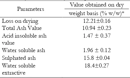
Table 2. Physicochemical analysis of C. phlomidis L. root
Parameters Value obtained on dry weight basis (% w/w)* Loss on drying 12.21±0.16 Total Ash Value 10.94 ±0.23 Acid insoluble ash
value
1.47 ± 0.37
Water soluble ash 1.96 ± 0.12 Sulphated ash 15.8 ±0.04 Water soluble
extractive
18.4±0.27
Alcohol soluble extractive
9.6±0.04
*Average of three reading ± SEM
parameters. The informationse are considered significant for the acceptability of herbal drugs in the present scenario. Fluorescence study of the root powder helps in the qualitative study of ste-roids which can be used as a reference data for the identification of adulterations.
CONCLUSION
The detailed pharmacognostic study on C. phlomidis root would be of high value in the identification, quality control, and formulation development. These data obtained pooled together will help in the proper identification and authentication as well as differentiate from its related species.
ACKNOWLEDGEMENT
The authors express sincere thanks to UGC, New Delhi (39-955/2010 SR) for financially supported the study.
JTLS | J. Trop. Life. Science 100 Volume 4 | Number 2 | May| 2014
1. Matthew KM (1991) An Excursion Flora of Central Tamilnadu, India. New Delhi, India: Oxford and IBH Publishing Co.
2. Kritikar KR, Basu BD (2005) Indian Medicinal Plants. 3rd Vol. Dehradun: International Book
Distributors and Publication.
3. Nadkarni KM (1976) Indian Materia Medica. Bombay, India: Popular Prakashan Pvt. Ltd. 4. Meena AK, Rao MM (2010) Folk Herbal
Medicines Used by the Meena Community in Rajasthan. Asian J Tradit Med. 5: 1-13.
5. Rani S, Ahamed N, Rajaram S, et al (1999) Anti-diarrhoeal Evaluation of Clerodendrum phlomidis
Linn. Leaf Extract in Rats. J Ethnopharmacol. 68: 315-319.
6. Rajasekaran A, Ponnusamy K (2006) Antifungal activity of Clerodendron inerme (L) and Clerodendron phlomidis (L). Turk J Biol. 30: 139-142.
7. Joshi H, Megeri K (2008) Antiamnesic Evaluation of C. phlomidis Linn. Bark Extract in Mice. Braz J Pharm Sci. 44: 717-725.
8. Dhanabal SP, Mohan Marugaraja MK, Suresh B (2008) Antidiabetic Activity of Clerodendron
phlomoidis Leaf Extract in Alloxan-Induced
Diabetic Rats. Indian J Pharm Sci. 70: 841-844. 9. Verma A, Ahmed B (2009) Anti-hepatotoxic
Activity of Clerodendrum Phlomidis. Int J Pharm-Tech Res. 1: 1027-1031.
10. Gupta SK, Chandra S, Mahadevan V (1967) Chemical Examination of Clerodendron phlomidis. Indian J Pharm. 28: 102-106.
11. Subramanium SS, Nair AGR (1972) Scutellarein and Pectolinaringenin from the Leaves of
Clerodendrum phlomidis and Duranta repens.
Phytochemistry. 11, 3095-3096.
12. Joshi KC, Singh P, Mehra A (1979) Chemical Investigation of the Roots of Different
Clerodendron species. Planta Med. 37: 64-66. 13. Seth KK, Pandey VB, Dasgupta B (1982)
Flavonoids of Clerodendron phlomidis Flowers. Pharmazie. 37: 74-75.
14. Anam EM (1999) Novel Flavanone and Chalcone Glycosides from Clerodendron phlomidis
(Verbenaceae). Indian J Chem. 38B: 1307-1310.
15. Pandey R, Kaur, Malasoni R, et al (2008) Lupeol Ester from Clerodendron phlomidis. Indian J Chem. 47B: 470-472.
16. Khatoon S, Singh N, Kumar S, et al (2009) Authentification and Evaluation of an Important Ayurvedic Drug - Ashoka bark. Indian J Tradit Knowl. 68: 393-400.
17. Kokate CK (2010) Practical Pharmacognosy. 4th
ed. New Delhi: Vallabh Prakashan
18. Bisht A, Zaman K, Singh M, et al (2011) Pharmacognostical Studies on Oroxylum indicum
(Linn.) Vent. Stem Bark. Indian J Nat Prod Resour. 2: 472-478.
19. Ahmed F, Urooj A (2011) Pharmacognostical Studies on Ficus racemosa Stem Bark. Pharmacog J. 3: 19-24.
20. Bhide B, Acharya RN, Naria P, et al (2011) Pharmacognostic Evaluation of Cordia macleodii
Hook. stem bark. Pharmacog J. 3: 49-53.
21. Khandelwal KR (2011) Practical Pharmacognosy. 21th ed. Pune: Nirali Prakashan.
22. Kokashi C J, Kokashi RJ, Sharma M (1958) Fluorescence of Powdered Vegetable Drugs in Ultraviolet Radiation. J Am Pharm Assoc. 47: 715-717.
23. WHO/QCMMPM (1992) Quality Control
Methods for Medicinal Plant Materials. Geneva: Organisation Mondiale De La Sante.
24. Sandhya S, Venkatramana K, Vinod K R, et al
(2011) Pharmacognostical standardization of
Tephrosia maxima Pers root. Pharmacog J. 3: 25-33.
25. Chellappan DR, Joseph J, Balasubramanian P (2011) Pharmacognostical, Antioxidant and Antiulcer Screening of Cyclea peltata roots. Braz J Pharmacogn, 21: 1096-1103.
26. Johansen DA (1940) Plant microtechnique. New York: McGraw Hill Book Co.
27. Kahraman A, Celep F, Dogan M (2010) Anatomy, trichome morphology and palynology of Salvia chrysophylla Stapf (Lamiaceae). South Afr J Bot. 76: 187-195.
28. Musa KY, Katsayal AU, Ahmed A, et al (2006) Pharmacognostic investigation of the leaves of