Iranian Journal of Otorhinolaryngology, Vol.30(5), Serial No.100, Sep 2018 Original Article
The Antiapoptotic Effect of Curcumin in the Fibroblast of the
Cochlea in an Ototoxic Rat Model
*
Tengku-Siti-Hajar Haryuna
1, Agustinus-Hamonangan-Winston Purba
1,
Farhat Farhat
1, Widayat Alviandi
2Abstract
Introduction:
This study aimed to show the potency of curcumin as an antiapoptotic agent that decreases the apoptotic index in the cochlea lateral wall in ototoxic rat models.
Materials and Methods:
A total of 24 Rattus norvegicus were divided into eight groups: Group 1 (control group), Group 2 (gentamicin (+)), Group 3 (gentamicin + curcumin 20 mg/day), Group 4 (gentamicin + curcumin 40 mg/day), Group 5 (gentamicin + curcumin 20 mg/day for 7 days), Group 6 (gentamicin + curcumin 40 mg/day for 7 days), Group 7 (curcumin 20 mg/day for 3 days + gentamicin), and Group 8 (curcumin 40 mg/day for 3 days + gentamicin). After the division, the rats were terminated in order to measure the apoptotic index using a terminal deoxynucleotidyl transferase (TdT) dUTP nick-end labeling (TUNEL) assay in the fibroblasts of the cochlea lateral walls. The data were analyzed using analysis of variance (ANOVA), and P<0.05 was used as the cut-off for statistical significance.
Results:
Administration of gentamicin showed significant differences (P<0.05) in the apoptotic index. Groups undergoing curcumin treatment at a higher dose (200 mg/kg bw) and the prevention groups showed significant differences compared with groups not treated with curcumin
Conclusion:
This study concluded that the apoptotic index can be decreased by curcumin and has a preventive benefit toward ototoxic rat models. The administration of curcumin depended on the dose and duration.
Keywords:
Apoptosis, Curcumin, Cochlea, Gentamicin, Prevention, Rats.
Received date: 7 Oct 2017 Accepted date: 10 Jun 2018
1
Department of Otorhinolaryngology-Head and Neck Surgery, Faculty of Medicine, University Sumatera Utara, Medan 20155, Indonesia.
2Department of Otorhinolaryngology-Head and Neck Surgery, Faculty of Medicine, University Indonesia, Jakarta 10430, Indonesia.
*
Corresponding Author:
Introduction
Aminoglycosides are a popular group of antibiotics with considerable antimicrobial efficacy and toxic side effects on the kidneys and inner ears. Gentamicin is one example of an aminoglycoside antibiotic. The damage inflicted by gentamicin on the kidneys is reversible, but damage to the inner ears is irreversible. The ototoxic side effects may become evident days or weeks after systemic application, and the damage is bilateral (1). Ototoxic effects caused by aminoglycoside are cochleotoxic and vestibulotoxic(1,2).
The incidence ototoxicity induced by aminoglycosides is increasing in developing countries compared with industrialized countries(1). Aminoglycoside exposure leads to oxidative stress and radical oxygen species (ROS) formation, which leads to an apoptotic signalling pathway (3). Gentamicin induces ROS formation in cellular system and leads to the opening of mitochondrial permeability, ROS, and cytokines that may cause cell death through apoptosis or necrosis in a dose-dependent manner (4). Hearing loss by aminoglycosides on the cellular level is caused through damage of the cochlea hair cells, but the biochemistry and molecular mechanism are still hard to understand(5).Curcumin is a popular herb in South East Asia, and is used as a natural food spice(6,7) because of its color stability and low toxicity (6). Curcumin is a potential natural compound that impacts on different pathways, intracellular components, and important enzymes (8). Research with rat models has shown that curcumin has antiapoptotic activity against indomethacin, marked by a decreased expression of caspase-3 (9). The aim of this research was to show the potential of curcumin as an antiapoptotic agent in obstructing the apoptotic index in the cochlea lateral wall in ototoxic rat models, which could be the basis of future clinical trials.
Materials and Methods
1. Experimental treatments in animal models
This research used male rats, Rattus norvegicus, weighing 150–250 g. Thirty-two rats were used in this study, divided into eight groups of four rats. The curcumin given to the rats was extracted from Curcuma longa L. (turmeric). The level of the curcumin was (16.62 ± 0.14)% b/b, counted using a
thin-layer chromatographic (TLC)-densitometric method. The curcumin doses varied from 100 mg/kg to 200 mg/kg, and all doses were suspended in carboxy methyl cellulose (CMC) 0.5% and were administered through a nasogastric tube. The rats were first anesthetized with 10 mg/kg of xylazine and 90 mg/kg of ketamine, which were administered via an intraperitoneal injection (10).
After the rats were calmed following the effect of the prior injection, they were injected with 0.03–0.05 ml of 40 mg/ml gentamicin (11). The dose used was gentamicin 40 mg/ml 0.1 ml, injected into the anterosuperior rats of the same strain that were homogeneous with respect to gender and age, and were bred in the Biochemistry Laboratory, Faculty of Medicine, Airlangga University. Food for rats was provided ad libitum, and the room temperature in the laboratory was maintained between 20 and 26 °C. Lighting inside the cages during the light phase was maintained at an exposure below the reluctance threshold for rats, and the relative ambient humidity was 55±15%. Ethical permission to conduct this research was obtained from the Health Ethics Committee, Faculty of Medicine, University of North Sumatra/H. Adam Malik General Hospital, Indonesia. The ethics code was 433/KOMET/FK USU/2015.
given curcumin for 7 days at a dose of 40 mg per day, and were terminated 18 hours after the final curcumin dose; Group 7 was given 20 mg of curcumin per day for 3 days, then were injected with gentamicin 18 hours after the final curcumin dose, and were terminated 18 hours later; Group 8 was given 40 mg of curcumin per day for 3 days, then gentamicin injection 18 hours after they were given the last dose of curcumin, and were terminated 18 hours afterwards. Groups 3, 4, 5, and 6 were curative groups, in which curcumin was given after gentamicin. Groups 7 and 8 were preventive groups, in which curcumin was given before gentamicin. The rats were terminated and examined through necropsies. Temporal bone tissues from the head were taken and fixated with 10% formalin buffer
solution and decalcified with
ethylenediaminetetraacetic acid (EDTA) within 4 weeks. They were then tested in the laboratory to assess the apoptotic index on the fibroblasts of the cochlea lateral wall.
2. Apoptosis examination
The apoptosis examination was performed using an TACS 2 Tdt-DAB in situ apoptosis detection kit (Trevigen, Helgermen Ct. Gaithersburg). The procedures were performed through various steps. Samples were immerse hydrated, fixed, and immobilized in 1× phosphate-buffered saline (PBS) for 10 min, then the samples were covered with 50 μl of proteinase K solution for 15–30 min, and washed twice in deionized water for 2 min each time. The samples were immersed in quenching solution for 5 min, and washed in 1× PBS for 1 min. The samples were immersed in 1x Tdt labeling buffer for 5 min and covered with 50 μl of labeling reaction mix and incubated for 60 min at 37 °C in a humidity chamber. The samples were immersed in 1× Tdt stop buffer for 5 min, washed twice in deionized H2O, for 5 min each time. The samples were covered with 50 μl of Strep-HRP solution and incubated for 10 min at 37 °C in a humidity chamber to avoid evaporation. The samples were washed twice in 1× PBS, for 2 min each time, then immersed in 1% methyl green for 30 s up to 5 min. Slides were dipped 10 times each in two changes of deionized H2O, 95% and 100% ethanol. Each of them was dipped 10 times in two changes of o-xylene, with mount glass
coverslips using mounting medium.The apoptotic index was analyzed using an Olympus XC 10 microscope under 40× magnification, marked by brown-stained nuclear staining, and counted in a masked manner using terminal deoxynucleotidyl transferase (TdT) dUTP nick-end labeling (TUNEL) (+). The researchers counted two randomly selected fields (13).
3. Statistical analysis
Data were analyzed by one-way analysis of variance (ANOVA) using IBM SPSS statistical software, with a significance level of 0.05.
Results
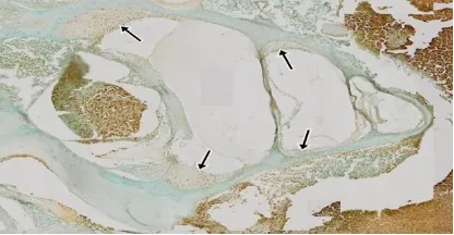
Figure 1 shows the lateral wall of the cochlea and Figure 2 shows the role of curcumin in the
lateral wall of the cochlea of Rattus norvegicus
gentamicin ototoxic models, observed using the TUNEL assay method, in which the researchers assessed the fragmentation of DNA in the cell nucleus that was brown.
Fig 1: Supporting tissues and lateral cochlea wall with TUNEL assay (10× magnification). The arrows indicate the lateral wall.
Fig 2: Apoptosis index in each group (40×
magnification). (A) Group 1; (B) Group 2; (C)
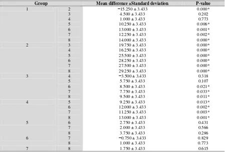
In Group 2, where the rats were only given decrease in the number of core cells undergoing apoptosis in the fibroblasts of the cochlea lateral walls were found in all groups that received curcumin (Groups 3- 8). Table 1 shows significant differences (P<0.05) between the control group and the other treatment groups, except for Groups 3 and 4. The
curcumin administration influenced the
apoptotic index in all groups, compared with Group 2. Table 1 also shows that the dose and
duration of the administration of curcumin affected the apoptotic index.
Fig 3: The Mean of Apoptotic Index in Cochlear Lateral Wall Fibroblasts
Table 1: ANOVA LSD test result in terms of apoptotic index
Group Mean difference ±Standard deviation P-value
1 2
The principal microscopic changes were found in the lateral wall spiral ligament, with degeneration of mitochondria, and location of endocochlear potential production. The research was performed by giving rats 3-nitropropionic acid directly into their round window membrane to evaluate the destruction
in the cochlea lateral wall (14). This information encouraged the researchers to conduct research in the lateral wall of cochlea.
In this study, the researchers analyzed the antiapoptotic role of curcumin as seen from
the index of apoptosis in the fibroblasts of
the lateral wall of the cochlea fibroblasts compared with the control group. This was also found in a study in Chinchillas that were given gentamicin and showed improvement in
cochlea cell death markers, including
cytochrome C, caspase-9 and caspase-3 (15). The same intratympanic gentamicin dose injection was once given to albino Wistar rats as controls to evaluate the effects of ototoxic intratympanic terbinafine in the middle ear (11). The rats were terminated 18 hours after the administration of gentamicin in order to follow the results of the round window membrane of Hartley strain guinea pigs by gelatin sponge soaked in gentamicin, where the representative photomicrograph showed TUNEL-positive nuclei, mostly in groups of 18 hours (12).
The results showed that the administration of curcumin decreased the apoptotic index in the lateral wall of the cochlea of the fibroblasts of rats that were seen in the comparison of Group 2 and Groups 3,4, 5, and 6. A similar finding was observed in the testicular tissues of the rats in which the administration of cadmium significantly increased testicular apoptosis. By administering curcumin, the reactivity and number of germinal cells of Leydig cells
undergoing apoptosis was significantly
reduced (16). groups in which rats were given a dose of 200 mg/kg of curcumin, the researchers investigated whether an increase in dose could give a better anti-apoptosis result. The same statistically significant, which means that the provision of curcumin at larger doses and longer duration is better in lowering the apoptotic index. The comparison between Groups 5 and 6 and Group 4 was statistically significant, which means that curcumin administration with a longer duration is better in lowering the apoptotic index. A single dose of curcumin was sufficient for the preventive
effect, as shown in the decreased the expression of caspase-3 that leads to the conclusion of antiapoptotic activity (9). In this research, curcumin was given for 3 days. The results showed that the ratio of the prevention groups (Groups 7 and 8) compared with Groups 1, 2, 3, and 4 showed statistically significant results, which means that curcumin has a preventive role in reducing the apoptosis index. This is consistent with a study in which rats were given acetaminophen, which reported 469% DNA fragmentation when compared with the control group (quantitative marker was considered the presumed predictor of apoptotic cell death), and decreased to 162% (almost 3×) in rats receiving curcumin as a preventive measure (7).
Apoptosis caused by aminoglycosides could occur through a number of mechanisms. First aminoglycosides may enter the cell by opening the cation channels directly in the cytosol.
Aminoglycosides that bind to ferric iron (FeIII)
(3,17) will then bond FeII-aminoglycoside
complexes in the cytosol and form ROS compounds, using arachidonic acid as a donor electron. ROS then activates Bax (protein pro-apoptotic), which then translocates to the membranes of the mitochondria, and apoptosis can be halted if the proteins and antiapoptotic agents (Bcl-2 and Bcl-XL) are successfully stopped. In the second possible mechanism, cytochrome C exits through the mitochondrial transitional pores made by a Bax-dependent mechanism, then activates caspase-9 and caspase-3 and leads to apoptosis. A third possible mechanism, a caspase-independent
mechanism, involves the release of
endonuclease G and AIF from the
caspase-9-induced cytochrome C released from the mitochondria to the cytosol. Caspase-9 activates caspase-3 (caspase executioner), which divides antiapoptotic proteins or block deoxyribonucleases and leads to cell death. The Bcl-2 family of proteins is made up of antiapoptotic Bcl-2 and Bcl-XL, namely the pro-apoptotic Bax and Bak. When apoptotic signals beat the inhibition/protection by Bcl-2 and Bcl-XL, Bax translocates from the cytosol
the mitochondrial permeability transition
pore (17). The role of curcumin in anti-apoptosis indicated by the decreased expression of Bax (12;18) in the ratio of Bax/Bcl-2 (18,19) decreased the activity of caspase-3 (18,19) as well as a decrease in the activity of caspase-9 (18), increased the concentration of the protein Bcl-2 (18) and the expression of Bcl-Xl (7).
In other research, curcumin has a protective effect against noise-induced hearing loss (NIHL), which is hypothesized to be through the inhibited the degradation of Ikβ in the cochlea and suppression formation of 4-hyroxynonenal (4-HNE) in the cochlea spiral ligament, which leads to suppression of the
NFκβ signal (20). In vivo research
demonstrated the protective effect of curcumin against cisplatin-induced ototoxicity. The mechanism is not yet clear, but curcumin acts as direct scavenging activity and indirect anti-oxidant action by induction of endogenous heme oxygenase-1 (HO-1) (21).
counteracting the ototoxic effects from the application of gentamicin in the lateral wall of the cochlea in the rats studied.
Acknowledgments
The research was funded by Direktorat Penelitian Pengabdian kepada Masyarakat Universitas Sumatera Utara (DIPA). There are no conflict of interest within the research, authorship, or publication.
References
1. Huth ME, Ricci AJ, Cheng AG. Mechanism of aminoglycoside ototoxicity and targets of hair cell protection. Int J Otolaryngol. 2011:1-19.
2. Petersen L, Rogers C. Aminoglycoside-induced hearing deficits of cochlea ototoxicity. South African Fam Pract. 2015;57:77–82.
3. Fetoni AR, Eramo SLM, Rolesi R, Troiani D, Paludetti G. Antioxidant treatment with coenzyme Q-ter in prevention of gentamycin ototoxicity in an animal model. Acta Otorhinolaryngol Ital. 2012; 32:103–110.
4. Alkahtani S, Alarifi SA, Al-Doaiss AA. Detection of apoptosis induced by gentamicin in rat hepatocytes. Int J Zool Res. 2009;5:161–70. 5. Selimoglu E. Aminoglycoside-induced ototoxicity. Curr Pharm Des. 2007; 13: 119–26. 6. Yu L, Fan Y, Ye G, Li J, Feng X, Lin K, et al. Curcumin inhibits apoptosis and brain edema induced by hypoxia-hypercapnia brain damage in rats models. Am J Med Sci. 2015; 6:521–5.
7. Bulku E, Stohns SJ, Cicero L, Brooks T, Halley H, Ray SD. Curcumin exposure modulates multiple pro-apoptotic and anti-apoptotic signaling pathways to antagonize acetaminophen-induced toxicity. Curr Neurovasc Res. 2012;9:58–71. 8. Ho C, Hsu YC, Lei CC, Mau SC, Shih YH, Lin CL. Curcumin rescues diabetic renal fibrosis by targeting superoxide-mediated wnt signaling pathways. Am J Med Sci. 2016;3:286–295.
9. Morsy MA, Moselhy MAE. Mechanism of the protective effects of curcumin against indomethacin-induced gastric ulcer in rats. Pharmacol. 2013;91:267–74.
10. Toydemir T, Kanter M, Erboga M, Oguz S, Erenoglu C. Antioxidative, antiapoptotic and proliferative effect of curcumin on liver regeneration after partial hepatectomy in rats. Toxicol Ind Health. 2015;31:162–72.
11. Sagit M, Somdas MA, Korkmaz F, Akcadag A. The ototoxic effect of intratympanic terbinafine applied in middle ear rats. J Otolaryngol Head Neck Surg. 2013;42(1):1-6.
12. Suzuki M, Ushio M, Yamasoba T. Time course of apoptotic cell death in guinea pig cochlea following intratympanic gentamicin application. Acta OtoLaryngol. 2008;128: 724–31.
13. Zhang W, Feng H, Gao Y, Sun L,Wang J, Li Y, et al. Role of pigment epithelium-derived factor (PEDF) in arsenic-induced cell apoptosis of liver and brain in a rat model. Biol Trace Elem Res. 2013; 151: 269–76.
15. Ding D, Jiang H, Salvi RJ. Mechanism of rapid sensory hair-cell death following co-administration of gentamicin and ethacrynic acid. Hearing Res. 2010;259:16–23.
16. Aktas C, Kanter M, Erboga M, Ozturk S. Anti-apoptotic effects of curcumin on cadmium-induced apoptosis in rat testes. Toxicolo Indust Health. 2012; 28:122–130.
17. Kurasawa T, Steyger PS. Intracellular mechanism of aminoglycoside-induced cytotoxicity. Integr Biol. 2011;3:879–86.
18. Motaghinejad M, Karimian M, Motaghinejad O, Shabab B, Yazdani I, Fatima S. Protective effect of various dosage of curcumin against morphine induced apoptosis and oxidative stress in rat isolated hippocampus. Pharmacol Rep. 2015; 67:230–5.
19. Fan J, Li X, Yan YW, Tian XH, Hou WJ, Tong H, et al. Curcumin attenuates rat thoracic aortic aneurysm formation by inhibition of the c-Jun N-terminal kinase pathway and apoptosis. Nutrition. 2012;28:1068–1074.
20. Yamaguchi T, Yoneyama M, Onaka Y, Imaizumi A, Ogita K. Preventive effect of curcumin and its highly bioavailable preparation on hearing loss induced by single or repeated exposure to noise: A comparative and mechanistic study. J Pharmacol Sci. 2017;134:225–33.