P-ISSN : 1978-225X; E-ISSN : 2502-5600 DOI: https://doi.org/10.21157/j.ked.hewan.v12i1.6892
CHARACTERISTIC OF SKIN MORPHOLOGY OF SUNDA PORCUPINE
(
Hystrix javanica
) WITH SPECIAL REFERENCE TO
THE CONNECTIVE TISSUE
Andhika Yudha Prawira1, Desrayni Hanadhita1, Anisa Rahma1, Supratikno1, Savitri Novelina1, and Srihadi Agungpriyono1*
1
Department of Anatomy, Physiology, and Pharmacology, Faculty of Veterinary Medicine, Bogor Agriculture University, Bogor, Indonesia
*Corresponding author: [email protected]
ABSTRACT
This study was conducted to investigate the histological characteristic, type, and distribution of connective tissue in Sunda porcupine skin. The investigation was carried out in three adult of sunda porcupines at microscopic level using hematoxylin eosin, Masson thrichrome, Verhoeffs van Gieson, alcian blue pH 2.5 and periodic acid Schiff staining methods. Skin consists of epidermis, dermis hypodermis, and subcutaneous muscle. Quill follicles were the main and dominant structure as well as the specific characteristic on Sunda porcupine skin. The connective tissue was distributed well in basal membrane, dermis, quill follicle, and hypodermis with various intensity and density. The collagen was the main fiber found in the skin while the elastin fiber was not observed. The acid carbohydrate was found distributed well in the skin while the neutral carbohydrate was not detected in this study. In addition the fibers of connective tissue associated with the adipose tissue which found plentifully in quill follicles and hypodermis. The present results showed that the wide distribution of connective tissue might have an important role on the wound healing physiology of Sunda porcupine skin.
____________________________________________________________________________________________________________________ Key words: collagen, connective tissue, Hystricidae, quill follicle, skin
ABSTRAK
Penelitian ini menggunakan sampel kulit tiga ekor landak jawa (Hystrix javanica) dewasa untuk mengetahui karakteristik histologi kulit serta jenis dan distribusi jaringan ikat dengan menggunakan metode pewarnaan hematoxylin eosin, Masson trichrome, Verhoeffs van Gieson, alcian blue pH 2,5, dan periodic acid Schiff. Kulit landak jawa tersusun atas epidermis, dermis, hipodermis, dan musculus subcutaneous. Folikel duri merupakan struktur yang dominan dan utama serta menjadi karakteristik khas pada kulit landak jawa. Jaringan ikat terdistribusi pada membran basalis, dermis, folikel duri, dan hipodermis dengan intensitas dan kepadatan yang bervariasi. Serabut kolagen merupakan serabut utama dalam kulit sedangkan serabut elastin tidak terdeteksi pada penelitian ini. Komponen jaringan ikat non serabut dengan kandungan karbohidrat asam ditemukan pada hampir semua bagian kulit sedangkan komponen non serabut dengan kandungan karbohidrat netral tidak terdeteksi pada penelitian ini. Jaringan ikat berasosiasi dengan jaringan lemak dan terdistribusi di folikel duri dan hipodermis. Hasil penelitian ini menunjukkan adanya kemungkinan peran penting luasnya distribusi jaringan ikat pada fisiologi penyembuhan luka pada kulit landak jawa.
____________________________________________________________________________________________________________________
Kata kunci: kolagen, jaringan ikat, Hystricidae, folikel duri, kulit
INTRODUCTION
The Sunda porcupine (Hystrix javanica) is an endemic Indonesian animal belonging to the family of Hystricidae (Myers, 2001). The porcupine body is covered by modified hair of quills. Previous research showed that quill in Sunda porcupine skin consist of four types with varying sizes, i.e. flat spines, true quills, transitional quills, and rattling quills (Sheila, 2011). In addition, fine hairs still grow between quills of porcupine. Observation of the healing of wounds of Sunda porcupine showed the process of healing wound on the skin occurred in a fairly fast time without causing scarring of the skin, which was about 32 days (unpublished data).
There are several intrinsic components that affect the wound healing process, including connective tissue and immune system. The connective tissue plays a role in modulation of interactions between cells to cells and between cells to matrices, thus affecting the activity of cells in the wound healing process (Shock and Laurent, 1996). The immune system plays a major role in maintaining the success of wound healing process by initiating physiological processes such as inflammation and cytokine regulation among cells (Clark, 1996).
Connective tissue is the largest component of the skin and comprises fibrous and non-fibrous components. The connective tissue composition in the skin determines properties and characteristics of the skin. Connective tissues such as collagen, elastin (fibrous components) and proteoglycans, and glycoproteins (non-fibrous components) play a role in strength, stress, elasticity, flexibility, and skin viscosity (Kent and Carr, 2001; Akers and Denbow, 2008). The composition of connective tissue in Sunda porcupine is thought to be different from general mammals and also related to the process of healing wounds without scars observed on the skin of Sunda porcupine.
This study aimed to determine the characteristic distribution of connective tissue on the skin of Sunda porcupine microscopically. Information from the research is expected to be the basis in further research on various physiological processes on Sunda porcupine skin, such as wound healing process.
MATERIALS AND METHODS
Animals were anesthetized using a combination of ketamine and xylazine with a dose of 5 mg/kg and 2 mg/kg, respectively (Morin and Berteaux, 2003). Then, quills in 6 dorsal skin regions (dorsal thoracic, thoracolumbal, lumbar dorsal, lumbosacral, caudal radix, apex caudal) were removed and the skin was taken as sample. The skin was incised up to the subcutaneous muscle layer using a knife then fixed into 10% neutral buffered formalin (NBF) for three days. Fixed skin was cross-cut to observe the structure macroscopically and skin thickness measurements were made using a sliding range. Measurement of thickness was divided into 3 categories namely muscle thickness, subcutaneous, epidermal skin thickness to hypodermis, and full skin thickness.
Then, the skin sample was processed for histological examination by dehydration process in alcohol from concentration 70% to absolute then clarified in xylol solution, before blocked in paraffin. The tissue block was cut in 4-5 μm thickness using microtom. Furthermore, the tissue was stained with several staining methods i.e. hematoxylin-eosin, Masson's thrichrome modified Goldner, Verhoeff's van Gieson, alcian blue (AB) pH 2.5 and periodic acid Schiff (PAS). The samples were observed using light microscope which was equipped with digital camera.
RESULTS AND DISCUSSION
General Structure of the Skin
The porcupine skin composed of epidermis, dermis, hypodermis, and subcutaneous muscle, but in the apex caudal region the skin muscles were inseparable because they were attached to the underlying tail bone (os vertebrae caudalis) (Figures 1 and 2). In general, the skins are the largest organ in the body of mammals (Gawkrodger, 2002) and consist of 3 layers, namely epidermis, dermis, and hypodermis (Kent and Carr, 2001). Epidermis is a superficial layer of skin that acts as a protector of physical and chemical trauma between the body and the environment. The dermis is a layer under the epidermis and serves to provide a support structure for the skin. The most profundal layer of hypodermis which is the place of fat reserves (Gawkrodger, 2002). The inside of the skin showed the follicular structure of the quills that dominate the skin (Figure 1).
Skin thickness from epidermal layer to m. subcutaneous varies in each region. The thinnest layer was the apex caudal region whereas the thickest was in the dorsal lumbar. The skin layer of the lumbosacral region appeared quite thin compared to other regions. This was influenced by the size of the quill follicles in
the dermis. M. subcutaneous was found to be thickest in the caudal radix region while the thinnest was the apex caudal region. This possibility related to the type of quill and its function (Cobert and Jones, 1956).
The thickest component of the skin was found in the dorsal lumbar region whereas the thinnest was in the lumbosacral region. This is related to the structure of quill follicles and the distance between clusters of quills in the skin. The dorsal lumbar skin of the dorsal region has a true type of quill that was the largest size compared to other types of quill whereas the lumbosacral region skin has a true quill that was small but has a relatively longer distance between one cluster with another cluster. Table 1 and Figure 1 showed the size of skin thickness in each region. In general, mammalian skin exhibits structural resemblance to all parts of the body, but skin thickness varies greatly in different regions of the body and age (Akers and Denbow, 2008).
Follicle of quill was a very dominant part in the dermis of Sunda porcupine. The quill follicle shaped like a long white capsule on the outside and yellow on the inside that extends diagonally from the hypodermis and m. subcutaenous to epidermis in the dorsal thoracic region to the caudal radix. The quill follicle in the apex caudal region looked similar to other regions but diagonally extends from the epidermis to the profundal has no clear capsule form. The thorn follicle was wrapped by a white dense connective tissue membrane that could be seen macroscopically. This connective tissue macroscopic characteristic could be seen clearly in the dorsal lumbar region. The yellowish tissue, which was an adipose tissue found inside the follicle of
the thorn, except the apex caudal region scattered among the skin tissues.
Connective Tissue Distribution
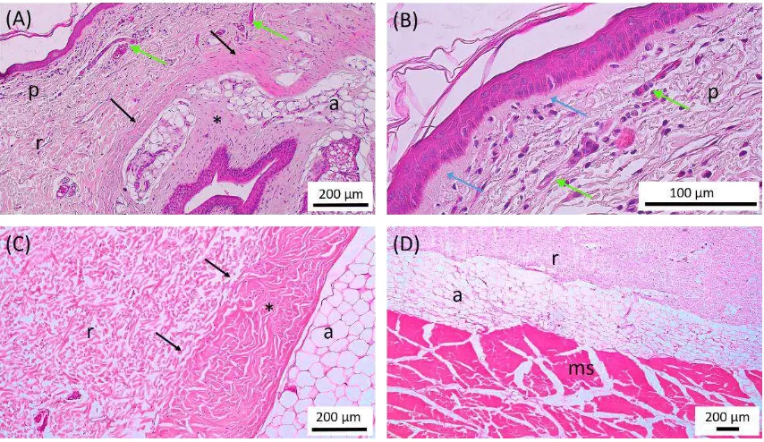
The connective tissue of the porcupine was spread over the basal membrane, the dermis layer and the hypodermis (Figure 2). The connective tissue composed of collagen fibers, adipose tissue, and non-fibrous components containing acidic carbohydrates (Figure 3). The connective tissue known as the extracellular matrix (ECM) contains a large number of collagen proteins and proteoglycans that have different structures and functions. Some parts of the proteoglycan molecule can bind, activate or inhibit cytokine activity and affect cell behavior with direct contact. The connective tissue composition is essential in regulating the biological function of a tissue (Clark, 1996).
Elastin fibers were not detected in all preparations stained by van Gieson method (Figure 3b). It showed that the porcupine's connective tissue did not have and/or had very small amount of elastin fibers. According to Kielty (2006), generally elastin fibers consist of three types of oxytalan fibers in the dermo-epidermal junction; elaunin fibers in the papillary layer of the dermis; and thick elastin fibers in the same position with the reticular layer of the dermis.
Woven collagen fibers were a major component in porcupine skin. Collagen was spread over the basal membrane, the papillary and reticular layers of the dermis, and the follicle of the thorns. Collagen fibers in the papillary layer in the dorsal thoracic region, thoracolumbal, dorsal lumbar, lumbosacral, and caudal
radix were composed of loose connective tissue whereas in the apex caudal region composed of thicker fibers containing many small blood vessels as well as fibroblast and fibrocyte cells (Figure 2b). The papillary layer and the epidermal basal membrane formed the dermo-epidermal junction. The basal membrane of the porcupine was generally thick except in the apex caudal region which was visibly attached to the collagen fibers in the papillary dermis layer. These results differ from those of common mammals, characterized by thin basal membranes. Basal membranes were known to be composed of various fiber and non-fibrous connective tissue components including collagen IV, glycoprotein and proteoglycans, including laminin, nidogen and perlecan (Breitkreutz et al., 2009).
Basal membrane plays a role in the regulation of cell movement and bioactive molecules between layers of the skin and binds to various cytokines and growth factors that play a role during wound healing, especially in the remodeling and repairing stages (Iozzo, 2005; Breitkreutz et al., 2009). The skin of Sunda porcupine is presumed to have similar connective tissue composition or might be other types of collagen component or other type of fibers in the structure of the basal membrane of the Sunda porcupine associated with its thickness. A thick and well-developed basal membrane was thought to be due to a larger basal membrane role in the skin of Sunda porcupine.
The dermo-epidermal junction layer appeared flat in most parts of the skin (Figure 2b) except in the apex caudal region and around quill orificium. Skin
characteristics with dermo-epidermal junctions that were flat, regular and have no clear dermal papilla were commonly found in mammals that do not have thick hair and non-hairy portions (Pough et al., 1996).
It was related to the condition of porcupine skin that had quills as a major derivative compared to in mammal hairs in general. In addition, this character also characterized skin that has a thin epidermis that generally had large blood vessels and nerves found in the profundal layer of the dermis which then branched to form capillary webbing in the papilary layer (Pough et al., 1996).
Dermo-epidermal junctions in the epidermis in the apex caudal region and around the orificium of quill follicles formed dermal papillae which had a great deal of elongation to the epidermis. This structure was similar to the skin, which had a thick epidermis and dermo-epidermal junction characteristics were generally irregular and formed a clear boundary to the epidermis and involved the formation of many dermal papillae (Pough et al., 1996).
The arrangement of collagen fibers in the reticular layer of the dermis differed in some of the body's regions. The dorsal thoracic region had a loose woven collagen fiber in all parts of the reticular layer of the dermis (Figures 2a and 2c). The thoraco-lumbal region up to the caudal radix had a graded woven from a loose to quite dense. The apex caudal region had woven collagen fibers that were relatively denser and larger against the thickness of the skin when compared to other regions. The thorn follicle had the most densely packed fibers in the casing and was visibly white in
macroscopic terms. These fibers had a clear boundary with the dermal reticular layer in the dorsal thoracic region to the caudal radix and were visibly attached to the apex caudal region (Figure 2a and 2c). The density of woven fibers on the sheath of the thorn follicles in Sunda porcupine was similar to that of the follicle sheath structure of the Erethizon dorsatum (porcupine) consisting of a dense collagen bundle network in a circular arrangement (Chapman and Roze, 1997). The thickness of the connective tissue of the follicle of the thorns varies depending on the region and the type of thorn. The thickest follicle sheath was found in the dorsal lumbar region, while the thinnest was found in the dorsal thoracic region. Collagen is a protein that is commonly found in body shaped like a thread with varying degrees of density. Collagen is generally white in fresh preparations and is eosinophilic. The more collagen found in the extracellular matrix, the stronger the tissue (Suvarna et al., 2013). The connective tissue collagen of the follicle of thorns in the apex caudal region does not form the capsule but is dispersed and fused with the connective tissue in the dermis.
The woven collagen fibers in the proximal part of the thorn follicle form elongation into the channel portion of the follicle channel forming a round compartment filled with fatty tissue (Figure 3). In contrast to the distal, collagen fibers did not form elongation. Collagen fibers located inside the follicle look straighter, loose and contain more blood vessels there are 28 types of collagen that have been identified, but the skin comprises about 90% of collagen type I and about 10% of collagen type III, and about 2% of collagen type V, while the rest is composed by other connective tissue types. The possibility of fibers composing the follicle of thorns in Sunda porcupines is the association and aggregation of various types of collagen. Type I, II, III, V, and XI collagen fibers are fibers that have the ability to associate large fibers with large molecular weights and have diameters ranging
from 25 to 400 nm (Hulmes and Miller, 1981). Collagen types I and III are major components in the reticular tissue in the interstitial space in some organs, including the skin. Collagen type I and III associate to form fibers in the reticular layer of the skin (Fleschmajer et al., 1990). The composition or ratio between certain types of collagen fibers can affect fibroblast function. Research conducted by Volk et al. (2011) reported that in mice with collagen III deficiency showed large myofibroblast differentiation and further increased wound contraction. In addition, the same mice also showed abnormalities of collagen I fibrillogenesis in the skin and other organs that showed that collagen III may have a role in the regulation of collagen I synthesis (Liu et al., 1997). This correlates that wound-free wound healing in fetuses is associated with high collagen III ratios of collagen I in some small and large mammal model animals (Merkel et al., 1988; Burd et al., 1990; Cuttle et al., 2005; Carter et al., 2009). The presence of varying distribution of collagen fibers in porcupine skin may be related to regulation of collagen fiber production, including certain physiological processes such as wound healing. connective tissue of the porcupine contained acidic carbohydrates (Figure 3c). The dosage stained with PAS showed a negative reaction in all skin regions studied (Figure 3d) which indicated that Sunda porcupines did not contain neutral carbohydrates (Table
2). Jӓrvelӓinen et al. (2009) stated that the acid
carbohydrate component (proteoglycans and
glycosaminoglycans) fill most of the interstitial space between the tissues and protein fibers. Negative charge and hydrophilic properties of this component play a role in hydration, buffering, and spreading pressure within the network. The most abundant proteins in the skin are hyaluronic acid, decorin, versican and dermatopontin (Carrino et al., 2000; Okamoto and Fujiwara, 2006). The hyaluronic acid component is known to have an important role in wound healing such
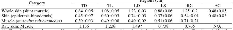
Table 1. Result of measurement of leather thickness of Sunda porcupine (Hystrix javanica)
Category Regions (cm)
TD TL LD LS RC AC
Whole skin (skint+muscle) 0.84±0.05 1.08±0.05 1.23±0.03 0.88±0.06 1.25±0.2 0.48±0.05 Skin (epidermis-hipodermis) 0.45±0.07 0.60±0.03 0.74±0.03 0.37±0.06 0.54±0.01 0.48±0.05 Muscle (musculus sub-cutaneous) 0.39±0.03 0.49±0.08 0.49±0.02 0.51±0.06 0.71±0.21 -
Rate skin: Muscle 1.136 1.226 1.497 0.738 0.765 N/A
(-)= The value could not be measured because the muscle layer was very thin and attached to the os. caudal vertebrae. The skin ratio if >1 then the skin component was thicker than musculus subcutaneous whereas if <1 then the skin component was thinner than musculus subcutaneous. N/A= Value ratio could not be calculated because one of the values was not met. TD= Thorax dorsal. TL= Thoracolumbar, LD= Lumbar dorsal, LS= Lumbosacral, RC= Radix caudal, AC= Apex caudal
Table 2. AB and PAS reactivity scores on Sunda porcupine leather connective tissue
Staining Regions
MB PD RD SFD SPFD FR
AB pH 2,5 + ++ + ++ +++ +++
PAS - - - -
as facilitating inflammatory processes, cell migration, proliferation, and collagen deposition (Chen and Abatangelo, 1999). Further research is felt necessary to know the type of acid carbohydrates in the porcupine skin tissue network.
The collagen-bound connective tissue of the skin had associations with adipose tissue, as distributed in thorn follicles, hypodermis, and dermis (Figure 2 and Figure 3). Adipose tissue was most commonly found in thorn follicles in the dorsal thoracic region up to caudal radix. In addition, adipose tissue was also present between the connective tissue of the follicle sheath. Adipose tissue in the apex caudal region was spread over the reticular layer of the dermis. Adipose tissue is an important component in porcupine skin. Adipose tissue plays a major role in energy reserves. This becomes an important part in the process of tissue proliferation, especially in hair and thorn cycles. In addition, fat cells are known to have the ability as stem cells. Various studies have been conducted to determine and develop the potential of fat cells as one source of stem cells (Zuk et al., 2001; Zuk et al., 2002). The ability of fat cells to be differentiated into other cells depends on the induction (Zuk, 2013). These multi-potential characteristics are thought to be related to the regeneration process or wound healing in porcupine skin.
CONCLUSION
The connective tissue of the porcupine is composed of collagen fibers, acidic non-fiber components and adipose tissue. The distribution of connective tissue in porcupines forms an important functional structure as a medium of interaction between cells and extracellular matrix that plays a role in various physiological processes such as skin regeneration and wound healing processes.
ACKNOWLEDGMENT
We are grateful to the Ministry of Research and Higher Education for funding research through the Master of Education scholarship program to Doctorate for Excellence (PMDSU) under contract number 330/SP2H/LT/ DRPM/IX/ 2016. We are also grateful to the Ministry of Environment and Forestry (KLHK) for granting permits to use animals from natural catches with the agreement letter of SK.284/KSDAE/SET/KSA.2/9/2016
REFERENCES
Akers, R.M. and D.M. Denbow, 2008. Anatomy and Physiology of
Domestic Animals. Blackwell publishing, Iowa, USA.
Barrientos, S., O. Stojadinovic, M.S. Golinko, H. Brem, and M. Tomic-Canic. 2008. Growth factors and cytokines in wound healing. Wound Repair Regen. 16:585-601.
Breitkreutz, D., N. Mirancea, and R. Nischt. 2009. Basement membranes in skin: Unique matrix structures with diverse functions? Histochem. Cell. Biol. 132:1-10.
Burd, D.A., M.T. Longaker, N.S. Adzick, M.R. Harrison, and H.P. expression of procollagen genes between midand late-gestational fetal fibroblasts. J. Surg. Res. 156:90-94.
Chapman, D.M. and U. Roze. 1997. Functional histology of quill erection in the porcupine, Erethizon dorsatum . Can. J. Zool. 75:1-10. Chen, W.Y.J. and G. Abatangelo. 1999. Function of hyaluronan in
wound repair. Wound Repair Regen. 7(2):79-89
Clark, R.A.F. 1996. Wound Repair: Overview and General Considerations. In The Molecular and cellular Basis of Wound
Repair. Clark, R.A.F. (Ed.). Plenum Press, New York.
Cobert, G.B. and L.A. Jones. 1965. The specific characters of the crested porcupines, subgenus Hystrix. J. Zool. 144:285-300. Cuttle, L., M. Nataatmadja, J.F. Fraser, M. Kempf, R.M. Kimble, and
M.T. Hayes. 2005. Collagen in the scarless fetal skin wound: detection with picrosirius-polarization. Wound Repair Regen.
13:198-204.
Fleschmajer, R., E.D. MacDonald, J.S. Perlish, R.E. Burgeson, and L.W. Fisher. 1990. Dermal collagen fibrils are hybrids of type I and type III collagen molecules. J. Struct. Biol. 105:162-169 Gawkrodger, D.J. 2002. Dermatology, an Illustrated Colour Text.
3rd ed. Churchill Livingstone, Edinburgh.
Hulmes, D.J. and A. Miller. 1981. Molecular packing in collagen.
Nature. 293:234-239.
Iozzo, R.V. 2005. Basement membrane proteoglycans: From cellar to ceiling. Nat. Rev. Mol. Cell. Biol. 6:646-656.
Jӓrvelӓinen, H., A. Sainio, M. Koulu, T.N. Wight, and R. Penttinen. 2009. Extracellular matrix molecules: Potential targets in pharmacotherapy. Pharmacol. Rev. 61:198-223.
Kent, G.C. and R.K. Carr. 2001. Comparative Anatomy of the
Vertebrates. 9th ed. McGrawl-Hill Companies, London, USA
Kielty, C.M. 2006. Elastic fibres in health and disease. Expert. Rev.
Mol.Med 8:1-23.
Liu, X., H. Wu, M. Byrne, S. Krane, and R. Jaenisch. 1997. Type III collagen is crucial for collagen I fibrillogenesis and for normal cardiovascular development. Proc. Natl. Acad. Sci. USA
94:1852-1856.
Merkel, J.R., B.R. DiPaolo, G.G. Hallock, and D.C. Rice. 1988. Type I and type III collagen content of healing wounds in fetal and adult rats. Proc. Soc. Exp. Biol. Med. 187:493-497.
Morin, P. and D. Berteaux. 2003. Immobilization of North American porcupines (Erethizon dorsatum) using ketamine and xylazine. J. Wildlife. Dis. 39(3):675-682.
Myers, P. 2001. “Hystricidae” (on-line), Animal Diversity Web.
http://animaldiversity.org/accounts/hystricidae.
Okamoto, O. and S. Fujiwara. 2006. Dermatopontin, a novel player in the biology of the extracellular matrix. Connect. Tissue Res.
47:177-189.
Pough, F.H., J.B. Heiser, and W.N. McFarland. 1996. Vertebrate Life. 4th ed. Prentice-Hall International, London.
Sheila. 2011. Klasifikasi Duri Landak Jawa Hystrix javanica
Berdasarkan Morfologi dan Pola Distribusi. Skripsi. Institut Pertanian Bogor. Bogor.
Shock, A. and G.J. Laurent. 1996. Cell Adhesion in Wound Healing and Pulmonary Fibrosis. In Lung Biology in Health and
Disease. Adhesion Molecules and the Lung. Ward, P.A. and
J.C. Fantone (Eds.). Marcel Dekker Inc., New York.
Suvarna, S.K., C. Layton, and J.D. Bancroft. 2013. Theory &
Practice of Histological Technique. 7th ed. Churdchill
Livingstone, New York.
Volk, S.W., Y. Wang, E.A. Mauldin, K.W. Liechty, and S.L. Adams. 2011. Diminished type III collagen promotes myofibroblast differentiation and increases scar deposition in cutaneous wound healing. Cells Tissues Organs. 194:25-37.
Zuk, P.A. 2013. Adipose-Derived Stem Cells in Tissue Regeneration: A Review. ISRN Stem Cells. Article ID713959.
http://dx.doi.org/10.1155/2013/713959.
Zuk, P.A., M. Zhu, H. Mizuno, J. Huang, J. W. Futrell, A. J. Katz, P. Benhaim, H. P Lorenz, and M.H. Hedrick, 2001. Multilineage cells from human adipose tissue: Implications for cell-based therapies. Tissue Eng. 7:211-228.