THESIS
RADIOLOGY BASED DETECTION OF
OSTEOPOROSIS IN CASTRATED CAT (
Felis catus
)
By:
IGNASIA PUTRI MISHA HAPSARI S.I.N. 061211133073
FACULTY OF VETERINARY MEDICINE UNIVERSITAS AIRLANGGA
iv Has been assessed in Result Seminar Date: August 10th, 2016
RESULT SEMINAR ASSESSMENT COMMITTEE
Head : Prof. Mas’ud Hariadi, drh., M.Phil., Ph.D. Secretary : Ira Sari Yudaniayanti, drh., MP.
vi
RADIOLOGY BASED DETECTION OF OSTEOPOROSIS IN CASTRATED CAT (Felis catus)
Ignasia Putri Misha Hapsari ABSTRACT
The aim of this research is to know the changes of bone density and the possibility of osteoporosis in castrated cats. Twenty cats were used and grouped according to how long castration had been performed and intact cats. The first, second and third groups were castrated for ± 3, 4 and 5 years respectively. The changes of bone density in cats were observed based on radiological image interpretation focused on proximal end of the femur. Data were scored using Singh Index. Kruskal-Wallis H Test showed a significant difference (p < 0.05) on bone density loss. Further analysis using Mann Whitney (Independent Sample T- Test) showed that cats that have been castrated for ± 3 years and for ± 5 years, ± 4 years and intact cats and ± 5 years and intact cats had significance different. In addition, cats that had been castrated for ± 3 years and intact cats, ± 3 years and ± 4 years and ± 4 years and ± 5 years had no significance different. It could be concluded that there was no sign of osteoporosis in castrated cats, only decreasing radiopacity but still in normal stage.
Key words: radiology, castration, osteoporosis, cats .
vii
ACKNOWLEDGEMENT
First of all, praise and thank God for all His blessing and guidance for author to finish the thesis entitled:
RADIOLOGY BASED DETECTION OF OSTEOPOROSIS IN CASTRATED CAT (Felis catus)
In this occasion, author would like to thank:
Prof. Dr. Pudji Srianto, drh., M.Kes. as the dean of Faculty of Veterinary Medicine Universitas Airlangga, for giving the opportunity to study in Faculty of Veterinary Medicine Univeritas Airlangga.
Rudy Sukamto Setiabudi, drh., M.Sc. as the supervisor and Suzanita Utama, drh., M.Phil., Ph.D as co-supervisor for the patience, advice, time, and guidance in the process of making this thesis.
The examiner committee, Prof. Mas’ud Hariadi, drh., M.Phil., Ph.D. as the chair of examiner, Ira Sari Yudaniayanti, drh., MP. as the secretary of examiner, and Hardany Primarizky, drh., MVM. as the member of examiner for the guidance and advice in the process of making this thesis.
All of the lecturer and entire staff of Faculty of Veterinary Medicine, Universitas Airlangga. To the academic advisor, Prof. Nunuk Dyah Retno Lastuti, drh., M.Si. for all the scientific insight during the learning process in Faculty of Veterinary Medicine, Universitas Airlangga. And to Dr. Soeharsono, drh., M.Si. for the guidance in the process of data analysis.
All of staff in Veterinary Clinical Department of Faculty of Veterinary Medicine, Universitas Airlangga. Also for my acquaintances and friends in
viii
Surabaya Save Paws for their help in the process of the research.
For my mother Irene Primiyanti for the love, prayer, care and support. To my grandmother, Caecilia Sutardjiah, my sisters Angela Kezia Damayanti and Brigitta Lintang Saura, My uncles, aunts and cousins for the support for the love and motivation during the research process.
For my best friends Ruth Tyas Advendtine, Yoana Jun, Sasi Kirana, Fithria Nisa Hanifah, Gamelita Rizkawandi, Annisa Karimah, Alfonsus Arianto Wibowo, Samuel Anugerah, Gowri Marriapan, Tiofani and M. Hadi S., who always love and support me whenever I need motivation.
And also all of my friends in Small Class of 2012, KMPV Pet and Wild Animal, SKK FKH UA and all of my friends in Faculty of Veterinary Medicine of Universitas Airlangga that cannot be mentioned one by one, who always support and help author wholeheartedly in the preparation of this thesis. Author realize that this writing is still far from perfection. Therefore, criticism and constructive suggestions are very expected. Author hope this paper can be one of the useful information source for the veterinary medicine field.
Surabaya, August 2016
Author .
TABLE OF CONTENTS
ACKNOWLEDGEMENT ... vii
TABLE OF CONTENTS ... ix
LIST OF TABLES ... xi
LIST OF FIGURES ... xii
LIST OF APPENDICES ... xiii
ABBREVIATIONS AND SYMBOLS ... xiv
CHAPTER I INTRODUCTION ... 1
2.4.1 Bone Development/Osteogenesis ... 10
2.4.2 Composition and Structure of Bone ... 11
2.5 Osteoporosis ... 13
2.6 Radiology ... 15
2.6.1 Image Generation ... 16
2.6.2 Principle of Interpretation ... 16 .
x
CHAPTER III MATERIALS AND METHOD ... 18
3.1 Time and Location of Research ... 18
3.2 Materials of Research ... 18
3.2.1 Experimental Animal ... 18
3.2.2 Research Materials ... 18
3.2.3 Research Equipments ... 18
3.3 Methods of Research ... 19
3.3.1 Preparation of Experimental Animal ... 19
3.3.2 Radiology Interpretation ... 19
3.4 Research Variable ... 19
3.5 Experimental Design and Data Analysis ... 19
3.6 Research Framework ... 23
CHAPTER IV RESEARCH RESULT ... 24
CHAPTER V DISCUSSION ... 26
CHAPTER VI CONCLUSSION AND RECOMMENDATION ... 29
SUMMARY ... 30
REFERENCES ... 34
LIST OF TABLES
Page Table 3.1 Singh grades and signifies ... 21 Table 3.2 Scoring table used for data analysis ... 22 Table 4.1 Scoring result analyzed with Mann-Whitney (Independent Sample
T-Test) ... 25
xii
LIST OF FIGURES
Page Figure 2.1 Domestic cat ... 7 Figure 2.2 The reproductive system of the male cat ... 9 Figure 2.3 A typical long bone shows the gross anatomical
characteristics of bone ... 12 Figure 2.4The trabecular architecture of normal bone (left) is lost in
osteoporotic bone (right) ... 14
Figure 3.1 Structure pattern of the proximal femur ... 20 Figure 4.1 Representative of radiology imaging and scoring result of
castrated and intact cats... 24
LIST OF APPENDIX
Page
1. Data Identification of Cats ... 40
2. Data Scoring ... 42
3. SPSS Data Analyzing ... 43
4. Research Documentation ... 54
xiv
ABBREVIATIONS AND SYMBOLS
AR : Androgen Receptor C18 : 18 carbon
cAMP : Cyclic Adenosine Monophosphate COLIA1 : Collagen Type 1 alpha 1 chain CT : Computed Tomography CYP : Cytochrome P 450 DHT : Dihydrotestosterone
DMPA : Depot Medroxyprogesterone Acetate DXA : Dual-energy X-ray absorptiometry E2 : Estradiol
ERα : Estrogen Receptor-α ERβ : Estrogen Receptor-β
FSH : Follicle-Stimulating Hormone IGF-II : Insulin-like Growth Factor 2 GnRH : Gonadotropin-Releasing Hormone LH : Luteinizing Hormone
mRNA : messenger Ribonucleic Acid pg : picogram
PTH :Parathyroid Hormone rRNA : ribosomal Ribonucleic Acid Sp1 : Specificity protein 1
TGF- β : Transforming Growth Factor beta 1 .
± : more or less < : less than % : percent
α : alpha
β : beta
1
CHAPTER I INTRODUCTION
1.1 Research Background
Domestic cats are considered to be prolific breeders. Female cats are seasonally polyoestrous with prolonged anoestrus resulting from decreasing or short day length (Johnston et al., 1996). The onset and duration of ovarian activity
is also linked closely to day length. In the northern hemisphere, female cats cycle between January and September with peaks of sexual activity in February, May and June, and occasionally in September (Ptaszynska, 2009).
Cats usually become sexually mature between 5-12 months of age for both male and females. The gestation period lasts 60 to 65 days (Foster and Smith, 2016). Females can have one to two litters a year and most cats give birth to four to six kittens a litter, with the first litter at five to six months of age (Clutton-Brock, 2014). There can be high neonatal and juvenile mortality; causes include infectious disease, trauma from road accidents, malnutrition and predator attacks (Nutter et al., 2004).
Longevity of stray cats is estimated at 4-5 years, while domestic cats can live 15-17 years as house pets (Ogen and Jurek, 1997). Every year in the United States an estimated 6 to 8 million lost, abandoned or unwanted dogs and cats enter animal shelters. According to the American Humane Association, the most common reasons why people relinquish or give away their cats is because their place of residence does not allow pets (29%), not enough time, divorce/death, health and behavior issues (10% each) (ASPCA, 2015).
The objectives of a program to control the cat population may include improve health and welfare of owned and stray cat population and prevent spread of disease to human (LaCroix, 2006). The more effective way is to sterilized male cats, because male cats contribute to the companion animal overpopulation crisis even more than females do. Just one unsterilized male can impregnate dozens of females, creating dozens upon dozens of unwanted offspring (PETA, 2013).
There are methods for the non-surgical control of reproduction to control cat population. In chemical sterilization method, GnRH is one target for fertility inhibitors. GnRH controls the release of the pituitary gonadotropins, LH and FSH, which in turn control the production of sex hormones and ultimately ovulation, spermatogenesis, and sexual behavior (Miller, 2013).
At present the most promising methods are the development of immunocontraception/sterilization vaccines or cytotoxin conjugates because both can be delivered as a single dose. Other methods currently rely on repeated dosing for long-term suppression of reproduction (Tasker, 2008).
In surgical method, castration on male cat is necessary as it is causing permanent infertility and can significantly reduce sexual behavior. Castration or orchidectomy is the surgical removal of a male cat's testicles for feline population control, medical health benefit, genetic-disease control and behavioral modification (Mohan-Gibbons and Zawistowski, 2015). During the procedure, each of the cat's testis and testicular epididymis are removed along with sections of the cat's testicular blood vessels and spermatic ducts (Schneck, 2009).
The testis performs two basic functions, sperm production and testosterone secretion (Shalet, 2009). Testosterone provides the necessary substrate for
aromatization to estradiol in the testes and in peripheral tissues, including locally in bone (Khosla et al., 2001). Orchidectomy not only reduces testosterone but also
estradiol serum levels (Erben, 2001).
In a research using orchidectomized male beagle dogs, the bone volume, mean trabecular thickness, and the fraction of labeled trabecular surface decreased significantly compared with the pre-orchidectomy values. These findings indicate an imbalance in bone metabolism ( i.e. bone resorption > bone formation). These results indicate that a loss of bone volume accompanied the fall in sex hormone levels following orchidectomy (Fukuda and Iida, 1999).
In a research using male rats as research material, there are a few reports of decreasing bone mass starting 4 weeks after orchidectomy. This led to considerable bone mass loss at 8 and 10 weeks. There are studies which found decreasing bone mass after orchidectomy and reports of osteoporosis (Ryu et al.,
2015).
Estradiol levels are strongly associated with bone mass density, bone turnover and bone loss in adult men (Vandenput and Ohlsson, 2009). Estradiol slows the rate of bone remodeling and protect against bone loss. Conversely, loss of estradiol leads to increased rate of remodeling and tilts the balance between bone resorption and formation (Manolagas et al., 2002).
Osteoporosis occurs when there is an imbalance between new bone formation and old bone resorption (Feng and McDonald, 2011). Osteoporosis is characterized by reduced bone mass, deterioration of bone micro-architecture and increased risk of low-trauma fractures. Fragility fractures are the endpoint of
osteoporosis and represent the major cause of morbidity and mortality (Marini and Brandi, 2009).
1.2 Problem Statement
1. What is the effect of length of time of castration in bone density? 2. Is there any possibility of osteoporosis in castrated cats?
1.3 Theoretical Basis
Surgical sterilization has been the cornerstone of efforts to curb pet overpopulation. In males, each testis with attached epididymis is removed in a procedure commonly referred to as neutering or castration. In cats, both gonads are usually removed through a single incision made just anterior to the scrotum and the incision is sutured closed. Estradiol in males produced more in testicles, orchidectomy not only reduces testosterone but also estradiol serum levels (Erben, 2001).
Both gonadal and adrenal testosterone can be converted into estrogens (C18 steroids) by the P 450 aromatase, encoded by CYP 19, which is present in many peripheral tissues, including bone. Bone cells express androgen receptor (AR) as well as estrogen receptor-α (ERα) and – β (ERβ). Therefore, androgen action on male bone may be explained by AR activation or, alternatively, activation of ERα and –β (Sinnesael, 2011). Testosterone, dihydrotestosterone (DHT) and non-aromatizable androgens increase the proliferation of osteoblast-like cells in culture, and induce osteoblast differentiation. The mechanism of action is believed to be through a complex endocrine, paracrine and autocrine fashion. DHT increases the expression of TGF-β mRNA and increases the mitogenic response to fibroblast growth factor and to IGF-II. Testosterone and
DHT reduce cAMP response and prostaglandin E2 production in bone cultures
exposed to stimulation with parathyroid hormone (PTH). The effects of androgen on PTH action suggest that androgen could modulate bone turnover in response to PTH (Manolagas and Kousteni, 2012).
In sex steroid deficiency, loss of transcriptional effects may be responsible for the increased osteoclastogenesis and osteoblastogenesis and thereby the increased rate of bone remodeling. Loss of nongenotropic anti-apoptotic effects on mature osteoblasts and osteocytes, in combination with an opposite effect on the lifespan of mature osteoclasts, may be responsible for the imbalance between formation and resorption and the progressive loss of bone mass and strength (Compston, 2001).
The radiologic appearances of osteoporosis are essentially the same no matter what the cause. Despite the advent of newer and highly accurate and precise quantitative techniques such as DXA and quantitative CT, osteoporosis is still most commonly diagnosed at conventional radiography. Moreover, variability in technical factors, such as radiographic exposure, film development, and soft-tissue thickness in patients, could make the diagnosis difficult. The main radiographic features of generalized osteoporosis are increased radiolucency and cortical thinning. Increased radiolucency is the result of resorption and thinning of the trabeculae, some of which may be lost. As a consequence, the term
osteopenia is used as a generic designation for radiographic signs of decreased
bone density (Guglielmi et al., 2010)
1.4 Aim of Research
To find out the changes of bone density and possibility of osteoporosis in castrated cats.
1.5 Outcomes of Research
1. This research gives information about the bone density of cats that underwent castration surgery.
2. This research gives information about the possibility of osteoporosis in castrated cats.
1.6 Hypothesis
Castration may increases risk of loss in bone density and osteoporosis in male cats. The longer the cats castrated, higher the risk of decreased bone density.
7
CHAPTER II LITERATURE REVIEW
2.1 Classification and Characteristics of Cat
Cats are very sensitive to sound, with a range of hearing both above and below the range of frequencies that can be detected by people. Feline hearing also acts as a direction finder, which is useful for hunting purposes. The ear canal of cats is deeper and more tapered than in people. The semicircular canals, which are found within the inner ear, are filled with fluid and are important for maintaining balance. These are highly developed in cats, accounting for their agility and excellent sense of balance (Shaw and Martin, 2015).
Figure 2.1 Domestic cat (Hogan, 2015)
The sense of smell is less developed in cats than in dogs. Like people, odor is an extremely important part of taste and enjoyment of food for cats. Cats that have lost their sense of smell due to illness often stop eating completely. The bottom of the paw in cats is covered by thick, resilient pads that cushion the foot and help provide a secure grip on many types of surfaces. Feline claws are retractable, very sharp and curved, which makes it easier to grasp prey while hunting or to slash during fights over territory (Bukowski, 2011).
Cat taxonomy is as follows (Wilson and Reeder, 2005): Kingdom : Animalia
Phylum : Chordata Class : Mammalia Order : Carnivora Family : Felidae Genus : Felis
Species : Felis catus 2.2 Reproduction of Cat
2.2.1 Male Reproductive System
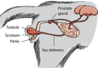
In the male, the important structures of the reproductive system are the testicles, ductus or vas deferens, prostate gland, and penis. Sperm production and storage occurs within the testicles. Upon ejaculation, sperm is transported to the prostate gland by the vas deferens. Within the prostate, additional fluids are added to the sperm to nourish it and aid in its transport from the penis and through the uterus (Johnson, 2011).
The penis is located below the scrotum and directed backward. A major portion of the feline penis is covered with spines,facing the body of the cat. The male cat’s testicles are descended into the scrotum at birth, and by three and a half months of age the testicles are developed enough to produce testosterone. Testosterone initiates the growth of penile spines, which reach mature size when the car is six to seven months old (Whiteley, 2006)(Figure 2.2).
Figure 2.2 The reproductive system of the male cat (Johnson, 2011)
2.2.2 Sexual maturity
The male cat reaches sexual maturity and begins to produce sperm varies from 6 to 18 months old. Ideally, a male cat should not be bred before he is 18 months. In male cats, there is no seasonal period of increased or decreased sexual activity. Rather, they can be stimulated at any time by nearby females that are in season (heat) (Elderedge et al., 2008).
2.3 Castration on Cat
Castration or orchidectomy is the surgical removal of a male cat's testicles for feline population control, medical health benefit, genetic-disease control and behavioral modification (Mohan-Gibbons and Zawistowski, 2015).
During the procedure, both testicles are removed by cutting carefully through the skin just in front of the scrotum, and through the various layers which cover the testicle. The very large blood vessels and the spermatic cord have to be tied carefully before cutting, allowing removal of the testicle. The layers are then closed with sutures, which may be visible on the surface or may be buried. Further drugs can be given as needed. This surgery requires general anesthesia (Little, 2015).
The main advantages of castrating a male cat are prevention of breeding, prevention of testicular cancer, reduction in the risk of prostate problems (including prostate cancer) and modification of certain behaviours. Testosterone greatly affects aggression in cats. One of the most important behavioral advantages of castration is that as adults, these neutered cats will tend to be less aggressive toward other cats (Foster & Smith, 2013).
2.4 Bones
Bone is the substance that forms the skeleton of the body. It is composed chiefly of calcium phosphate and calcium carbonate. It protects the vital organ, provides an environment for marrow (both blood forming and fat storage), acts as a mineral reservoir for calcium homeostasis and a reservoir of growth factors and cytokines and also takes part in acid-base balance (Taichman, 2005).
2.4.1 Bone Development/Osteogenesis
Bone is composed of support cells, osteoblasts and osteocytes; remodeling cells, osteoclasts; and non-mineral matrix of collagen and non-collagenous proteins called osteoid, with inorganic mineral salts deposited within the matrix (Mackie, 2003). Osteoblasts is mononucleated “bone forming” cells found near the surface of bones. They are responsible for making osteoid, which consists mainly of collagen. The osteoblasts then secrete alkaline phosphatase to create sites for calcium and phosphate deposition, which allows crystals of bone mineral to grow at these sites. The osteoid become mineralized, thus forming bone. Osteocytes are osteoblasts that are no longer on the surface of the bone, but are instead found in lacunae between the lamellae in bone. Their main role is homeostasis i.e. maintaining the correct oxygen and mineral levels in the bone.
And the osteoclasts is multinucleated cells responsible for bone resorption. They travel to specific sites on the surface of bone and secrete acid phosphatase, which unfixes the calcium in mineralized bone to break it down (McCloskey and Furnival, 2013).
The development of the vertebrate skeletal elements may take place through two different mechanisms: osteoblastogenesis and osteochondrogenesis, and relies on the differentiation of the required cell types: osteoblasts and chondrocytes (intra-membranous and endochondral ossification), which are derived from common mesenchymal stem cells.
Intra-membranous ossification takes place in the mesenchymal membrane, where osteoblast progenitor cells differentiate directly from embryonic condensed mesenchyme, mature to osteoblasts and begin to secrete type I collagen, proteoglycans and intercellular substance of the organic component of bone – osteoid. These mechanisms are responsible for the development of skull bones, facial bones and parially colarbone. Endochondral ossification takes place in long bones starting with the condensation of skeletal precursors, proceeding to the formation of a cartilagenous template. A cartilaginous template is initially formed and subsequently mineralized and replaced bone (Witkowska-Zimny, 2012). 2.4.2 Composition and Structure of Bone
The biochemical structure of bone is as follows: organic substances, 20 to 40%; inorganic (mineral) substances, 50 to 70%; water, 5 to 10% and <3% lipids. The organic component of bone includes collagen, proteoglycans, matrix proteins, cytokines and growth factors. Inorganic components include calcium hydroxyapatite and osteocalcium phosphate (Clarke, 2008).
The functional component of the bone includes growth factors and cytokines. The hardness and rigidity of bone is due to the presence of mineral salt in the osteoid matrix, which is a crystalline complex of calcium and phosphate (hydroxyapatite). Calcified bone contains about 25 % organic matrix, 5 % water and 70 % inorganic mineral (hydroxyapatite) (Brodsky and Persikov, 2005).
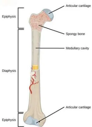
Figure 2.3 A typical long bone shows the gross anatomical characteristics of bone (Betts, 2013)
The end region of the bone is called the epiphysis and the middle region is called the diaphysis or bone shaft. The region between is called the metaphysis. Between the metaphysis and epiphysis is the epiphyseal disk or plate, which is responsible for longitudinal bone growth in childhood. It is at the epiphysis where one bone contacts another in a joint to allow for movement. Each epiphysis is coated with an articular cartilage. The articular cartilage is simply a coating of hyaline cartilage, which reduces friction and absorbs shocks at freely moveable joints (Snell, 2008)(Figure 2.3).
All bones are covered by a thin membrane called a periosteum. The periosteum is made of two layers of a dense connective tissue. The outer fibrous
layer consists of fibroblasts and collagen fibres. Long bones have a hollow region called the medullary cavity in the middle of the diaphysis. The perimeter of the medullary cavity is covered with an endosteum. The cavity itself is filled with marrow. Marrow can be either red or yellow depending on its function and composition. Red marrow is responsible for generation of blood cells and yellow marrow stores fat (Clarke, 2008).
There are two types of bone tissue: spongy and compact, also known as dense. Spongy bone makes up most of the tissue of epiphyses. It consists of lamellae arranged in an irregular latticework of thin plates of bone called trabeculae. The spaces between trabeculae are filled with red bone marrow. Compact bone structure is based on Haversian systems. Haversian systems are located in the diaphysis. They also cover spongy bone in the epiphyses. The functions of Haversian systems are to protect, support, and resist stress (Sjastaad
et al., 2010).
2.5 Osteoporosis
Osteoporosis occur when there is an imbalance between the amount of bone resorbed and that formed at each remodeling site due to decrease in the functional capacity of osteoblasts or to a decrease in the number of osteoblasts recruited to resorption sites (Drife and Studd, 2012). Osteoporosis is a disease marked by loss of bone tissue and low bone mass, which can lead to bone weakness and fragile bones (Thompson, 2004).
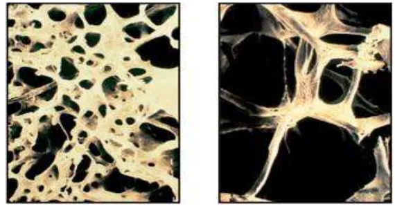
The main cause of osteoporosis is a lack of specific hormones; in particular, estrogen in women and androgen in men. Hypogonadism contributes to bone loss in 20–30% of elderly men, and in both sexes hyperparathyroidism secondary to calcium malabsorption increases remodelling, loss of trabecular bone, leading to vertebral crush fractures and Colles' fractures (Khosla, 2010) (Figure 2.4).
Figure 2.4The trabecular architecture of normal bone (left) is lost in osteoporotic
bone (right)(Kalpakcioglu et al., 2008)
Testosterone, however, is converted into estradiol via aromatization in many tissues including male bone. The importance of estrogen receptor alpha activation as well of aromatization of androgens into estrogens was highlighted by a number of cases of men suffering from an inactivating mutation in the estrogen receptor alpha or in the aromatase enzyme. All these men typically had low bone mass, high bone turnover and open epiphyses. In line with these findings, cohort studies have confirmed that estradiol contributes to the maintenance of bone mass after reaching peak bone mass, with an association between estradiol and fractures in elderly men (Sinnesael et al., 2011).
In human, bone mass peaks during the third decade of life. After this point, the amount of bone in the skeleton typically begins to decline slowly as removal of old bone exceeds formation of new bone. By age 65 or 70, however, men and women are losing bone mass at the same rate, and the absorption of calcium, an essential nutrient for bone health throughout life, decreases in both sexes (NIH, 2015).
The genetic component is characterized by variations in bone mineral density (BMD), bone quality, bone geometry, bone turnover and bone fragility in both men and women. The heritability of BMD is between 50 and 85%. Recent studies have focused on type I collagen, the major structural protein of bone, and especially, the COL1A1 (Collagen Type 1 alpha 1 chain) gene which encodes the major constituent of Type I collagen. Variations in the structure of the α1 (I) chain of type I collagen may result in an alteration of bone quality and mass and predispose the patient to osteoporotic fractures. A mutation at the Sp1 site on the COL1A1 gene is associated with an increased risk for osteoporotic fracture (Specialty Laboratories, 2002).
Other conditions that may lead to osteoporosis include overuse of corticosteroids (Cushing syndrome), thyroid problems, lack of muscle use, bone cancer, certain genetic disorders, use of certain medications, and problems such as low calcium in the diet (Shiel, 2015).
2.6 Radiology
Radiology is a medical specialty in which a variety of radiologic methodologies are used to diagnose and treat diseases. Diagnostic radiology encompasses a variety of diagnostic and image guided therapeutic techniques,
including all aspects of radiological diagnosis (nuclear radiology, diagnostic ultrasound, magnetic resonance, computed tomography, interventional procedures, and the use of other forms of radiant energy). The radiologist's role has grown not only through great improvements in diagnosis, but also through the technological developments that permit numerous interventional radiology procedures. A diagnostic radiologist is the eye of medicine, helping the primary care physician diagnose and treat diseases (AAMC, 2003).
2.6.1 Image Generation
An x-ray (radiograph) is a noninvasive medical test that helps physicians diagnose and treat medical conditions. When irradiating matter, the quanta (photons) of the X-ray radiation interact with atoms of the matter. When passing through matter, the X-ray radiation generated primarily for radiographic imaging is attenuated to a greater or lesser extent depending on the matter density and thickness, recording an image on photographic film or a special detector
Dense bone absorbs much of the radiation while soft tissue, such as muscle, fat and organs, allow more of the x-rays to pass through them. As a result, bones appear white on the x-ray, soft tissue shows up in shades of gray and air appears black (Hertrich, 2005).
2.6.2 Principle of Interpretation
Radiographic imaging is possible because x-rays penetrate matter, x-rays absorption is a function of tissue and thickness and an image of the pattern if x-ray emergence from a patient can be created. Therefore a radiograph is the image of the number and distribution of x-rays that pass through the patient. The ability of a radiograph, whether analog or digital, to display subtle differences in x-ray
absorption is limited. This concept, termed contrast resolution, is what allows
adjacent structures to be discriminated from each other in the radiographic image (Thrall, 2013).
Radiopacity refers to the relative inability of electromagnetic radiation, particularly x-rays, to pass through a particular material. Radiolucency indicates greater transparency to x-ray photons. Materials that inhibit the passage of
electromagnetic radiation are called radiodense or radiopaque, while those that allow radiation to pass more freely are referred to as radiolucent. The term refers to the relatively opaque white appearance of dense materials or substances on radiographic imaging studies, compared with the relatively darker appearance of less dense materials (Novelline, 1997).
The relatively poor contrast of radiographs also means that the range of opacities visible radiographically can be described, in general terms, according to one of five general terms-air opacity, fat opacity, soft tissue opacity, mineral opacity and metal opacity (Thrall, 2013).
CHAPTER III MATERIALS AND METHODS
3.1
Time and Location of Research
This research was performed in Hospitalization Room of Veterinary Teaching Hospital Universitas Airlangga Surabaya. The duration of this research was about one month. It started from the middle of June until the end of July 2016.
3.2 Materials of Research
3.2.1 Experimental Animal
Experimental animals used in this study were 20, five to seven years old domestic male cats (Felis catus) consisted of 15 castrated and 5 intact cats.
Experimental animals that fulfilled criteria, obtained from the owners. Cats were healthy, not consuming any drugs contains substances that influenced bone health and bone mass such as calcium supplement, glucocorticoids, heparin, DMPA (depot medroxyprogesterone acetate), aromatase inhibitor, thiazolidinediones, proton pump inhibitors, loop diuretics, cyclosporine and antiretroviral therapy (Weng and Lane, 2007).
3.2.2 Research Materials
Research materials used rontgen film for X-ray imaging, developer liquid and image fixer liquid as the part of radiology imaging process.
3.2.3 Research Equipments
The equipments used in this study were conventional x-ray unit equipment, intensifying, cassette, grid, anti-radiation cloak, markers, dark room, illuminator, illuminator hanger, digital camera, paper label and notes.
3.3 Methods of Research
3.3.1 Preparation of Experimental Animal
All cats were taken for radiology diagnostic. This radiology diagnostic was mainly performed to determine the trabecular structural system from cancellous bone of the proximal femur of cat. Cats were categorized according to the distance of castration. First category is a group of cats castrated for more or less 3 years. Second category is a group of cats castrated for more or less 4 years. Third category is a group of cats castrated for more or less 5 years. Fourth category is a group of intact cats as control.
3.3.2 Radiology Interpretation
Proximal femur had been chosen for the site of radiology diagnostic. Results of radiology diagnostic were interpreted by a lecturer of Veterinary Clinical Department, Faculty of Veterinary Medicine, Universitas Airlangga. 3.4 Research Variable
Variables observed the description of trabecular structural system in the cancellous bone of the proximal femur of cat by radiology appearance interpretation. Dependent variable in this study was the compactness of cancellous bone of proximal femur mass. Independent variable was the length of castration. 3.5 Experimental Design and Data Analysis
According to Hasan (2005), descriptive statistics is only to elaborate or give particulars of the data or phenomena and conclusion on descriptive statistic that only aimed to the existing collection.
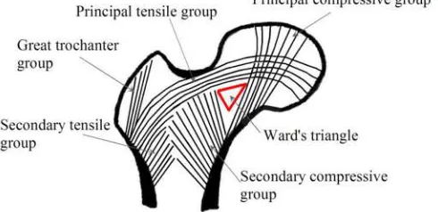
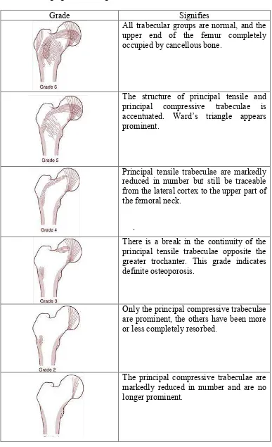
Data that obtained in this research was from radiology interpretation and analyzed with scoring based by Singh Index. Singh index is a method of grading trabecular bone loss in human. Based on the assessment of five anatomic group in the proximal end of the femur, which has been used to stratify the risk of femoral neck fracture in senile osteoporosis. The severity of bone loss can be graded on a scale from six (normal) to one (osteopenia) (Biswas, 2013).
Figure 3.1 Structure pattern of the proximal femur (Kwon, 2012) .
Table 3.1 Singh grades and signifies (Hoda, 2010; Wheeless, 2016)
Grade Signifies
All trabecular groups are normal, and the upper end of the femur completely occupied by cancellous bone.
The structure of principal tensile and principal compressive trabeculae is accentuated. Ward’s triangle appears prominent.
Principal tensile trabeculae are markedly reduced in number but still be traceable from the lateral cortex to the upper part of the femoral neck.
There is a break in the continuity of the principal tensile trabeculae opposite the greater trochanter. This grade indicates definite osteoporosis.
Only the principal compressive trabeculae are prominent, the others have been more or less completely resorbed.
The principal compressive trabeculae are markedly reduced in number and are no longer prominent.
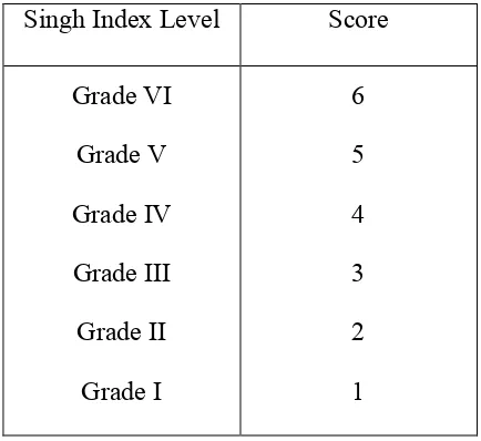
Table 3.2 Scoring table used for data analysis
Singh Index Level Score Grade VI
Grade V Grade IV Grade III Grade II
Grade I
6 5 4 3 2 1
Data that had been obtained was processed with SPSS program by computer. Data was analyzed by Kruskal-Wallis H Test and Mann-Whitney test (Independent Sample T-Test) for further analysis to know the changes of bone density and possibility of osteoporosis resulted from by castration surgery.
3.6 Research Framework
20 Cats
5 Cats Intact
Radiology Examination
Interpretation
Scoring
Analysis 5
Cats Castrated ± 3 years
5 Cats Castrated
± 4 years
5 Cats Castrated ± 5 years
CHAPTER IV RESULTS
This research used 20 cats that were divided into 4 groups according to how long castration had been performed (3, 4 and 5 years) and intact cats (control). All cats had their identification recorded (Appendix 1) and their X-ray image taken, interpreted and scored (Appendix 2).
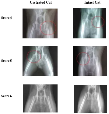
Castrated Cat Intact Cat
Score 4
Score 5
Score 6
Figure 4.1 Representative of radiology imaging and scoring result of castrated and intact cats; Score 4 = principal tensile trabeculae are markedly reduced in number but still be traceable from the lateral cortex to the upper part of the femoral neck; Score 5 = the structure of principal tensile and principal compressive trabeculae is accentuated. Ward’s triangle appears prominent; Score 6 = normal bone density
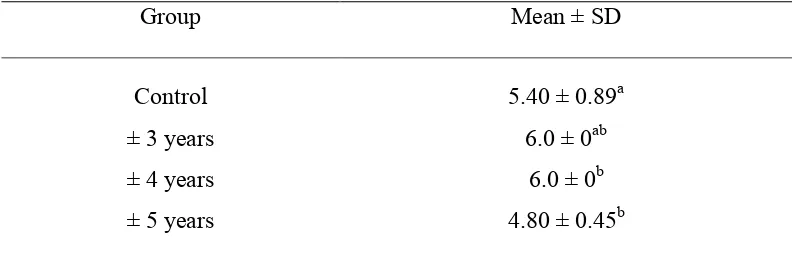
Based on the scoring result, data were analyzed with Kruskal-Wallis H Test showed a significance difference (p < 0.05). For further analysis, data were analyzed with Mann-Whitney (Independent Sample T- Test) for further analysis (Appendix 3).
Table 4.1 Scoring result analyzed with Mann-Whitney (Independent Sample T-Test)
a, ab, b: different superscript in the same column shows significant difference; ±3, 4, 5 years = cats have been castrated for ±3, 4, 5 years; Replicates = 5
The result showed that cats that castrated for more or less 3 years and intact cats have no significance different. Cats that castrated for more or less 3 years and for more or less 4 years, the statistical analysis showed that the result has no significance different. Cats that castrated for more or less 3 years and for more or less 5 years, the statistical analysis showed that the result has significance difference. Cats that castrated for more or less 4 years and for more or less 5 years, the statistical analysis showed that the result has no significance difference. Cats that castrated for more or less 4 years and intact cats, the statistical analysis showed that the result has significance different. Cats that castrated for more or less 5 years and intact cats, the statistical analysis showed that the result has significance different (Table 4.2).
CHAPTER V DISCUSSION
The result of this study that was analyzed by Kruskal-Wallis H Test showed significant difference (p < 0.05). Further analysis using Mann-Whitney (Independent Sample T- Test) showed cats that had been castrated for more or less 3 years and intact cats have no significance different. Cats that castrated for more or less 3 years and for more or less 4 years, the statistical analysis showed that the result has no significance different. Cats that castrated for more or less 3 years and for more or less 5 years, the statistical analysis showed that the result has significance difference. Cats that castrated for more or less 4 years and for more or less 5 years, the statistical analysis showed that the result has no significance difference. Cats that castrated for more or less 4 years and intact cats, the statistical analysis showed that the result has significance different. Cats that castrated for more or less 5 years and intact cats, the statistical analysis showed that the result has significance different.
All cats that used in this research fulfilled to the criteria that has been appointed. But, there is another factors that probably affecting the results such as food and compensatory action of another source of androgen. In this research, there are 2 cats (1 castrated cat and 1 intact cats) that showed decreasing bone density with Singh grade 4.
Decreasing in bone density could be caused by the loss of estradiol that leads to the increase of remodeling rate and tilts the balance between bone resorption and formation (Manolagas et al., 2002). In sex steroid deficiency, loss
of transcriptional effects may be responsible for the increased osteoclastogenesis .
and osteoblastogenesis and thereby the increased rate of bone remodeling. Loss of nongenotropic anti-apoptotic effects on mature osteoblasts and osteocytes, in combination with an opposite effect on the lifespan of mature osteoclasts, may be responsible for the imbalance between formation and resorption and the progressive loss of bone mass and strength (Compston, 2001).
Another factor that may affect the result is the consumption of mujair fish. Both of the cats that showed decreasing in bone density, have been identified diet with mujair fish. Mujair fish contains of 95 mg calcium and 190 mg phosphorus (Bogard et al., 2015). It is essential for animal health that provision be made for
the ratio and quantities of calcium and phosphorus required for development. The ideal ratio of calcium to phosphorous in the feline diet is approximately 1.1:1 (Pitcairn and Pitcairn, 2005).
High phosphorous intake leads to a chronically elevated serum parathyroid hormone (PTH) concentration (Adatorwovor et al., 2015). The elevation in PTH
results in increased intestinal reabsorption of calcium by stimulating the production of calcitriol, increased osteoclastic bone resorption, and increased renal tubular reabsorption of calcium (Moe, 2008).
Compensatory action of another source of androgen may affect the result. Other important site of aromatization is the abdominal fat tissues, which have been considered to be significant extra-gonadal sources of estrogens (Nimrod and Ryan, 1975). Androgen also secreted by the adrenal glands and it is well-known that prostatic tissue is capable of metabolizing adrenal androgens into testosterone and DHT (Van Weerden et al., 1990).
The age of cat when castration was done, may not have effect on bone density. But early neutering removes sex hormones, this delays maturation of osteoclasts resulting in the delayed closing of the growth plates of the long leg bones creating leggy taller bones (Hart et al., 2014).
29
CHAPTER VI CONCLUSION AND RECOMMENDATION
6.1. Conclusion
Based on the research that has been done, it can be concluded that there is no sign of osteoporosis in castrated cats, only decreasing radiopacity but still in normal stage.
6.2. Recommendation
1. Further research using feed categories.
2. Further research about osteoporosis in castrated cats based on other study focus is needed.
SUMMARY
Ignasia Putri Misha Hapsari, research with title “Radiology Based Detection of Osteoporosis in Castrated Cat (Felis catus)” under the supervision of
Rudy Sukamto Setiabudi, drh., M.Sc., as the first supervisor, Suzanita Utama. drh., M.Phil., Ph.D. as the co-supervisor, Prof. Mas’ud Hariadi, drh., M.Phil., Ph.D. as the head of examiners, Ira Sari Yudaniayanti, drh., MP. as the secretary of examiner, and Hardany Primarizky, drh., MVM as the member of examiner.
The aim of this research was to find out the changes of bone density and possibility of osteoporosis in castrated cats. This research was expected to be used as a reference to knowing the bone density of cat underwent castration surgery and as a literature to study about bone density and possibility of osteoporosis through cat that underwent castration surgery.
All cats were taken for radiology diagnostic. This radiology diagnostic was mainly performed to determine the trabecular structural system from cancellous bone of the proximal femur of cat.
Cats (Felis catus) that used in this study are divided into four groups by
its catagories. Proximal femur had been chosen for the site of radiology diagnostic.
Data that has been obtained analyzed by Kruskal-Wallis H Test and Mann-Whitney test (Independent Sample T- Test). The software used for data analysis is Statistical Program for Social Science (SPSS) 22.0 for Windows to know the changes of bone density and possibility of osteoporosis through cat that treated by castration surgery.
After radiology examination and interpretation, the result showed no osteoporosis detected in castrated and intact cats. The result was decreasing bone density in both castrated and intact cats, but still in normal stage. Cats with grade 4 were found in one castrated cat and one intact cat. Based on Singh index, it signifies principal tensile trabeculae are markedly reduced in number but still be traceable from the lateral cortex to the upper part of the femoral neck.
Based on the scoring result, data were analyzed with Kruskal-Wallis H Test showed there is a significance difference (p < 0.05).For further analysis, data were analyzed with Mann-Whitney (Independent Sample T- Test) for further analysis.
The result showed that cats that castrated for more or less 3 years and intact cats have no significance different. Cats that castrated for more or less 3 years and for more or less 4 years, the statistical analysis showed that the result has no significance different. Cats that castrated for more or less 3 years and for more or less 5 years, the statistical analysis showed that the result has significance difference. Cats that castrated for more or less 4 years and for more or less 5 years, the statistical analysis showed that the result has no significance difference. Cats that castrated for more or less 4 years and intact cats, the statistical analysis showed that the result has significance different. Cats that castrated for more or less 5 years and intact cats, the statistical analysis showed that the result has significance different.
Decreasing in bone density could be caused by the loss of estradiol that leads to the increase of remodeling rate and tilts the balance between bone resorption and formation (Manolagas et al., 2002). In sex steroid deficiency, loss
of transcriptional effects may be responsible for the increased osteoclastogenesis and osteoblastogenesis and thereby the increased rate of bone remodeling. Loss of nongenotropic anti-apoptotic effects on mature osteoblasts and osteocytes, in combination with an opposite effect on the lifespan of mature osteoclasts, may be responsible for the imbalance between formation and resorption and the progressive loss of bone mass and strength (Compston, 2001).
Another factor that may affect the result is the consumption of mujair fish. Both of the cats that showed decreasing in bone density, have been identified diet with mujair fish. Mujair fish contains of 95 mg calcium and 190 mg phosphorus (Bogard et al.,2015). It is essential for animal health that provision be
made for the ratio and quantities of calcium and phosphorus required for development. The ideal ratio of calcium to phosphorous in the feline diet is approximately 1.1:1 (Pitcairn and Pitcairn, 2005).
High phosphorous intake leads to a chronically elevated serum parathyroid hormone (PTH) concentration (Adatorwovor et al., 2015). The elevation in PTH
results in increased intestinal reabsorption of calcium by stimulating the production of calcitriol, increased osteoclastic bone resorption, and increased renal tubular reabsorption of calcium (Moe, 2008).
Compensatory action of another source of androgen may affect the result. Other important sites of aromatization is the abdominal fat tissues, which have been considered to be significant extra-gonadal sources of estrogens (Nimrod and Ryan, 1975). Androgens also secreted by the adrenal glands and it is well-known that prostatic tissue is capable of metabolizing adrenal androgens into testosterone and DHT (Van Weerden et al., 1990).
The age of castration may not have effect on bone density. But early neutering removes sex hormones, this delays maturation of osteoclasts resulting in the delayed closing of the growth plates of the long leg bones creating leggy taller bones (Hart et al., 2014).
Based on the research that had been done, it could be concluded that there was no sign of osteoporosis in castrated cats, only decreasing radiopacity but still in normal stage.
REFERENCES
Adatorwovor, R., K. Roggenkamp and J. J. B. Anderson. 2015. Intakes of Calcium and Phosphorus and Calculated Calcium-to-Phosphorus Ratios of Older Adults: NHANES 2005–2006 Data. Nutrients. 7(11): 9633–9639. ASPCA. 2015. Pet Statistics.
http://www.aspca.org/animal-homelessness/shelter-intake-and-surrender/pet-statistics [May 1, 2016]
Biswas, S.K. 2013. Orthopedics: A Postgraduate Companion. JP Medical Ltd. New Delhi, India.
Bogard, J.R., S. H. Thilsted, G. C. Marks, M. A. Wahab, M. A.R. Hossain , J. Jakobsen and J. Stangoulis. 2015. Nutrient Composition of Important Fish Species in Bangladesh and Potential Contribution to Recommended Nutrient Intakes. Journal of Food Composition and Analysis 42: 120–133 Brodsky, B. and A.V. Persikov. 2005. Molecular structure of the collagen triple
helix. Adv Protein Chem. 70:301–339
Challa, S. and J. Satyaprasad. 2013. Reliability of Singh Index in Grading of Osteoporosis Using Digital Radiographs in Elderly Patients with Proximal Femoral Fractures. International Journal of Development Research. Andhra Pradesh, India. 3(11): 159-161
Clarke, B. 2008. Normal Bone Anatomy and Physiology. Clin. J. Am. Soc. Nephrol. 3(3): S131–S139.
Clutton-Brock, J. 2014. DK Eyewitness Books: Cat. Dorling Kindersley Limited. London, UK.
Compston, J. E. 2001. Sex Steroids and Bone. Department of Medicine, University of Cambridge School of Clinical Medicine. Cambridge, United Kingdom. 81(1): 419-447.
Damilakis, J., J.E. Adams, G. Guglielmi and T.M. Link. 2010. Radiation Exposure in X-Ray-Based Imaging Techniques in Osteoporosis. Euro Radio. Foggia, Italy. 20: 2707-2714.
Drife, J.O. and J.W.W. Studd. 2012. HRT and Osteoporosis. Springer Science & Business Media. London, U.K.
Eldredge, Debra M., D. G. Carlson, L. D. Carlson and J. M. Giffin. 2008. Cat Owner's Home Veterinary Handbook. John Wiley & Sons, Inc. Hoboken, New Jersey.
Erben, R.G. 2001. Skeletal Effects of Androgen Withdrawal. J Musculoskel Neuron Interact 2001; 1(3):225-23.
Falahati-Nini, A., B.L. Riggs, E.J. Atkinson, W.M. O’Fallon, R. Eastell and S. Khosla. 2000. Relative contributions of testosterone and estrogen in regulating bone resorption and formation in normal elderly men. J. Clin. Invest. Minnesota, USA. 106: 1553–1560
Feng, X. and J.M. McDonald. 2011. Disorders of Bone Remodeling. Annual Review of Pathology: Mechanisms of Disease. Birmingham, Alabama. 6: 121-145.
Fink, H.A. , S.K. Ewing., K.E. Ensrud., E. Barrett-Connor., B.C. Taylor., J.A. Cauley and E.S. Orwoll. 2006. Association of Testosterone and Estradiol Deficiency with Osteoporosis and Rapid Bone Loss in Older Men. The Journal of Clinical Endocrinology & Metabolism. Oregon, USA. 91(10): 3908–3915.
Fukuda, S. and H. Iida. 1999. Effects of Orchidectomy on Bone Metabolism in Beagle Dogs. Journal of Veterinary Medical Science. Chiba, Japan. 62(1): 69–73
Gennari, L., R. Nuti and J. P. Bilezikian. 2004. Aromatase Activity and Bone Homeostasis in Men. The Journal of Clinical Endocrinology & Metabolism. Siena, Italy. 89(12): 5898–5907.
Gilbert, S.F. 2000. Developmental Biology, 6th edition. Sinauer Associates. Sunderland, Massachusetts.
Guglielmi, G., S. Muscarella and A. Bazzocchi. 2010. Integrated Imaging Approach to Osteoporosis: State-of-the-Art Review and Update. The Journal of Continuing Medical Education in Radiology. Bologna, Italy. 31(5): 1343-1364.
Hart, B. L. , L. A. Hart, A. P. Thigpen and N. H. Willits. 2014. Long-Term Health Effects of Neutering Dogs: Comparison of Labrador Retrievers with Golden Retrievers. PLoS One. 9(7): e102241.
Hertrich, P. 2005. Practical Radiography. John Wiley & Sons. Erlangen, Germany.
Hess, R.A. 2003. Estrogen in The Adult Male Reproductive Tract : A Review. Reproductive Biology and Endocrinology. Urbana, USA. 1: 52.
Hoda, S. 2010. Orthopedics Notes: Singh index for femoral neck fractures. http://orthopedicnotes.blogspot.co.id/2010/05/singh-index-for-femoral-neck-fractures.html [May 3, 2016]
International Companion Animal Management Coalition. 2013. Humane Cat
Population Management Guidance.
http://www.ifaw.org/sites/default/files/Cat%20Pop%20Management.pdf [May 19, 2016]
Johnston, S.D., M.V. Root. and P. N. S. Olson. 1996. Ovarian and Testicular Function in the Domestic Cat: Clinical Management of Spontaneous Reproductive Disease. Anim Reprod Sci. 42: 261-274.
Kalpakcioglu, B.B., S. Morshed , K. Engelke and H.K. Genant. 2008. Advanced Imaging of Bone Macrostructure and Microstructure in Bone Fragility and Fracture Repair. J Bone Joint Surg Am 90 (1): 68-78
Khosla, S., L. J. Melton III and B.L. Riggs. 2002. Estrogen and the Male Skeleton. The Journal of Clinical Endocrinology & Metabolism. Minnesota, USA. 87(4): 1443–1450
Khosla, S. 2010. Update in Male Osteoporosis. The Journal of Clinicalogy and Metabolism. 95(1):3-10.
Kini, U. and B. N. Nandeesh. 2012. Physiology of Bone Formation, Remodeling, and Metabolism. Department of Pathology , St. John’s Medical College and Hospital , Bangalore, India. Springer-Verlag Berlin Heidelberg 2012 Kutzler, M and Wood, A. 2006. Non-surgical methods of contraception and
sterilization. Theriogenology. 66:514-525.
Kwon, J. Y, H. Naito, T. Matsumoto and M. Tanaka. 2012. Osteocyte Apoptosis-Induced Bone Resorption in Mechanical Remodeling Simulation - Computational Model for Trabecular Bone Structure. In: Ntuli, T. M. Biochemistry, Genetics and Molecular Biology. Apoptosis and Medicine. Intech. Rijeka, Croatia
LaCroix, A.E. 2006. Overview of Feral Cat Population Control. Animal Legal and Historical Center. Michigan State University College of Law.
Little, Susan. 2015. August's Consultations in Feline Internal Medicine, Volume 7. Elsevier Health Sciences. St. Louis, Missouri.
Manolagas S.C., S. Kousteni and R.L. Jilka. 2002. Sex Steroids and Bone. Recent Progress in Hormone Research. Little Rock, Arkansas. 57: 385-409. McCloskey, C and Furnival, T. 2013. Formation and Remodelling of Bone.
Dissemination of IT for the Promotion of Materials Science. University of Cambridge. http://www.doitpoms.ac.uk/tlplib/bones/formation.php [April 14, 2016]
Moe, S. M. 2008. Disorders Involving Calcium, Phosphorus, and Magnesium. Prim Care. 35(2): 215–vi.
Mohan-Gibbons, H. and S. Zawistowski. 2015. Animal Behavior for Shelter Veterinarians and Staff. John Wiley & Sons. Pondicherry, India.
Nasu, M., T. Sugimoto, H. Kaji and K. Chihara. 2000. Estrogen Modulates Osteoblast Proliferation and Function Regulated by Parathyroid Hormone in Osteoblastic SaOS-2 cells: Role of Insulin-Like Growth Factor (IGF)-I and IGF-Binding Protein-5. Journal of Endocrinology. Kobe, Japan. 167: 305–313.
NIH. 2015. Osteoporosis in Men. National Institutes of Health Osteoporosis and Related Bone Diseases National Resource Center. Maryland, USA.
Nimrod, A. and Ryan, K. J. 1975. Aromatization of androgens by human abdominal and breast fat tissue. J. Clin. Endocrinol. Metab. 40: 367-372. Novelline, R. 1997. Squire's Fundamentals of Radiology. 5th Edition. Harvard
University Press.
Nutter, F. B., J. F. Levine and M. K. Stoskopf. 2004. Reproductive Capacity of Free-Roaming Small Animals Domestic Cats and Kitten Survival Rate. In: Journal of the American Veterinary Medical Association. 225 (9): 1399-1402
Ogan, C.V. and R.M. Jurek. 1997. Biology and ecology of feral, free-roaming and stray cats. Pages 87-92 in J.E. Harris, and C.V. Ogan, (eds.), Mesocarnivores of northern California: Biology, management and survey techniques, workshop manual. August 12-15, 1997, Humboldt State University, Arcata, CA. The Wildlife Society, California North Coast Chapter, Arcata, CA 127 p.
Ohlsson, C. and L. Vandenput. 2009. The Role of Estrogen for Male Bone Health. European Journal of Endocrinology. Gothenburg, Sweden. 160: 883-889 Penido, M. G. M. G. and U. S. Alon. 2012. Phosphate homeostasis and its role in
bone health. Pediatr Nephrol. 2012 Nov; 27(11): 2039–2048.
PETA. 2013. Spay and Neuter. http://www.peta.org/issues/companion-animal-issues/overpopulation/spay-neuter/ [March 18, 2016]
Pitcairn, R. H. and S. H. Pitcairn. 2005. Dr. Pitcairn's New Complete Guide to Natural Health for Dogs and Cats. Rodale Inc. USA.
Ptaszynska, M. 2010. Compendium of Animal Reproduction. Intervet International. Boxmeer, Netherlands.
Raisz, L. G. 1999. Physiology and Pathophysiology of Bone Remodeling. Clinical Chemistry. 45(8): 1353–1358.
Ralston, S. H. and De Crombrugghe, B. 2006. Genetic Regulation of Bone Mass and Susceptibility to Osteoporosis. Genes and Development. 30(9): 2492-2506.
Ryu, JS, DS Ryu, JY Kim, JY Park, KH Kim, DK Chin, KS Kim, YE Cho and SU Kuh. 2015. Bone Mineral Density Changes after Orchiectomy using a Scrotal Approach in Rats. Korean J. Spine. Seoul, Korea. 12(2):55-59
Salter, R.B. 1999. Textbook of Disorders and Injuries of the Musculoskeletal System: An Introduction to Orthopaedics, Fractures, and Joint Injuries, Rheumatology, Metabolic Bone Disease, and Rehabilitation. Lippincott Williams & Wilkins. Baltimore, Maryland.
Shalet, S.M. 2009. Normal Testicular Function And Spermatogenesis. Pediatr Blood Cancer. 53(2):285-8.
Shaw, J. and Martin, D. 2015. Canine and Feline Behavior for Veterinary Technicians and Nurses. John Wiley & Sons, Inc. Oxford, UK.
Shneck, P.A. 2009. Saunders Comprehensive Review of the NAVLE. Elsevier Health Sciences. Missouri, USA.
Sinnesael, M., S. Boonen, F. Claessens, E. Gielen and D. Vanderschueren. 2011. Testosterone and the Male Skeleton: A Dual Mode of Action. Journal of Osteoporosis. 2011(12):240328
Sjaastad, O. V., K. Hove and O. Sand. 2010. Physiology of Domestic Animals. Scandinavian Veterinary Press. Oslo, Skandinavia.
Taichman, R.S. 2005. Blood and Bone: Two Tissues whose Fates Are Intertwined to Create The Hematopoeitic Stem Cell Niche. Blood. 105: 2631-2639. Tasker, L. 2008. Non-Surgical Methods For Controlling The Reproduction Of
Dogs And Cats. Internal Document: Guidance for WSPA Staff and Member Societies.
Thrall, D. E. 2013. Textbook of Veterinary Diagnostic Radiology. Elsevier Health Sciences. Raleigh, North Carolina.
Turner, D. C. and P. Bateson. 2013. The Domestic Cat: The Biology of its Behaviour. Cambridge University Press. UK.
Vandenput, L. and C. Ohlsson. 2009. Estrogens as regulators of bone health in men. Nature Reviews Endocrinology. 5: 437-443.
Van Weerden, W. M., G. J. Van Steenbrugge, A. Van Kreuningen, E. P. C. M. Moerings, F. H. De Jong And F. H. Schröder . 1990. Effects Of Low Testosterone Levels And Of Adrenal Androgens On Growth Of Prostate Tumor Models In Nude Mice. J. Sterom Biochern. Molec. Biol. 37(6): 903-907
Weng, M.Y. and N.E. Lane. 2007. Medication-Induced Osteoporosis. Curr Osteoporos Rep. 5(4):139-145.
Wheeless, C.R., J. A. Nunley and J. R. Urbaniak. 2016. Wheeless' Textbook of Orthopaedics. Duke University. Medical Center. Division of Orthopaedic Surgery. North Carolina, USA
Whiteley, H.E. 2006. Understanding and Training Your Cat or Kitten. Sunstone Press. Santa Fe, USA.
Wilson, D.E. and D.M. Reeder. 2005. Mammal Species of the World: A Taxonomic and Geographic Reference, 3rd ed., vols. 1 & 2. Johns Hopkins University Press. Maryland, USA.
APPENDIX 1. DATA IDENTIFICATION OF CATS
No Cat’s Name Age Information Disease Record Medicine Record Feed Owner Address
1 Popon 5 Castrated - - Dry and canned food Puspanita Surabaya
2 Nino 5 Castrated - - Dry food and boiled fish Ratna Surabaya
3 Abang 5 Castrated - - Dry food and pindang Dian Surabaya
4 Srigala 5 Castrated - - boiled chicken Dry food and Nimas Sidoarjo
5 Goldy 5 Castrated - - boiled chicken Dry food and Nimas Sidoarjo
6 Ilo 5 Castrated - - Dry food, mujair, pindang Febrico Surabaya
7 Bimbim 6 Castrated - - Dry and canned food Puspanita Surabaya
8 Bubu 6 Castrated Flu Kanamycin Vicilin, milkfish, rice, Dry food,
tongkol Krisnoadi Surabaya
9 Oliver 5 Castrated - - Dry food and pindang Yanto Surabaya
10 Romeo 6 Castrated - - Dry food and Yanto Surabaya
40
41
11 Utung 7 Castrated - - Dry food and mujair Ayu Surabaya
12 Iwet 6 Castrated - - Dry food, mujair and pindang Ajeng Sidoarjo
13 Kepi 7 Castrated - - Dry food and pindang Alin Surabaya
14 Ago 6.5 Castrated Flu - Dry food, mujair and pindang Ajeng Sidoarjo
15 Juki 6 Castrated - - pindang and Dry food,
chicken Ade Surabaya
16 Bidu 6 Intact Flu Acetromycin Rhinos, Dry food, mujair and pindang Dewi Surabaya
17 Nemo 6 Intact - - Dry food, mujair and pindang Dewi Surabaya
18 Nebo 6 Intact - - Dry food, mujair and pindang Dewi Surabaya
19 Nebi 6 Intact - - Dry food, mujair and pindang Dewi Surabaya
20 Tedy 5 Intact - - Dry food and fish Hira Sidoarjo
43 APPENDIX 3. SPSS DATA ANALYZING
Kruskal-Wallis Test
Ranks
groups N Mean Rank
Groups CONTROL 5 9.70
3 yrs 5 14.00
4 yrs 5 14.00
5 yrs 5 4.30
Total 20
Test Statisticsa,b
Groups
Chi-Square 12.783
df 3
Asymp. Sig. .005
a. Kruskal Wallis Test
b. Grouping Variable: groups
NPAR TESTS
/M-W= Groups BY groups(4 1)
/MISSING ANALYSIS.
Mann-Whitney Test
Ranks
groups N Mean Rank Sum of Ranks
Groups CONTROL 5 6.70 33.50
5 yrs 5 4.30 21.50
Total 10
Test Statisticsa
Groups
Mann-Whitney U 6.500
Wilcoxon W 21.500
Z -1.361
Asymp. Sig. (2-tailed) .174
Exact Sig. [2*(1-tailed Sig.)] .222b
a. Grouping Variable: groups
b. Not corrected for ties.
NPAR TESTS
/M-W= Groups BY groups(2 1)
/MISSING ANALYSIS.
Mann-Whitney Test
Ranks
groups N Mean Rank Sum of Ranks
Groups CONTROL 5 4.50 22.50
3 yrs 5 6.50 32.50
Total 10
Test Statisticsa
Groups
Mann-Whitney U 7.500
Wilcoxon W 22.500
Z -1.491
Asymp. Sig. (2-tailed) .136
Exact Sig. [2*(1-tailed Sig.)] .310b
a. Grouping Variable: groups
b. Not corrected for ties.
NPAR TESTS
/M-W= Groups BY groups(2 3)
/MISSING ANALYSIS.
Mann-Whitney Test
Ranks
groups N Mean Rank Sum of Ranks
Groups 3 yrs 5 5.50 27.50
4 yrs 5 5.50 27.50
Total 10
Test Statisticsa
Groups
Mann-Whitney U 12.500
Wilcoxon W 27.500
Z .000
Asymp. Sig. (2-tailed) 1.000
Exact Sig. [2*(1-tailed Sig.)] 1.000b
a. Grouping Variable: groups
b. Not corrected for ties.
NPAR TESTS
/M-W= Groups BY groups(2 4)
/MISSING ANALYSIS.
Mann-Whitney Test
Ranks
groups N Mean Rank Sum of Ranks
Groups 3 yrs 5 8.00 40.00
5 yrs 5 3.00 15.00
Total 10
Test Statisticsa
Groups
Mann-Whitney U .000
Wilcoxon W 15.000
Z -2.887
Asymp. Sig. (2-tailed) .004
Exact Sig. [2*(1-tailed Sig.)] .008b
a. Grouping Variable: groups
b. Not corrected for ties.
NPAR TESTS
/M-W= Groups BY groups(3 4)
/MISSING ANALYSIS.
Mann-Whitney Test
Ranks
groups N Mean Rank Sum of Ranks
Groups 4 yrs 5 8.00 40.00
5 yrs 5 3.00 15.00
Total 10
Test Statisticsa
Groups
Mann-Whitney U .000
Wilcoxon W 15.000
Z -2.887
Asymp. Sig. (2-tailed) .004
Exact Sig. [2*(1-tailed Sig.)] .008b
a. Grouping Variable: groups
b. Not corrected for ties.
NPAR TESTS
/M-W= Groups BY groups(1 3)
/MISSING ANALYSIS.
Mann-Whitney Test
Ranks
groups N Mean Rank Sum of Ranks
Groups CONTROL 5 4.50 22.50
4 yrs 5 6.50 32.50
Total 10
Test Statisticsa
Groups
Mann-Whitney U 7.500
Wilcoxon W 22.500
Z -1.491
Asymp. Sig. (2-tailed) .136
Exact Sig. [2*(1-tailed Sig.)] .310b
a. Grouping Variable: groups
b. Not corrected for ties.
Case Processing Summarya
Cases
Included Excluded Total
N Percent N Percent N Percent
Grade * groups 20 100.0% 0 0.0% 20 100.0%
a. Limited to first 100 cases.
Case Summariesa
Grade
groups Control 1 6
2 6
3 5
4 4
5 6
Total N 5
Median 6.00
3 yrs 1 6
2 6
3 6
4 6
5 6
Total N 5
Median 6.00
4 yrs 1 6
2 6
3 6
4 6
5 6
Total N 5
Median 6.00
5yrs 1 4
2 5
3 5
4 5
5 5
Total N 5
Median 5.00
Total N 20
Median 6.00
a. Limited to first 100 cases.
Case Processing Summarya
Cases
Included Excluded Total
N Percent N Percent N Percent
Singh Index * Group 20 100.0% 0 0.0% 20 100.0%
a. Limited to first 100 cases.
Case Summariesa
Singh Index
Group Control 1 6
2 6
3 5
4 4
5 6
Total N 5
Mean 5.40
Std. Deviation .894
3yrs 1 6
2 6
3 6
4 6
5 6
Total N 5
Mean 6.00
Std. Deviation .000
4yrs 1 6
2 6
3 6