Tendon injury healing with G-90 in a rabbit model: biomechanical
and histopathological evaluation
Amin Bigham-Sadegh1*, Mehran Arabi2, Effat Karimi3, Iraj Karimi4, and Ahmad Oryan5
1Department of Veterinary Surgery and Radiology, Faculty of Veterinary Medicine,
Shahrekord University, Iran
2Department of Biology, Faculty of Basic Sciences, Shahrekord University, Iran
3Department of Veterinary Surgery and Radiology, Faculty of Veterinary Medicine, Shiraz University, Iran 4Department of Veterinary Pathobiology, Faculty of Veterinary Medicine, Shahrekord University, Iran
5Department of Veterinary Pathology, School of Veterinary Medicine, Shiraz University, Shiraz, Iran
________________________________________________________________________________________ ________________________________________________________________________________________ BIGHAM-SADEGH, A., M. ARABI, E. KARIMI, I. KARIMI, A. ORYAN: : Tendon injury healing with G-90 in a rabbit model: biomechanical and histopathological evaluation.. Vet. arhiv 86, 407-420, 2016.
ABSTRACT
Tendon injuries are one of the most common and disabling acute orthopedic disorders. Several techniques are used to restore the mobility of patients, but all have signifi cant limitations. In some studies utilization of biomaterials has been investigated in the healing of tendons, skin and nerves. The present study was designed to investigate the effects of G-90, as a stimulating factor agent, on the healing of the superfi cial digital fl exor tendon (SDFT) of rabbits after 35 days post tenotomy and surgical repair. Twenty male rabbits, 12 months old and weighing 2.0 ± 0.5 kg were used in this study. All rabbits were anesthetized, and the superfi cial fl exor tendons of both hind limbs were cut transversely and then sutured with a Bunnel-Mayer suture pattern. After suturing tendons and skin, 0.5 mL of normal saline was injected in the injured tendon area of the left leg and 0.5 mL earthworm extract (EW) G-90 was injected at a concentration of 10 mg/mL into the injured tendon area of the right leg. Every two weeks 4 rabbits were euthanized, and samples were collected and sent for histopathological and biomechanical evaluation. In the histopathological evaluation less infl ammation, more maturity of fi brocytes and more aggregation of collagen fi bers were observed in tendons treated with G-90 in comparison with untreated tendons. In the biomechanical evaluation the ultimate strength of tendons treated with G-90 was superior to untreated tendons. The fi ndings of the present experiment clearly show that administration of G-90 could enhance the structural and biomechanical properties of the experimentally tenotomized SDFT in rabbits.
Key words: earthworm extract, G-90, tendon healing, rabbit model
________________________________________________________________________________________ ________________________________________________________________________________________
*Corresponding author:
Amin Bigham-Sadegh, DVM, DVSc, Department of Veterinary Surgery and Radiology, School of Veterinary Medicine, Shahrekord University, Postal code: 8818634141, PO Box: 115, Shahrekord, Iran, E-mail: dr.bigham@gmail.com
Introduction Introduction
Tendons are anatomic structures interposed between muscles and bones that transmit the force created in the muscle to the bone, and make joint movement possible (KANNUS, 2000). Tendons vary in form, and may be rounded cords, strap-like bands or fl attened ribbons. When healthy they appear brilliant white and have a fi broelastic texture. Structurally, a tendon is composed of tenoblasts and tenocytes, lying within the network of an extracellular matrix (ECM) (SHARMA and MAFFULLI, 2006). When tendons are subjected to high physiological loads, these tissues are commonly injured and fail to heal optimally, due to their low cellularity and vascularity (STRAUSS et al., 2007). Tendon injuries produce substantial morbidity, and at present there are only a limited number of scientifi cally proven management modalities (SHARMA and MAFFULLI, 2005). Tendons are at the highest risk for rupture if tension is applied quickly and obliquely, and the highest forces are seen during eccentric muscle contraction (SHARMA and MAFFULLI, 2005). Growth factors are short sequences of amino acids, which usually transmit signals between cells and thereby modulate their activities (CHAN et al., 1997; McGEACHIE and TENNANT, 1997). They regulate cell activity by a number of mechanisms, such as: mitogenic activity, cell differentiation, cell migration and gene regulation, and they play important roles in cell chemotaxis, proliferation, matrix synthesis, and differentiation. The use of growth factors to enhance tendon healing remains largely experimental and has been restricted to in vitro studies and animal models (SHARMA and MAFFULLI, 2005; McGEACHIE and TENNANT, 1997).
G-90 was obtained from the tissue homogenate of the earthworm Eisenia foetida (phylum Annelida, family Lumbricidae). Earthworms, which possess antibacterial activity, have been widely used in traditional Chinese medicine (HRZENJAK et al., 1992; POPOVIC et al., 2001; EL-KAMALI, 2000). It has been shown that G-90 is neither an allergen nor a toxin, and it possesses antibacterial activity which aids wound healing (POPOVIC et al., 1998; HRZENJAK et al., 1992; HRZENJAK et al., 1993; HRZENJAK et al., 1998). The G-90 mixture contains the growth factors of the insulin superfamily, adhesion of the immunoglobulin superfamily, and proteolytic enzymes of the trypsin family (GRDISA et al., 2001). Therefore, the present study was designed to investigate the effects of G-90, as a stimulating factor agent, on the healing of the superfi cial digital fl exor tendon (SDFT) of rabbits, after 35 days post tenotomy and surgical repair.
Materials and methods
Animals. Twenty male rabbits, 12 months old and weighing 2.0 ± 0.5 kg, were used in this study. Before the experiment, they were kept in their new location for 20 days for adaptation and stress removal. The rabbits were kept in individual standard rabbit cages and were maintained on a standard rabbit diet, with no limitation of access to food
or water. The experimental protocol was approved by the Animal Care and Experiment Committee of the University, in accordance with the ethics standards of the “Principles of Laboratory Animal Care”.
Preparation of G-90 complex.To prepare this compound, Hrzenjak et al.’s method was used (HRZENJAK et al., 1992). 200 earthworms of the species Eisenia foetida were washed with warm water several times to remove impurities, and then the worms were immersed in 10 % sodium chloride solution for one hour at room temperature until they expired. After this period, the worms were washed again, cut into pieces with scissors and homogenized with homogenizer machine. The mixture was transferred to a beaker, ethanol and chloroform in a 1:1 ratio were added to the solution, and it was left at 4 °C overnight, after which distilled water was added to produce a fi nal volume of 200 mL of mixture. After stirring, it was fi ltered several times, until the mixture was light brown in appearance. The mixture was centrifuged at 4000 rpm for at least 10 minutes in 50 mL Falcon® tubes (Corning Life Sciences, Corning, NY, USA). After centrifugation, 3
layers developed in each tube: The top layer was a clear, light brown-colored liquid, the middle layer was a brown-colored solid, and the bottom layer was a straw colored liquid. The solid middle layer was placed on a fi lter paper until the remaining liquid slowly evaporated, the pellet dried, and the brown color was clearly visible. The discs were transferred into 1000 mL balloons and freeze dried at -50 °C. The resultant powder (G-90) was placed under UV light for 30 minutes.
Surgical procedure. All the rabbits in the present study were sedated using acepromazine (0.02 mg/kg i.m., Alfasan, Woerden, Holland), the caudal parts of both hind limbs between the stifl e and hock joint were clipped and prepared aseptically, and the limb was draped with sterile drapes. Anesthesia was induced using ketamine (30 mg/kg, IM, Alfasan, Woerden, Holland). An incision was made directly over the Achilles tendon, the superfi cial digital fl exor tendon was exposed, cut transversely and then sutured with nylon (Supa, Tehran, Iran) 2/0 in a Bunnel-Mayer suture pattern. The skin was sutured routinely with silk suture. After suturing the tendon and skin, 0.5 mL of normal saline was injected in the injured tendon area of the left leg and 0.5 mL earthworm extract G-90 was injected at a concentration of 10 mg/mL into the injured tendon area of the right leg. Postoperatively, the antibiotic [enrofl oxacin(Bytril®, Bayer, Germany) 10 % at a dose of
5 to 10 mg/kg body weight] was injected subcutaneously for 3 days.
Sampling. At the 1st, 2nd, 3rd, 4th and 5th postoperative weeks 4 rabbits were euthanatized
with anesthetic overdosing for pathological and biomechanical evaluation. The treated tendons were excised and removed.
Biomechanical evaluation. Freshly harvested specimens were submitted to tensile strength measurement using a biomechanical analyzer (Instron, Canton, MA). The harvested tendons were clamped in the upper jaw and in the distal jaw. Each tendon
was loaded by elongating it at a displacement rate of 10 mm/s until a 50 % decrease in load was detected. During tensile testing no slippage was noted. Load and cross-head displacement data were recorded at 1500 Hz, and load-deformation and stress-strain curves were generated and biomechanical markers, including ultimate strength, stiffness and stress, were measured.
Histopathological evaluation. Immediately after the biomechanical tests, samples were fi xed using formalin solution (10 %) and transported to the pathology laboratory. The formalin solution was changed after 24 hours and then after 10 days, tissue samples were sectioned, stained with the H&E method, and observed under light microscopy. For histopathological evaluation, samples were scored qualitatively and semi-quantitatively based on a modifi ed Rosenbaum et al and Oryan et al scoring system (Table 1) (ROSENBAUM et al., 2010).
Table 1. Histopathological scoring system
Marker Scores
Infl ammation degree 0, 1, and 2 (qualitative) Fibroblast maturation 0, 1, and 2 (qualitative) Aggregation of connective fi bers 0, 1, and 2 (qualitative)
Connective fi bers alignment 1, 2, 3, and 4 (semi-quantitative)(1-25 %: 1), (25-50 %: 2), (50-75 %: 3), and (75-100 %: 4) Vasularization rate 1, 2, and 3 (semi-quantitative)Average number of vascular sections in 5 microscopic fi elds
(×40). (0-5: 1), (5-10: 2), and (>10: 3)
Statistical analysis. Biomechanical test driving data were analyzed by the t-student test (P<0.05 was considered signifi cant). Histopathological driving data were analyzed by Mann-Whitney U test and P<0.05 was considered signifi cant (SPSS version 20 for Windows, SPSS Inc, Chicago, USA).
Results
There was no intraoperative and postoperative death during the study. None of the rabbits sustained any tendon rupture in the injured area. The tendons treated with G-90 on the right legs showed less peritendinous adhesions, less hyperemia and better general appearance in comparison with the other legs at the time of necropsy.
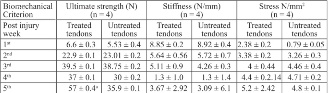
Biomechanical fi ndings. Biomechanical data are presented in Table 2 as Mean ± standard deviation (M ± SD). There was no signifi cant difference between biomechanical markers, except for ultimate strength, which was statistically higher in the tendons treated with G-90 in comparison with the untreated tendons (P<0.05).
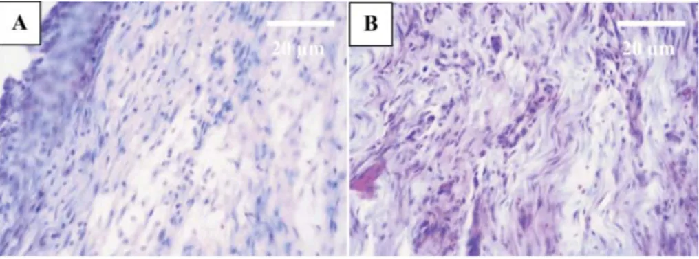
Histopathological fi ndings. In histopathological evaluation, some criteria such as: severity of infl ammation, maturation of fi brocytes, the rate of aggregation of connective fi bers, connective fi ber alignment and vascularization rate, were studied. Histopathological evaluation after the fi rst week showed signifi cantly less infl ammation in the tendons treated with G-90 in comparison with the untreated tendons (Fig. 1, Table 3, P = 0.03). Moreover, there were signifi cant differences (Table 3, P = 0.013) between the two groups in the rate of maturation of fi brocytes, where tendons treated with G-90 were superior to the untreated tendons after the fi rst week (Figs 2 and 3). Table 2. Results of biomechanical assessment (Mean ± SD)
Table 2. Results of biomechanical assessment (Mean ± SD) Biomechanical
Criterion Ultimate strength (N)(n = 4) Stiffness (N/mm)(n = 4) Stress N/mm 2 (n = 4) Post injury
week Treated tendons Untreated tendons tendonsTreated Untreated tendons tendonsTreated Untreated tendons 1st 6.6 ± 0.3 5.53 ± 0.4 8.85 ± 0.2 8.92 ± 0.4 2.38 ± 0.2 0.79 ± 0.05 2nd 22.9 ± 0.1 23.01 ± 0.2 5.64 ± 0.56 5.72 ± 0.7 3.38 ± 0.2 3.26 ± 0.3 3rd 39.5 ± 0.1 38.75 ± 0.2 5.11 ± 0.9 4.26 ± 0.3 4 ± 0.44 4.46 ± 0.4 4th 37 ± 0.1 30 ± 0.2 1.3 ± 1.0 1.3 ± 1.4 4.4 ± 0.2.14 4.71 ± 0.2 5th 57 ± 0.4a 35.9 ± 0.1 3.67 ± 2.92 3.09 ± 6.1 5.2 ± 2.42 4.8 ± 0.1 a Tendons treated with G-90 showed signifi cant difference (P<0.05), in comparison with untreated tendons
Table 3. The results of histopathological evaluation at postoperative week intervals (n = 4) Histopathological
criteria Med (Min-Max)
Postoperative
week
Infl ammation maturationFibroblast
Aggregation connective
fi bers Connective fi ber alignment Vasularization rate Treated
tendons Untreated tendons tendonsTreated Untreated tendon Treated tendons Untreated tendons Treated tendons Untreated tendons Treated tendons Untreated tendons 1st 1 (0-2) 2 (2-2)a 1 (1-2) 1 (1-1)b 2 (1-2) 1 (1-2) 1 (1-1) 1 (1-1) 2 (1-2) 2 (2-2) 2nd 1 (1-2) 1 (1-2) 2 (1-2) 2 (2-2) 2 (2-2) 2 (2-2) 2 (1-2) 2 (1-2) 2 (1-2) 2 (1-2) 3rd 0 (0-2) 1 (0-2) 2 (2-3) 2 (1-2) 3 (3-3)c 2 (1-2) 3 (2-3) 1 (1-3) 1 (1-1) 1 (1-2) 4th 0 (0-0) 0 (0-0) 3 (2-3) 3 (2-3) 3 (3-3) 3 (2-3) 3 (3-4) 3 (3-3) 1 (1-1) 1 (1-1) 5th 1 (0-1) 0 (0-0) 3 (2-3) 2 (2-3) 3 (2-4) 2 (2-3) 3 (2-3) 2 (2-3) 1 (1-1) 1 (1-1) a signifi cantly less infl ammation in tendons treated with G-90 in comparison with untreated tendons (P = 0.03); b there were signifi cant differences (P = 0.013) between the two groups in the rate of maturation of fi brocytes, where tendons treated with G-90 were superior to untreated tendons after the fi rst week; c signifi cant differences in the rate of aggregation of connective fi bers between tendons treated with G-90 and untreated tendons where treated tendons showed superior aggregation of connective fi bers in comparison of the untreated tendons (P = 0.013).
After the third postoperative week there was a signifi cant difference in the rate of aggregation of connective fi bers between tendons treated with G-90 and untreated tendons, where the treated tendons showed superior aggregation of connective fi bers in comparison with the untreated tendons (Figs 4 and 5, Table 3, P = 0.013).
Fig. 1. Note the connective tissue formation with large numbers of fi broblasts and fi brocytes in the treated tendon (A) and untreated tendon (B) after the fi rst week. Note the large number of
infl ammatory cells in the untreated tendon (H&E, ×40)
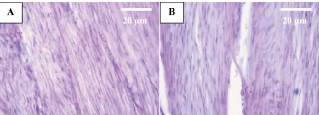
Fig. 2. Incision space fi lled by strands of connective tissue with more fi broblasts and proper orientation of connective fi bers in the treated tendon (A) and untreated tendon (B) at the second
Fig. 3. The incision space occupied by strands of connective tissue with proper orientation of connective fi bers in the treated tendon (A) compared with untreated tendon (B) after the third
week (H&E, ×40)
Fig. 4. Aggregation of connective fi bers with proper orientation of connective fi bers and increased number of fi brocytes to fi broblasts in the treated tendon (A) compared with the untreated tendon
(B) after the fourth week (H&E, ×40)
Fig. 5. The organization of the connective fi bers to the tendon at day 35 in the treated tendon was more pronounced than in the untreated tendon (H&E, ×40)
Discussion
This experiment was designed on the basis of the theory that G-90 can stimulate maturation of tenoblasts, initiate earlier collagen formation and maturation, and result in the improved biomechanical performance of the treated injured tendon. These hypotheses were morphologically and biomechanically tested 35 days post-injury on a completely sectioned superfi cial digital fl exor tendon in rabbits. The proximal section of the SDFT has been selected as the tissue of choice by many investigators as an extra-synovial model because of its accessibility and easier exposure (BIGHAM et al., 2011; ORYAN et al., 2008; ORYAN et al., 2011; ORYAN et al., 2009; ORYAN et al., 2010). In addition, it simulates the hand fl exor tendon of humans and SDFT injuries in horses (OSULLIVAN, 2007; SHARIFI et al., 2009). Superfi cial digital fl exor tendonitis is a common career injury in sport horses, and when severe or with recurrence it can result in early retirement or destruction of the affected horse (DYSON, 2004).
Tendon injuries are an important clinical issue for orthopedic surgeons. There are obstacles on the path to healing tendon injuries as follows: 1. Low blood supply: the tendon healing period is considerably longer than other types of connective tissue such as bones (CHAN et al., 2000; YOUNG, 2012). 2. Healing does not lead to a normal histological structure, i.e. healing occurs by forming scar tissue, whose quality is less than that of normal tendon. Therefore, a healed tendon does not have normal function and may suffer recurrent injury (GULOTTA et al., 2011; LACITIGNOLA et al., 2008; LEEA and HUI, 2006). 3. These tissues are susceptible to adhesion due to excess fi brous tissue formation (GULOTTA et al., 2011).
Despite progress in surgical techniques and rehabilitation, early complications, such as rupture of the repaired area and postoperative adhesions, may occur (KHANNA et al., 2009; SHARMA and MAFFULLI, 2005). Adhesion formation after intrasynovial tendon injury poses a major clinical problem. Synovial sheath disruption at the time of injury or surgery allows granulation tissue and the tenocytes from surrounding tissue to invade the repair site. Exogenous cells are predominant over endogenous tenocytes, allowing the surrounding tissue to attach to the repair area and thus create adhesion (SHARMA and MAFFULLI, 2006). The most common complications, after repair of fl exor tendon rupture, are tendon adhesion and joint contracture (ROUHANI et al., 2013). Flexor tendon healing leads to complications in the form of soft tissue adhesions to the surrounding sheath (HAKIMI et al., 2012). In the present study, necropsy evaluation showed that there were fewer tendon adhesions to the surrounding tissues in tendons treated with G-90 in comparison to the untreated tendons. This phenomenon in the treated tendons may be related to the fi brinolytic property of G-90 which was mentioned in previous studies. The fi rst extract of fi brinolytic enzymes from the earthworm Eisenia foetida, was reported in 1983 (MIHARA et al., 1983). Similar enzymes extracted from different species, such
as Eisenia foetida and Lumbricus bimastus, indicate that these earthworm species have fi brinolytic properties (HU et al., 2005; ZHAO et al., 2005; MIHARA et al., 1990; LI et al., 2011).
In the present study, histopathological evaluation showed less infl ammatory response after the fi rst postoperative week in tendons treated with G-90 in comparison with the untreated tendons. This fi nding is in accordance with the previous study by BALAMURUGAN et al. (2009), where the anti-infl ammatory and anti-pyretic properties of G-90 were proven in a hind paw infl ammation model in Wistar albino rats (BALAMURUGAN et al., 2009). In addition, histopathological evaluation in our study showed superior fi broblast maturation and superior aggregation of connective fi bers in the tendons treated with G-90 in comparison with untreated tendons. Earlier investigations of a G-90 glycolipoprotein mixture showed that this mixture is capable of stimulating various activities, such as mitogenesis, as well as stimulating the synthesis of transforming growth factor and epithelial growth factor, which could all contribute to the speed of wound healing (HRZENJAK et al., 1993). The mitogen activity of G-90 (HRZENJAK et al., 1993; GRDISA et al., 2004) could be responsible for the proliferation of fi broblasts cells, contributing in that manner to the celerity of the wound-healing process (POPOVIC et al., 2005).
The infl uence of G-90 on the structural organization of the tendon, including improved tissue alignment and crimp formation, enhanced cell maturation, increased collagen fi bril differentiation and maturation with decreased peritendinous adhesion, are possibly the most signifi cant effects of G-90 on the tendon healing. Therefore, due to the enhanced hierarchical organization, improved biomechanical parameters were expected to be seen in the treated animals in the present study. The tensile strength of the tendons is co-related to the total collagen content, type of collagen, diameter and unimodal or multimodal distribution pattern of the collagen fi brils, the quality of the cross links of the collagen fi brils, and the quantity and quality of the non-collagenous material of the ground substance (ORYAN and MOSHIRI, 2011). The fi ndings of the present study are in agreement with those of HAMADA et al. (2006) who showed that, after placement of a nylon monofi lament coated with bFGF in the injured area of the fl exor tendon of rabbits, the treated animals showed signifi cantly enhanced ultimate strength, compared to those of the control, after 21 days post injury (HAMADA et al., 2006). TANG et al. (2008) also found that injured SDFT treated with bFGF showed a higher tensile strength at 2, 4, and 8 weeks post injury (TANG et al., 2008). On the other hand, it has also been reported that bFGF affected the initial events of tendon healing on the cell proliferation, but had no signifi cant effect on the ultimate stress of the injured tendon during the fi rst two weeks post injury, in a rat patellar model (CHAN et al., 2000).
Previous studies have shown that G-90 could enhance epithelialization recovery, collagen production, fi broblast proliferation and angiogenesis in the wound area (GRDISA et al., 2004). The most common growth factors are EGF and FGF, which are also involved in the wound healing process. GRDISA et al. (2004) measured EGF and FGF activity on intact skin, physiological wounds or healing and wounds treated with G-90. In the second and third groups after the fi rst 24 hours, EGF and FGF activity was measured, and in both cases, in the fi rst 6 hours of restoration, enhancement was observed. Compared with normal skin, the concentration of EGF was ten times higher, and the concentration of FGF was 5 times higher in the group treated with G-90. In physiological conditions, EGF doubled and FGF increased one point fi ve (1.5) times. The macromolecules of earthworms stimulate the process of creating EGF and FGF which assumes the role of epithelization during mouse skin wound healing. EGF stimulates proliferation of epithelial cells and causes rapid healing of the wound. FGF is effective in the angiogenesis and induction of fi broblast growth (GRDISA et al., 2004).
The fi ndings of the present experiment clearly show that administration of G-90 could enhance the structural and biomechanical properties of experimentally tenotomized SDFT in rabbits. Many factors, such as: less peritendinous adhesion, less infl ammation, maturity and proper organization of the collagen fi bers, and good biomechanical performance of the healing tendon, were observed in this study.
Conclusion
In the present study, G-90 treatments of tendons led to signifi cant differences in some biomechanical factors and enhanced tissue strength. Histopathology showed that G-90 reduces the severity of infl ammation and increases aggregation of connective fi ber.
Further biochemical and molecular studies are needed to elucidate other aspects of the mechanism of the action of this reagent on the structural and functional performance of tendon injuries.
Confl ict of Interest
There are no confl icts of interest related to this study _______
Acknowledgements
The authors would like to thank Shahrekord University for fi nancial support and cooperation.
References
BALAMURUGAN, M., K. PARTHASARATHI, E. L. COOPER, L. S. RANGANATHAN (2009): Anti-infl ammatory and anti-pyretic activities of earthworm extract - Lampito mauritii
BIGHAM, A. S., M. SHADKHAST, H. NOURANI, H. R. SHAHBAZKIA, S. KOSHA, S. ASGHARZADEH, Z. SHAFIEI (2011): Fresh autogenous and allogenous tendon graft in rabbit model. Comp. Clin. Pathol. 20, 109-113.
CHAN, B. P., K. M. CHAN, N. MAFFULLI, S. WEBB, K. K. H. LEE (1997): Effect of basic fi broblast growth factor; an in vitro study of tendon healing. Clin. Orthop. Relat. Res. 342,
239-247.
CHAN, B. P., S.-C. FU, L. QIN, K.-M. LEE, C. G. ROLF, K.-M. CHAN (2000): Effects of basic fi broblast growth factor (bFGF) on early stages of tendon healing: a rat patellar tendon model. Acta Orthopaedica 71, 513-518.
DYSON, S. J. (2004): Medical management of superfi cial digital fl exor tendonitis: a comparative study in 219 horses (1992-2000). Equine. Vet. J. 36, 415-419.
EL-KAMALI, H. H. (2000): Folk medicinal use of some animal products in Central Sudan. J. Ethnopharmacol. 72, 279-282.
GRDISA, M., M. POPOVIC, T. HRZENJAK (2001): Glycoprotein extract of Eisenia foetida exerts
some antioxidative activity. Comp. Biochem. Physiol. 128A, 821-825.
GRDISA, M., M. POPOVIC, T. HRZENJAK (2004): Stimulation of growth factor synthesis in skin wounds using tissue extract (G-90) from the earthworm Eisenia foetida. Cell. Biochem.
Funct. 22, 373-378.
GULOTTA, L. V., S. CHAUDHURY, D. WIZNIA (2012): Stem cells for augmenting tendon repair. Stem Cells Int. 2012, 1-7.
HAKIMI, O., R. MURPHY, U. STACHEWICZ, S. HISLOP, A. J. CARR (2012): An electrospun polydioxanone patch for the localisation of biological therapies during tendon repair. Eur. Cell Mater. 24, 344-357.
HAMADA, Y., S. KATOH, N. HIBINO, H. KOSAKA, D. HAMADA, N. YASUI (2006): Effects of monofi lament nylon coated with basic fi broblast growth factor on endogenous intrasynovial fl exor tendon healing. J. Hand. Surg. 31, 530-540.
HRZENJAK, M., L. KOBREHELD, S. LEVANAT, M. JURIN, T. HRZENJAK (1993): Mitogenicity of the earthworm’s (Eisenia foetida) insulin-like proteins. Comp. Biochem. Physiol B. 104,
723-729.
HRZENJAK, T., M. HRZENJAK, V. KAUBA, P. EFENBERGER-MARINCULIC, S. LEVANAT (1992): A new source of biologically active compounds-earthworm tissue (Eisenia foetida,
Lumbricus rubelus). Comp. Biochem. Physiol A. Physiol. 102, 441-447.
HRZENJAK, T., M. POPOVIC, T. BOZIC, M. GRDISA, D. KOBREHEL, L. TISKA-RUDMAN (1998): Fibrinolytic and anticoagulative activities from the earthworm Eisenia foetida. Comp.
Biochem. Physiol. Biochem. Mol Biol. 119, 825-832.
HU, Y., X.-L. MENG, J.-P. XU, W. LU, J. WANG (2005): Cloning and expression of the novel gene PV242 of earthworm fi brinolytic enzyme. Protein. Expr. Purif. 43, 18-25.
KANNUS, P. (2000): Structure of the tendon connective tissue. Scand. J. Med. Sci. Sports. 10, 312-320.
KHANNA, A., M. FRIEL, N. GOUGOULIAS, U. G. LONGO, N. MAFFULLI (2009): Prevention of adhesions in surgery of the fl exor tendons of the hand: what is the evidence? Br. Med. Bull. 90, 85-109.
LACITIGNOLA, L., A. CROVACE, G. ROSSI, E. FRANCIOSO (2008): Cell therapy for tendinitis, experimental and clinical report. Vet. Res. Commun. 32, 33-38.
LEEA, E. H., J. H. HUI (2006): The potential of stem cells in orthopaedic surgery. J. Bone. Joint. Surg. (British) 88, 841-851.
LI, W., C. WANG, Z. SUN (2011): Vermipharmaceuticals and active proteins isolated from earthworms. Pedobiologia 54, S49-S56.
McGEACHIE, J., M. TENNANT (1997): Growth factors and their implications for clinicians: A brief review. Aust. Dent. J. 42, 375-380.
MIHARA, H., H. SUMI, K. AKAZAWA, T. YONEDA, H. MIZUMOTO (1983): Fibrinolytic enzyme extracted from the earthworm. J. Thromb. Haemost. 50, 258-258.
MIHARA, H., H. SUMI, T. YONETA, H. MIZUMOTO, R. IKEDA, M. SEIKI, M. MARUYAMA (1990): A novel fi brinolytic enzyme extracted from the earthworm, Lumbricus rubellus. Jpn.
J. Physiol. 41, 461-472.
ORYAN, A., A. E. GOODSHIP, I. A. SILVER (2008): Response of a collagenase-induced tendon injury to treatment with a polysulphated glycosaminoglycan (Adequan). Connect. Tissue. Res. 49, 351-360.
ORYAN, A., A. MOSHIRI (2011): A long term study on the role of exogenous human recombinant basic fi broblast growth factor on the superfi cial digital fl exor tendon healing in rabbits. J. Musculoskelet. Neuronal. Interact. 11, 185-195.
ORYAN, A., A. MOSHIRI, A. H. MEIMANDIPARIZI (2011): Effects of sodium-hyaluronate and glucosamine-chondroitin sulfate on remodeling stage of tenotomized superfi cial digital fl exor tendon in rabbits: a clinical, histopathological, ultrastructural, and biomechanical study. Connect. Tissue. Res. 52, 329-339.
ORYAN, A., I. A. SILVER, A. E. GOODSHIP (2009): Effects of a serotonin S2-receptor blocker on healing of acute and chronic tendon injuries. J. Invest. Surg. 22, 246-255.
ORYAN, A., I. A. SILVER, A. E. GOODSHIP (2010): Metrenperone enhances collagen turnover and remodeling in the early stages of healing of tendon injury in rabbit. Arch. Orthop. Trauma. Surg. 130, 1451-1457.
OSULLIVAN, C. B. (2007): Injuries of the fl exor tendons: focus on the superfi cial digital fl exor tendon. Clin. Tech. Equin. Prac. 6, 189-197.
POPOVIC, M., M. GRDISA, T. M. HRZENJAK (2005): Glycolipoprotein G-90 obtained from the earthworm Eisenia foetida exerts antibacterial activity. Vet. arhiv. 75, 119-128.
POPOVIC, M., T. M. HRZENJAK, T. BABIĆ, J. KOS, M. GRDISA (2001): Effect of earthworm (G-90) extract on formation and lysis of clots originated from venous blood of dogs with cardiopathies and with malignant tumors. Pathol. Oncol. Res. 7, 197-202.
POPOVIC, M., T. HRZENJAK, M. GRDISA, S. VUKOVIC (1998): Adhesins of immunoglobulin-like superfamily from earthworm (Eisenia foetida). Gen. Pharmacol. 30, 795-800.
ROSENBAUM, A. J., J. F. WICKER, J. S. DINES, L. BONASSER, P. RAZZANO, D. M. DINES, D. A. GRANDE (2010): Histologic stages of healing correlate with restoration of tensile strength in a model of experimental tendon repair. HSS J. 6, 164-170.
ROUHANI, A., A. TABRIZI, E. GHAVIDEL (2013): Effects of non-steroidal anti-infl ammatory drugs on fl exor tendon rehabilitation after repair. Arch. Bone. Jt. Surg. 1, 28-30.
SHARIFI, D., D. KAZEMI, H. LATIFI (2009): Evaluation of tensile strength of the superfi cial digital fl exor tendon in horses subjected to transcutaneous electrical neural stimulation therapeutic regimen. Am. J. Appl. Sci. 6, 816-819.
SHARMA, P., N. MAFFULLI (2005): Tendon injury and tendinopathy: healing and repair. J. Bone. Joint. Surg. 87, 187-202.
SHARMA, P., N. MAFFULLI (2006): Biology of tendon injury: healing, modeling and remodeling. J. Musculoskelet. Neuronal. Interact. 6, 181-190.
STRAUSS, E. J., C. ISHAK, L. JAZRAWI, O. SHERMAN, J. ROSEN (2007): Operative treatment of acute Achilles tendon ruptures: an institutional review of clinical outcomes. Injury 38, 832-838.
TANG, J. B., Y. CAO, B. ZHU, K.-Q. XIN, X. T. WANG, P. Y. LIU (2008): Adeno-associated virus-2-mediated bFGF gene transfer to digital fl exor tendons signifi cantly increases healing strengthen in vivo study. J. Bone. Joint. Surg. 90, 1078-1089.
YOUNG, M. (2012): Stem cell applications in tendon disorders: a clinical perspective. Stem Cells Int. 2012, 1-10.
ZHAO, J., S.-P. QI, J. WU, L. LI, R.-Q. HE (2005): Earthworm fi brinolytic enzyme. Stud. Nat. Prod. Chem. 30, 825-847.
Received: 7 February 2015 Accepted: 12 April 2016
________________________________________________________________________________________ BIGHAM-SADEGH, A., M. ARABI, E. KARIMI, I. KARIMI, A. ORYAN: : Cijeljenje tetivnih ozljeda uporabom G-90 na modelu kunića: biokemijska i histopatološka procjena.. Vet. arhiv 86, 407-420, 2016.
SAŽETAK
Ozljede tetiva predstavljaju jedan od najčešćih akutnih ortopedskih poremećaja što dovode do njihove funkcionalne oslabljenosti. Razvijeno je nekoliko postupaka za ponovnu uspostavu pokretnosti pacijenata, ali svi imaju znatna ograničenja. U nekim istraživanjima opisana je uporaba biomaterijala za cijeljenje ozljeda tetiva, kože i živaca. U ovom su radu istraženi učinci G-90, kao stimulacijskog čimbenika cijeljenja tetive površinskog digitalnog fl eksora kunića 35 dana nakon tenotomije i kirurškog liječenja. U pokus je bilo uzeto 20 kunića u dobi od 12 mjeseci, tjelesne mase 2,0 ± 0,5 kg. Svim su kunićima pod anestezijom bile poprečno prerezane tetive površinskog digitalnog fl eksora stražnjih nogu te potom spojene Bunnel-Mayer-ovim šavom. Nakon šivanja tetive i kože, 0,5 mL fi ziološke otopine bilo je ubrizgano u područje tetivne ozljede lijeve noge, a 0,5 mL iscrpka G-90 kišne gujavice u koncentraciji od 10 mg/mL bilo je ubrizgano u područje tetivne ozljede desne noge. Svaka dva tjedna bila su eutanazirana 4 kunića te su im bili uzeti uzorci za patohistološke
i biokemijske pretrage. Patohistološkom pretragom tetiva kunića kojima je bio primijenjen G-90 ustanovljena je slabija upala, veća zrelost fi brocita i veće nakupljanje kolagenih vlakana u usporedbi s tetivama kojima nije bio primijenjen G-90. Kod biokemijske procjene konačna čvrstoća tetiva obrađenih s G-90 bila je veća u odnosu na one neobrađene. Rezultati ovog pokusa jasno su pokazali da primjena G-90 može osnažiti strukturu i biokemijska svojstva tetiva kunića nakon tenotomije i kirurškog liječenja.
Ključne riječi: iscrpak kišne gujavice, G-90, cijeljenje tetiva, kunić