Role of Isoflavones on Lipid Peroxidation, Superoxide Dismutase
in Lymphocytes under Oxidative Stress Conditions
I Nyoman Suarsana1*, I Nyoman Mantik Astawa2, I Made Kardena3
1Laboratory Biochemistry, 2Laboratory Virology and immunology, 3Laboratory Pathology
Faculty of Veterinary Medicine, University of Udayana, Bali, Indonesia *E-mail of the corresponding author: [email protected] Abstract
Oxidative stress conditions forms free radicals in cells. Free radicals react continuously and cause cell membranes damage including lymphocytes, thus decreasing cell function. Therefore, there were efforts need to be done to overcome abnormalities and decreased lymphocyte cell function under conditions of oxidative stress. One of them using bioactive compounds that have antioxidant activity, such as isoflavones. Isoflavones have been reported to have antioxidant activity and the potential to protect cells of oxidative stress. This study aims to determine the role of isoflavones as antioxidative in protecting lymphocytes under conditions of oxidative stress, be reviewed of CD4, CD8, malondialdehyde levels and superoxide dismutase activity. This study was used rat lymphocyte cell cultures in two conditions, i.e: oxidative stress and without oxidative stress. The cell culture were treated with 20 ml of 2 mM paraquat and isoflavone at dose 0, 1000, 2000, and 3000 ppm, respectively. The parameters were observed: (1) malondialdehyde (MDA) levels, (2) superoxide dismutase (SOD) activity, and (3) percentage of CD4 and CD8 lymphocytes cell using a monoclonal anti-rat CD4 and CD8 FITC-conjugate. The results showed paraquat administration in cell culture on oxidative stress conditions could reduce the number of CD4 and CD8 lymphocytes cels, increased of malondialdehyde (MDA) and superoxide dismutase (SOD) activity. Administration of isoflavones on culture that treated without oxidative stress condition could increase percentage number of CD4, CD8, MDA levels, and SOD activity. Whereas, administration of isoflavones on oxidative stress condition prevented the decrease percentage number of CD4, CD8, MDA levels, and SOD activity. Administration of isoflavone at dose 3000 ppm in cell culture that treated on oxidative stress condition provided optimal results in preventing the decrease number of CD4, CD8 lymphocytes cell, MDA levels, and SOD activity.
Keywords: isoflavone, lymphocytes, CD4, CD8, malondialdehyde, superoxide dismutase
.
1. Introduction
Oxidative stress can be defined as an imbalance between reactive oxygen species (ROS) production and the antioxidant defense mechanisms of a biological organism that results directly or indirectly in cellular damage (Jenkins, 2000; Fisher, 2010; Fisher et al, 2011). Sources of ROS can be from exogenous or endogenous
origins of the body. ROS from exogenous origins (chemicals, radiation, air pollutants, intoxication by oxygen, smoke, and alcohol) and from endogenous origins such as oxidative phosphorylation within the mitochondria or extramitochondrial sources (Jezek and Hlavata, 2005; Fisher, 2010). ROS are recognized for harmful to living systems. At high concentrations, ROS can be important mediators of damage to cell structures, nucleic acids, lipids and proteins (Valko et al., 2007).
Exposure of the body from the toxic or harmful substances are carcinogenic, imunotoxic, and other toxicant compound which can harmful to health. According Krejsa and Schieven (2000), toxicant compounds can induce oxidative stress by various mechanisms. This toxicant compounds can block the electron transport chain thereby increasing leakage of free radicals such as superoxide anion (O2•-), from the mitochondria to the cytosol.
Paraquat (PQ) is one of the toxicant compound. It is a herbicide that works fast, it is not selective and it is toxic to all cells (Ogamba et al., 2011). According to Raina et al (2008), paraquat can inhibit the reduction of NADP
to NADPH. This disorder causes formation of Free radicals.
Free radicals can injury all cells inclusive lymphocytes by destructing which injury cellular lipids, proteins, or DNA (Zakaria et al., 1997a). Therefore, normal function of the cells is inhibited. Oxidative stress condition can
cause degenerative diseases, immune system disorders and cells premature aging.
The main components of the specific immune system are lymphocyte. Lymphocytes are an important immunological cell and have been played a significant role in acquired immune system (Gautam et al., 2010).
According to Tood (2001), there are three major types of lymphocytes are called B cells, T cells and NK (natural killer) cells. T cells expressing the surface proteins CD8 (helper T cells) or CD4 (cytotoxic cells), respectively. T cells consists of three subsets, namely helper T cells (TH), suppressor T cells (TS), and cytotoxic
T cells (TC). Each of these subsets of T cells expressing surface markers or cluster of differentiation (CD)..
Free radicals can damage cell membrane of lymphocytes which are characterized by the increasing lipid peroxidation products, decrease intracellular antioxidant activity (SOD) and lymphocyte loses its primary function, which is not able to eliminate a variety of agents that entry into the body. Various studies have shown that oxidative stress can impact on immune function (Shi et al., 2003). The immune system is affected by stress,
for instance: physical stress, chemical stress, and oxidative stress. Stress that occures chronically, may cause death (apoptosis) cell lymphocytes.
important factor an antioxidants and immunomodulator maintaining survival and function of lymphocytes in body's defense. This study aims were to determine role of isoflavones as antioxidative of the CD4, CD8, lipid peroxidation, superoxide dismutase in lymphocytes under conditions of oxidative stress.
2. Materials and Methods
Preparation of lymphocytes. Isolation of lymphocyte cells using method that modified by Mantik et al (2005). A total of 5 ml blood of rats inserted into a tube that had been contained anticoagulant (EDTA). Then the tube was centrifuged at a speed of 2500 rpm for 10 minutes. A white layer (buffy coat) in between the red blood cells and plasma was taken and suspended in medium without serum. Lymphocytes were separated from buffy coat with density gradient centrifugation, Ficoll-paque speed of 3000 rpm for 45 minutes. Lymphocyte layer was taken and washed three times with a medium without serum. Lymphocyte cells then were washed up to lymphocytes free of red blood cells. Finally, lymphocytes diluted with 5 ml RPMI 1640 and counted using a hemocytometer.
Lymphocyte cell culture. Rat is lymphocyte cells that had been counted then arranged into a cell containing 1 x 10⁵ cells/ml using RPMI medium which was equipped with a 10% Feotal Calf Serum (FCS), penicillin (100
IU/ml), streptomycin (100 µg/ml), gentamicin (50 µg/ml) and 2.5 µg/ml fungison. Every 100 ml of lymphocyte cells (1 x 10⁵ cells / ml) in complete medium inserted randomly into the microplate wells then successively
added to each well of a 20 μl isoflavone compounds that had been prepared with a concentration of 1000, 2000 and 3000 μg/ml and added 20 μl of 2mM paraquat. For a positive control in oxidative stress, other microplate wells were added lymphocyte cells containing 1 x 10⁵ cells/ml on complete medium, and than added of 20
μl in 2mM paraquat. For a positive control of isoflavone, each microplate wells was added 100 μl lymphocyte cells 1 x 10⁵ cells/ml in complete medium and then added 20 staining method based on the procedures in the KIT. Monoclonal antibodies conjugated with the types of dyes fluorescens was used for staining. A total of 100 μl of lymphocyte cell suspension containing of 1x10⁶ cells/ml
was placed into a tube (falcon tube). The tube was added 4 μl of monoclonal antibody anti-mouse CD4-FITC conjugated (or CD8-FITC) and vortexed slowly. After it mixed, the suspension incubation for 35 min at 4°C. A total of 20 μl of cell suspension dropped on the object glass (microscope slide) and
and viewed with a microscope. Total cells that emitted yellowish-green fluorescens were counted under a microscope fluorescens and the lymphocytes were counted by using phase contrast microscopy. The results were expressed in percent of total cells, namely the number of fluorescent cells to total lymphocyte cells
50 μl solution of xanthine oxidase and homogenized. Absorbance values was read using a spectrophotometer at
λ 550 nm.
Analysis of lipid peroxidation (malondialdehyde). Lipid peroxidation (malondialdehyde) levels were analyzed using method addapted from Capeyron et al. (2002). A total of 0.5 ml of the supernatant or standard plus with 2
ml of 0.25 N HCl (cold) mixture containing 15%TCA, TBA 0.38%, and 0.5% BHT. The solution mixture was heated to 80oC for 1 hour. After the cold, solution was centrifuged 3500 rpm for 10 minutes. Absorbance values
was read at λ 532 nm. As standard solution used TEP (1,1,3,3-tetraetoksipropana)
.
3. Results
The results analysis of CD4, CD8 T-cells, malondialdehyde levels and superoxide dismutase activity of lymphocyte cells under conditions of oxidative stress are shown in the Table 1-4.
The results of the percentage of CD4 T-cells in a different culture conditions and was given isoflavones are presented in Table 1. Analysis of variance showed that treatment of culture conditions (stress and without stress) and isoflavone treatment significantly effect (P <0.05) to amount of CD4 lymphocyte cells.
Table 1. The number of CD4 lymphocyte cells that cultured under oxidative stress and without oxidative stress condition
.
Culture condition CD4 percentage (%) at different doses of isoflavones (ppm)
0 1000 2000 3000 Average
Non-stress 19.64Aa 21.56Aab 23.91Ab 25.65Ac 22.69A Stress 11.42Ba 13.15Bab 15.58Bb 18.38Bc 14.63B
Average 15.53 17.36 19.74 22.01
Description: Values with the same letter towards column (uppercase) and the values with the same letter in the direction of the line (lowercase) indicates not significantly different (P (> 0.05)
The analysis indicated that the amount of CD8 T-cells condition oxidative stress was lower than the condition without oxidative stress and significantly different (P <0.05) at all doses of isoflavones (Table 2). This suggests that paraquat administration significantly reduced the amount of CD8 T cells. On the condition of oxidative stress, the use of isoflavones at dose up to 3000 ppm resulted the amount of CD8 T cells was 19.65 while the amount of CD4 T cells without stress was 21.15.
Table 2. The percentage of CD8 T-cells that cultured under oxidative stress and without oxidative stress condition.
Culture condition CD8 percentage (%) at different doses of isoflavones (ppm)
0 1000 2000 3000 Average
Non-stress 21.15Aa 22.45Aa 25.76Ab 27.42Ab 24.19A Stress 11.93Ba 13.79Ba 16.97Bb 19.65Bb 15.59B
Average 16.54 18.12 21.37 23.54
Description: Values with the same letter towards column (uppercase) and the values with the same letter in the direction of the line (lowercase) indicates not significantly different (P (> 0.05)
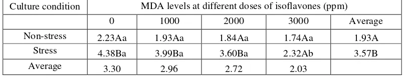
The analysis exhibited that MDA levels in the culture conditions stress and without stress were significant different (P <0.05) (Table 3) at all doses of isoflavones, except for dose at 3000 ppm. In adminitration of isoflavone at dose 3000 ppm, the MDA levels on stress condition was 1.74 while on without stress condition was 2.32 picomol/ml and both were no significant different (P> 0.05).
Table 3. The number of malondialdehyde in T-cells that cultured under oxidative stress and without oxidative stress conditions.
Culture condition MDA levels at different doses of isoflavones (ppm)
0 1000 2000 3000 Average
Non-stress 2.23Aa 1.93Aa 1.84Aa 1.74Aa 1.93A
Stress 4.38Ba 3.99Ba 3.60Ba 2.32Ab 3.57B
Description: Values with the same letter towards column (uppercase) and the values with the same letter in the direction of the line (lowercase) indicates not significantly different (P (> 0.05)
The analysis showed that SOD levels in oxidative stress condition was lower than the without oxidative stress condition and were significantly different (P <0.05), except for isoflavones at dose 3000 ppm, SOD levels was not significantly different (P> 0.05) (Table 4). This suggests that the administration of isoflavones can significantly increase the SOD levels.
Table 4. The average number of superoxida dismutase activities in T-cells that cultured under oxidative stress and without oxidative stress conditions
.
Culture condition Superoxida dismutase activities at different doses of isoflavones (ppm)
0 1000 2000 3000 Average
Non-stress 23.78Aa 25.63Aa 27.48Aa 31.19Aa 27.02A Stress 16.37Ba 17.61Ba 19.46Ba 26.25Ab 19.92B
Average 20.08 21.62 23.47 28.72
Description: Values with the same letter towards column (uppercase) and the values with the same letter in the direction of the line (lowercase) indicates not significantly different (P (> 0.05)
4. Discussion
Effect of isoflavone administration on percentage of CD4 T-cell is dependent on lymphocyte culture condition (condition stress and without oxidative stress). The number of CD4 lymphocyte cells on administration isoflavone at dose: 0, 1000, 2000, and 3000 ppm on culture that conditioned without oxidative stress were 19.64; 21.56; 23.91 and 25.64 respectively. These results indicate that the amount of CD4 lymphocyte cells increases in a line the increase doses of isoflavones. The Duncan test results showed that administration of isoflavones at dose 3000 ppm in without stress conditions was significantly different (P <0.05) compared with the other doses. Administration of isoflavones at dose 2000 ppm was no significant different (P> 0.05) compared with dose 1000 ppm. However, when they compared to administration of isoflavone at dose 0 ppm, there was a significant different(P<0.05).
In contrast, administration of isoflavones on oxidative stress conditions, resulting in percentage number of CD4 T cells was 11.42; 13.15; 15.58 and 18.38, respectively. When it counted the CD4 T-cells that under stress condition was lower than the without stress condition. This suggests that paraquat as initiated the oxidative stress is able to decrease the number of CD4 T-cells. Additionally, and administration of isoflavones has not been able to maintain and restore the number of CD4 T-cells even in the without stress condition.
In general, administration of isoflavones could increase amount of CD4 lymphocyte cells and this something to do with the isoflavone antioxidative capability in protecting T-cells from free radical or toxical effects of paraquat so that the amount of lymphocyte cells was not disrupted. Kartikawati (1999) showed that rats with paraquat administration caused decrease amount of CD3 and CD4 lymphocyte cells. In addition, administration of antioxidants, such as vitamin C and E was able to protect lymphocytes from the toxical effects of paraquat. Similar to CD4, CD8 T-cell count is dependent on the condition of lymphocyte culture. In without oxidative stress conditions and without administration of isoflavones (0 ppm) the amount of CD8 T-cells was 21.15, while administration of isoflavones at dose: 1000, 2000 and 3000 ppm, percentage number of CD8 T-cells was 22.45; 25.76, and 27.42, respectively (Table 2). Under oxidative stress conditions, isoflavones prevented the decrease amount of CD8 lymphocyte cells The increase of isoflavone gives better prevention capability, which is characterized by higher number of CD8 T-cells compared with the number of CD8 T-cells on under stress conditions without isoflavones administration. The involvement of amount of CD8 T-cells is important in the role of antioxidative isoflavones which is able to neutralize and destroy free radicals. Thus, they prevent damage to T-cells so that it does not impact to the number of CD8 T-cells.
product. Isoflavones at dose 3000 ppm was significantly decreased (P <0.05) the MDA levels compared to the doses of 1000 and 2000 ppm. This shows that the role of isoflavones as antioxidants can neutralize free radicals that prevent a peroxidation reaction. Thus, it prevents lipid peroxidation reaction so that MDA products are also decreased.
According to Rimbach et al., (2003), isoflavones have high antioxidant activity. As antioxidants, isoflavones are
able to decide by means of a chain reaction with of lipid radicals react and neutralize into a more stable product. Isoflavones play a role in eliminating free radicals which are normally produced in the body or free radicals that come from outside the body. The ability of isoflavones as antioxidants is complex and this can be seen from the structure owned by the isoflavone compounds. Apak et al. (2007) reported that, the structure of flavonoids,
including isoflavones, contributes to the antioxidative activity. The mechanism of antioxidant activity of isoflavones is closely related to the structure and highly dependent on the number and location of the OH group (Heijnen et al., 2002). Isoflavones have a hydroxyl group (-OH), the 4'-OH in the B ring (B-ring) and the OH
group on the ring AC (AC-ring). Thus, the mechanism action of isoflavones is to terminate the propagation site of free radicals (free radical chain breaking), in which all of the hydroxyl groups can donate an electron or hydrogen resulting in a clearance or interception against free radicals in order to form compounds that are relatively more stable (non-radical).
SOD activity is dependent on lymphocyte cell culture conditions and level doses of isoflavones. On the group of cell culture in without stress condition and untreated isoflavones (0 ppm), the SOD activity was of 23.78, while in the same conditions but treated with isoflavones doses of 1000, 2000, and 3000 ppm had increase SOD activity at 25.63; 27.48, and 31.19 U/ml, respectively. On the other hand, for the group of cell culture in stress conditions and without isoflavone treatment (0 ppm) gave SOD value at 16.37. While, giving isoflavne at dose 1000; 2000, and 3000 ppm affected SOD activity at 17.61, 19.46, and 26.25 U/ml respectively (Table 4)
Treatment of various concentrations of isoflavones in cell cultures without the stress condition affected increased SOD activity in a line with of isoflavones increase dose. Similarly, lymphocyte cell cultures on stress conditions and treated with isoflavones, SOD activity can be maintained and even increased a line with the increase dose of isoflavones treatment. However, SOD activity was much greater in the cell culture on without stress conditions compared to cell culture on stress condition although both were treated with isoflavones. The increased of SOD activity was associated with the ability of isoflavones as antioxidants (Rimbach, 2001). Superoxide dismutase (SOD) is an intracellular enzyme that is present in the cytosol. SOD plays an important role in catalyzing dismutation reaction of radical superoxide ions to produce hydrogen peroxide and molecular oxygen (Halliwell, 2006).
Isoflavones work synergistically with SOD, in helping to neutralize free radicals that generated by paraquat so that SOD activity did not decrease. Isoflavones can stimulate intracellular antioxidant enzymes by regulating gene expression as it reported by Badeau (2008). Genestein isoflavones can increase expression of glutathione peroxidase (GPx) and the mechanism of gene transcription through the activation of NRF (nuclear related factor). The similar research was reported by Borras et al (2006), isoflavones genestein is also able to increase
MnSOD expression through interaction with estrogen receptors in cells and subsequent activation of ERK1/2 (extracellular-signal-regulated kinase).
The SOD, glutathione peroxidase (GPx), and catalase enzymes are antioxidant cellular enzymes that function as the major defense in eliminating free radicals (Valko et al., 2007). This reaction is known as free radical
scavenger reaction (Free Radical Scavenger)
5. Conclusion
Treatment of isoflavones at dose 3000 ppm on lymphocyte cell culture on oxidative stress conditions may prevent decrease number of CD4, CD8 T-cells, malondialdehyde (MDA) levels, and superoxide dismutase (SOD) activities
.
Acknowledgement
References
Apak, R., Güçlü, K., Demirata, B., Özyürek, M., Çelik, S.E., Bektaşoğlu, B., Berker, K.I., and Özyurt, D.
(2007). Comparative evaluation of various total antioxidant capacity assays applied to phenolic compounds with the cupric assay. Molecules 12 :1496-1547.
Astuti, M., Bachelor, Meliala, A., Dalais, F.S., and Wahlqvist, M.L. (2000). Tempe, a nutritious and healthy food from Indonesia. Asia Pacific J Clin Nutr. 9(4): 322–325
Badeau, M. (2008). Genistein And 17 β-estradiol Fatty Acid Esters And The Structure related Antioxidant Activity Of Estrogens On Lipoproteins. [Dissertation]. Helsinki. The Faculty of Medicine, University of Helsinki. 57 Hlm.
Borra´s, C., Gambini, J., Go´mez-Cabrera, M.C., Sastre, J., Pallardo, F.V., Mann, G.E., and Vin˜a, J. (2006). Genistein, a soy isoflavone, up-regulates expression of antioxidant genes: involvement of estrogen receptors, ERK1/2, and NFкB. FASEB J Vol. 20:1476-1481.
Capeyron, M.F.M., Julie, C., Eric, B., Jean, P., Jean, M.R., Piere, B., Claude, L.L., and Benard, D. (2002). A diet cholesterol and deficient in vitamin E induces lipid peroxidation but does not enhace antioxidant enzyme expression in rat liver. J Nutr Biochem 13:296-301.
Fisher, G. (2010). Oxidative Stress and Antioxidant Defenses in Lymphocytes Following High Intensity Interval Training. A dissertation submitted to the Graduate Faculty of Auburn University. Auburn, Alabama. Fisher, G., Schwartz, D.D., Quindry, J., Barberio, M.D, Foster, E.B., Jones, K.W., and Pascoe , D.D. (2011).
Lymphocyte enzymatic antioxidant responses to oxidative stress following high-intensity interval exercise. J Appl Physiol 110: 730–737.
Gautam, N., Das, S., Mahapatra, S.K., Chakraborty, S.P., Kundu, P.K, and Roy, S. (2010). Age associated oxidative damage in lymphocytes. Oxidative Medicine and Cellular Longevity 3:4, 275-282.
Halliwell, B. (2006). Reactive spesies and antioxidants: Redox biology is a fudamental theme of aerobic life. Plant Phisiology 141:312-322.
Heijnen, C.G.M., Haenen, G.R.M.M., Oostveen, R.M., Stalpers, E.M., and Bast, A. (2002). Protection of flavonoid against lipid peroxidation: the structure activity relationship revisited. Free radic Res 36:575-581.
Jenkins R.R. (2000). Exercise and oxidative stress methodology: a critique. Amer J Clinical Nutr 72: 670S-674S. Jezek P, and Hlavata L. (2005). Mitochondria in homeostasis of reactive oxygen species in cell, tissues, and
organism. Int J Biochem Cell Bio 37: 2478-2503.
Kartikawati, D. (1999). Studi Efek Protektif Vitamin C dan E Terhadap Respon Imun dan Enzim Antioksidan pada Mencit yang di Papar Paraquat Thesis. Program Pascasarjana, IPB, Bogor.
Kelly, K. (2007). Understanding the Immune System: How It Works. Institute of Allergy and Infectious Diseases
U.S. Department Of Health And Human Services National Institutes Of Health. National. NIH Publication No. 07-5423. 113 pages
Krejsa, C.M., Schieven, G.L. (2000). Detection of Oxidative Stress in Lymphocytes Using Dichlorodihydrofluorescein Diacetate. Methods in Molecular Biology. Vol. 99:35-47
Lee, J., Renita, M., Fiorito, R.J., St.Martin, S.K., Schwartz, S.J., and Vodovotz, Y. (2004). Isoflavone characterization and antioxidant activity of ohio soybeans. J Agric Food Chem 52:2647-1651.
Mantik, A.N., Hartaningsih, N., Darma, D.M.N., Tenaya, W.M., Budiantono, dan Ekaana, W. (2005). Replikasi virus penyakit jembrana pada kultur limfosit darah tepi asal sapi bali. JVet. Vol 6 (4):135-142
Nebot, C., Moutet, M., Huet, P., Xu, J.Z., Yadan, J.C., and Chaudiere, J. (1993). Spectrophotometric assay of superoxide dismutase activity based on the activated autooxidation of a tetracyclic catechol. Analytical Biochem. 214:442-451.
Ogamba, E.N., Inyang, I.R., and Azuma, I.K. (2011). Effect of Paraquat Dichloride on Some Metabolic and Enzyme Parameters of Clarias gariepinus. Curr. Res. J. Biol. Sci., 3(3): 186-190.
Rimbach, G., De Pascual-Teresa, S., Ewins, B.A., Matsugo, S,. Uchida, Y., Minihane, A.M., Turner, R., VafeiAdou, K., Weinberg, P.D. (2003). Antioxidant and free radical scavenging activity of isoflavone metabolites. Xenobiotica 33:913-925.
Shi, Y., Devadas, S., Greeneltch, K.M., Yin, D., Mufson, R.A., and Zhoua, J. (2003). Stressed to death: Implication of lymphocyte apoptosis for psychoneuroimmunology. Brain, Behavior, and Immunity. 17:S18–S26.
Suarsana,N., Bintang, M., Priosoeryanto, B.P., dan Wresdiyati, T. (2010). The Effects of Tempe Isoflavone On Intracellular Antioxidant Enzymes And Malondialdehyde Level In Diabetic Rat Pancreatic Islets. Abstract International Conference, Exibition & Short Course On Nutraceuticals And Functional Foods. Sanur, Bali 11-15 October, 2010
Todd, I. (2001). Cells of the Immune System. Encyclopedia Of Life Sciences. Nature Publishing Group/www.els.net. 7 page.
Valko, M., Leibfritz, D., Moncol, J., Cronin, M.T.D., Mazur, M., and Telser, J. (2007). Review: Free radicals and antioxidants in normal physiological functions and human disease. Inter J Biochem & Cell Biol 39:44-84.
Zakaria, F., Khatib, A., Hartoyo, A. (1997). Bioactive Compounds of Food and Agriculture Products Having Immunomodulator Activities. Di dalam Hariyadi RD, Zakaria F, Editor. Seminar Proceeding Biomoleculer Reaction and Industrial Applications of Immunology in Food and Agriculture. Nopember 7, 1997 Bogor. Pusat Studi Pangan dan Gizi IPB dan Kedubes Perancis-Jakarta.