MUTATION ANALYSIS OF MITOCHONDRIAL DNA CONTROL REGIONS: A
STUDY ON ECTODERM TISSUES
YOHANIS NGILI1,*, YONI F. SYUKRIAN2, ADANG S. AHMAD3
AND A. SAIFUDDIN NOER1
1
Biochemistry Research Division, Institute Technology Bandung, Jalan Ganesha No.
10 Bandung, Jawa Barat, Indonesia (*e-mail: [email protected])
2
Forensic Laboratory, Hasan Sadikin Hospital/Faculty of Medicine, University of
Padjadjaran, Bandung, Jawa Barat, Indonesia
3
School of Electrical Engineering and Informatics, Jalan Ganesha 10 Bandung, Jawa
Barat, Indonesia
ABSTRACT
Mitochondrial DNA nucleotide sequence data are known and examined until the mutations
level. However, comparison of mutation that occurred in the D-loop and non-D loop in the
mtG on the origin of various different tissues on certain individuals who have not yet
reported. D-loop is a non-coding region, containing some important sequences such as the
promoter for heavy chain replication. On this research analyzed the diversity of D-loop
region mtG on the various tissues that different origin on a particular individual. The aim
of the research is to see if there is diversity of mtG that come from tissues that originated
from the ectoderm tissue and how the mutation pattern nucleotide D-loop mtG the original
came from tissue that is different. mtG analysis is done on some parts of the human body
cell, such as brain and skin cells. This research begins with the individual samples in the
different body tissue through an autopsy and to follow procedures / ethics applicable. Lysis
cells to get the template mtDNA using QIAamp DNA Mini Kit. Then, amplified with the
PCR technique using REPLI-g Mitochondrial DNA. PCR fragments of DNA D-loop
region sequenced with the Sanger Method. Comparative nucleotide sequence analysis
shows the diversity of mutation that occurred in the D-loop mtG on certain individuals.
authenticated and published in the International Nucleotide Sequence Database
Collaboration (INSDC). Research determining the sequence of mtG human nucleotide of
ectoderm in various tissues is involved and to contribute to frontier of knowledge. The
result is expected to give benefit the development of biotechnology. Research is expected
to generate future process of cooperation in the fields of biochemistry, molecular biology,
bioinformatics, and forensic medicine.
Keyword: mtDNA, Control Region, and Ectoderm
INTRODUCTION
DNA is one object of research in the field biotechnologi important today. Analysis of
DNA sequence can be of great benefit in various fields, including health. Various studies
conducted by experts have revealed a link between mutation of DNA with a number of
diseases. In addition, analysis of DNA can be utilized in the field of forensics.
Inside the cell, there are two types of DNA, ie nuclear DNA and mitochondrial DNA
(mitochondrial DNA, mtDNA). Nuclear DNA analysis performed by isolating DNA from
the cell nucleus, whereas mitochondrial DNA analysis conducted by collecting DNA
contained in mitochondria. In this study, reported the results of a study of nucleotide
sequence of the D-loop region of mitochondrial DNA. Mitochondrial DNA is unique
compared to nuclear DNA contained in the nucleus. The uniqueness in question include
the mitochondrial DNA of a specific pattern of inheritance through maternal lineages, a
high polymorphism and a system of genetic code that differ from the standard genetic code
system in the process of translation of codons into amino acids (Anderson et al, 1981.)
Analysis of mitochondrial DNA can provide solutions for scientists in overcoming the
problems encountered when researching nuclear DNA. Samples that have been long, have
no cell nucleus material again, thus not allowing nuclear DNA analysis of these samples.
However, mitochondrial DNA can still be obtained from these samples. Mitochondrial
DNA analysis was also conducted in the field of forensics. Maternal mitochondrial DNA is
Mitochondrial genome consists of 16,569 base pairs, and is composed of 37 genes
involved in energy production and storage in the form of ATP, which takes place in
mitochondria. Although most of the mitochondrial genome consists of genes, there are
areas noncoding region.
At first, people came from single cells that are the result of fertilization process. This
single cell has multiple copies of mitochondrial DNA with the same nucleotide sequence.
Furthermore, there has been growth in the number of cells become more numerous,
through the process of embryogenesis. When the number of cells that have reached a
certain level, these cells had differentiated into the three embryonic layers, namely layers
ectoderm, mesoderm, and endoderm. To date not yet known whether the mutation
occurred during embryogenesis in the mitochondrial DNA. To know this, should be an
analysis of mitochondrial DNA sequences in cells derived from human embryos that layer.
The cells in the lining of embryos experiencing differentiation and produce a variety of
tissues and organs. Research on the differentiation of tissues or organs of each layer of
these embryos can be done to determine if during the process of embryogenesis, ongoing
mutation of mitochondrial DNA. In this study reported the results of analysis of
mitochondrial DNA in skin and oral mucosal tissue obtained from the same individual.
Skin and oral mucosal tissue is the result of differentiation of human embryonic ectoderm
layer.
Unlike the genetic system of the cell nucleus consisting of chromosomal DNA, mtDNA
has no proofreading activity. Thus, replication errors can not be eliminated in this manner
and easy mutation occurs. A high mutation rate in mtDNA cause mtDNA sequence
differences between individuals can be easily observed. D-loop region is an area that has
the highest level of polymorphism in mtDNA. (Wallace, 1989).
In this study, carried out an analysis of the fragment loop and ATPase in mtDNA.
D-loop is a non-coding regions, while the ATPase is an area that mengode ATPase enzyme in
mitochondria. D-loops serve as controls for the process of mtDNA replication and
transcription. Therefore this is not a local area pengode, mutations in this region has no
effect on the function of any protein. Mutations in the gene ATPase would affect the
function of proteins (enzymes) are produced. Impaired function of this protein can cause
illness and even death. Research carried out on a network that is the result of
MATERIALS AND METHODS
Sample Preparation
This study aims to analyze mitochondrial DNA (mtDNA) in tissues that contained in the
ectoderm layer in humans. Therefore, in this study, the sample is obtained by taking tissue
samples of skin and oral mucosa. Both samples of skin and oral mucosal tissue were
obtained from a single individual. The second network is the result of differentiation of
human embryonic ectoderm layer. Tissue is taken using a surgical scissors that have been
sterilized with ethanol.
Cell lysis
Samples of tissue were prepared and then lysis to obtain mtDNA template. Lysis process
took place with the addition of lysis buffer, consisting of 0.5 M Tris-HCl pH 8.5, EDTA
0.01 M pH 8.0, Tween-20 5%. In addition, also added proteinase K and ddH2O. Sample
at this stage of lysis, 20 L been prepared, put into Eppendorf tubes 1.5 l, then, add 20
L proteinase K and ddH2O, and 10 L lysis buffer 10×, 170 l ddH2O, this mixture was
incubated in a waterbath at 50 oC for 1 hour. Furthermore, deactivation of the enzyme
performed by incubation at a temperature of 95 oC for 3 minutes. The mixture then
extracted using the tool sentrifugation 5415 type C with a speed 12.000 rpm for 3 minutes.
Supernatant obtained was used as a source of template DNA in the PCR process.
MtDNA PCR Amplification Method
Amplification carried out on the D-loop fragment located at position 16024 -576 in
mtDNA. PCR reaction for fragment D-loop performed in 200 L of reaction mixture
consisting of 25 L tube containing 2.5 L PCR buffer 2,5 mM MgCl2, 0.5 10 mM dNTP
mixture (AmershamPCR buffer, 2.5 Pharmacia 0,5 l of each forward and reverse
primers), 0.5 L of enzyme Taq DNA polymerase 5 l, 1 concentration of 20 pmol /L
and 0, 5 l, plus ddH2O up to volume 25 units / the lysis supernatant (source of template
DNA). Amplification primers used in the D Loop is the M1 and HV2R. Fragments are
PCR process is done by using GeneAmp PCR system 2400 machine (Perkin Elmer) by 30
cycles consisting of several stages. The first stage is initial denaturation at 94 °C (4
minutes), then go to program PCR cycles, with each cycle consisting of three stages:
denaturation step at 94 ° C (1 min), annealing phase, and phase extension or initial
polymerization at 72 oC. At the end of every cycle, the polymerization process was
extended by heating at 72 °C for 4 minutes to complete the polymerization reaction is not
perfect. In the process of amplification of the D-loop, use annealing temperature of 50 oC
for 1 minute with initial polymerization for 1 minute. DNA PCR product is then stored at
-20 °C before being processed further.
Agarose gel electrophoresis
Amplification results were analyzed by electrophoresis method, using 1% agarose gel. Gel
was prepared by mixing 0.3 g agarose, then added 30 ml solution of 1 x TAE (tris-acetate
0.04M, 0001 M EDTA pH 8). This mixture is heated until all the agarose dissolves, then
left at room temperature. After the agarose solution temperature reaches 50-60 °C, added 2
l into EtBr 10g/mL. This solution was then homogenized by turning the erlenmeyer flask slowly. Solution which is homogeneous, poured into the gel mold (tray) which has
been installed insulation at the tip and "comb" as forming the well (well).
After agarose gel freezes, comb and sealing the end of the tray is removed, then the tray
containing agarose gel is placed on the Mini subTM DNA Electrophoresis Cell (Biorad)
which was filled with TAE 1x buffer solution until the surface of the submerged agarose
gel. Into each well included 7 l loading buffer of PCR product and 2 l mixture
consisting of 5 L (50% sucrose, 0.1 M EDTA pH 8.0, 0.1% blue bromfenol pH 8.0) .
Electrophoresis process underway with 1x TAE as conductive medium flows, the voltage
of 80 volts for 45 minutes. Electrophoresis results of amplification performed in
conjunction with a marker or markers. Marker or DNA size marker used was pUC19/HinfI
that has 5 bands (each measuring 1419 bp, 517 bp, 396 bp, 214 bp and 75 bp).
Visualization of DNA with Ultraviolet Rays
Visualizing the results of electrophoresis performed using a series 9814-312 nm UV lamp
comparing the position and intensity of PCR bands with ribbon marker whose
concentration had been determined previously (Sambrook et al, 1989).
Nucleotide Sequence Determination and Analysis of Sequencing Results
Determination of the nucleotide sequence or sequencing performed by Macrogen Inc.,
Seoul, South Korea. The method used is dideoxy Sanger. For a one-time sequencing
reaction, put 800 to 1200 ng samples of PCR product and 2.5 l. Primer concentration of
10/3pmol /l. Primer sample and placed in a micro tube and wrapped with parafilm. The
data obtained are in the form of electropherogram abi.file. In the electropherogram,
alkaline nucleotide shown with different colors. Base A, indicated by the green color, base
G base with black, bases T are shown in red, and the base C is shown in blue.
Analysis of nucleotide sequence by using SeqMan program, which is one of the software
package DNASTAR program version 4.0.0. by using this program, conducted comparing
the sample sequence with the nucleotide sequence of nucleotides standard Cambridge
Reference Sequence (CRS). The program will mark the bases at certain positions that are
different bases on the standard CRS. In addition, also used EditSeq.
RESULTS AND DISCUSSION
DNA Template Preparation
Tissue samples of skin and oral mucosa obtained from the Forensic Laboratory, Hasan
Sadikin Hospital, Bandung. Both samples came from a single individual. Skin and oral
mucosa is the tissue lining the differentiation of human embryonic ectoderm. Preparation
of template was done by lysis of cells found in tissue samples. At this stage, 0.5 mg
samples of each tissue sample dilisis, by using lysis buffer, Proteinase K and Tween 20.
Tween 20 is non inonik detergents that form micelles in solution. Tween-20 structure
consists of the hydrophilic compound composed of esters and alcohols and the
hydrophobic compound is a hydrocarbon. Interaction of hydrophobic micelles of
Tween-20 with a compound of membrane phospholipids resulting in compounds soluble
membrane phospholipids to form mixed micelles with Tween-20 (Noer et al., 1994).
Mixed tissue samples, lysis buffer, Proteinase K and Tween-20 was heated at 55 oC. This
contained in this mixture can catalyze the degradation process DNAse enzymes and other
proteins. Enzyme activity was deactivated by heating at a temperature of 95 oC for 5
minutes. The results of this analysis be used as a template for PCR reaction (Noer et al.,
1994).
Amplification of Mitochondrial DNA D-loop
DNA amplification performed using PCR method. In the PCR process, takes place by
performing nucleotide polymerization temperature regulation. Amplification took place in
a specific region bounded by a pair of primers on the DNA strand. The process of PCR to
obtain fragments of the D-loop performed using M1 and HV2R primers. Expected
fragment size of 0.9 kb. The success of the PCR process is known to perform
electrophoresis of PCR results. Sample electrophoresis of PCR performed using agarose
gel 1% (w / v). In this agarose gel, add ethidium bromide (EtBr). EtBr is a flat molecule
which can insert between the DNA chain. When viewed under UV light, then EtBr which
inserts the DNA chain will produce luminescence (Dale, 2004). State is utilized to detect
the presence of DNA fragments after electrophoresed. The position of the electrophoresis
sample band compared with marker to evaluate the suitability of the results obtained with
the expected. Based on the electrophoresis is done, the D-loop fragment in the two samples
produced bands whose positions lie between the first and second bands of the marker pUC
/ HinfI. Both these bands each measuring 1419 bp and 517 bp. Ribbon amplified DNA
fragments against the D-loop region in between, measuring about 0.9 kb as expected.
Nucleotide Sequence Determination and Analysis
Determination of the nucleotide sequence performed by Macrogen Inc.. The data obtained
are in the form abi electropherogram files. Electropherogram and sequence of nucleotide
bases can be viewed by using the program SeqMan. EditSeq can be used to view the
sequence of nucleotide bases only. Sequencing the D-loop fragment in both samples is
done through two reactions by using primers HV2R and M1, in order to obtain two data
electropherogram for each sample of skin and oral mucosal tissue. Electropherogram
obtained help reading the nucleotide sequence of 932 bases. In the electrophoresis results
are shown in the example electropherogram data oral mucosal tissue samples, which were
in the appendix. This electropherogram data obtained in the form abi file., And can be
observed using SeqMan program. Observation of the base sequence of nucleotides,
without the electropherogram, it can be done using EditSeq.
In the picture looks the observed nucleotide sequence samples using EditSeq. Furthermore,
in silico analysis of this nucleotide sequence. Data complete nucleotide sequence of the
D-loop fragment samples of skin and oral mucosal tissue compared with standard nucleotide
sequence of D-loop. This analysis is accomplished using the SeqMan. Standard sequence
used is the Cambridge Reference Sequence (CRS). In this standard, there is a sequence of
1122 nucleotide base pairs segment of mtDNA D-loop positioned at nucleotide 16 024 to
576.
Figure 1. Nucleotide sequence of the CRS Standard D-loop region. D-loop region of
mitochondrial DNA are at nucleotide position 16024-576. DNA sequence was used as a
standard to observe a mutation of DNA in the sample.
Analysis of these data indicate different nucleotides or mutations in skin tissue samples
(Ku) with standard sequence of CRS. In tissue samples of oral mucosa (MM) also found
differences in the standard sequence of nucleotides with CRS. Followed by examination of
nucleotide differences electropherogram of each sample. Data analysis was performed
using a sample electropherogram SeqMan program. This program marks automatically if
Figure 2. Nucleotide Sequence Comparison Samples of CRS. CRS standard nucleotide sequence contained
in the first row. In the second and third rows are the nucleotide sequence of the D-loop fragment of my samples (skin) and MM samples (oral mucosa). SeqMan program marks a different base sequence of nucleotides compared.
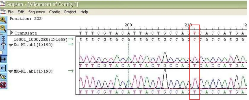
Figure 3. Sample electropherogram Data Analysis Tissues Skin and Oral Mucosal with CRS. Analysis of the
data sample electropherogram Ku (Skin) and MM (Mouth mucosa) of images showed a C insertion mutation at a specific position. The program automatically SeqMan mark different nucleotides.
Researchers who had previously conducted research on mitochondrial DNA have reported
a number of mutations they found. These mutations have been published in
www.mitomap.org. Data published mutation is updated if there are mutations that have not
been reported previously.
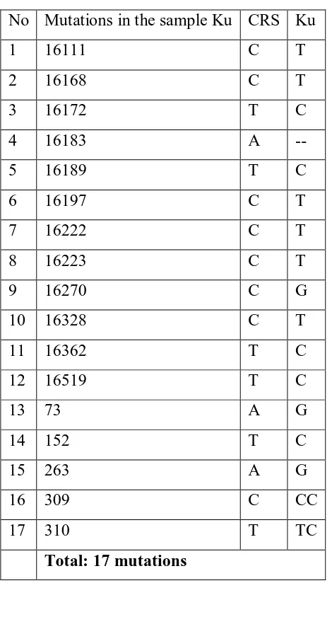
Among seventeen mutations observed in skin samples, there is a mutation that has not
Table 1. Mutation that is observed in Sample Skin Tissue.
No Mutations in the sample Ku CRS Ku
1 16111 C T
2 16168 C T
3 16172 T C
4 16183 A --
5 16189 T C
6 16197 C T
7 16222 C T
8 16223 C T
9 16270 C G
10 16328 C T
11 16362 T C
12 16519 T C
13 73 A G
14 152 T C
15 263 A G
16 309 C CC
17 310 T TC
Total: 17 mutations
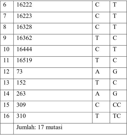
Table 2. Mutations Observed in Oral Mucosal Samples Tissue.
No Mutations in the sample MM CRS MM
1 16111 C T
2 16168 C T
3 16172 T C
4 16183 A --
6 16222 C T
7 16223 C T
8 16328 C T
9 16362 T C
10 16444 C T
11 16519 T C
12 73 A G
13 152 T C
14 263 A G
15 309 C CC
16 310 T TC
Jumlah: 17 mutasi
In this analysis, also carried mutations comparison between the two samples. The
comparison showed that among both samples is observed fifteen identical mutation. These
mutations occur in the same type and same nucleotide position in both samples. However,
the results of this comparison also shows that there are nucleotide differences in the two
samples. Mutation C (16 197), T and C (16 270), G is only observed in samples of skin
tissue, and not termati in oral mucosa samples. Mutation C (16 444), T is only observed in
samples of oral mucosa and absent in samples of skin tissue.
The two samples analyzed showed mutation T to C substitution at nucleotide position
16189 which resulted in a poly-C in both samples. These mutations may disrupt the
process of determining the nucleotide sequence through direct sequensing, so that the
resulting nucleotide sequence is incomplete. Therefore, bidirectional sequencing
electropherogram results using M1 as the primary forward primer and reverse primer
HV2R as very helpful reading of the nucleotide sequence of mtDNA samples.
Table 3. Differences Fragments of D-loop mtDNA Samples Skin and Oral Mucosal
No Mutations in the sample Ku Mutations in the sample MM
1 C(16111)T C(16111)T
3 T(16172)C T(16172)C
4 delesi A (16183) delesi A (16183)
5 T(16189)C T(16189)C
6 C(16197)T -
7 C(16222)T C (16222)T
8 C(16223)T C(16223)T
9 C(16270)G -
10 C(16328) C(16328)
11 T(16362)C T(16362)C
12 - C(16445)T
13 T(16519)C T(16519)C
14 A(73)G A(73)G
15 T(152)C T(152)C
16 A(263)G A(263)G
17 C(309)CC C(309)CC
18 T(310)TC T(310)TC
Jumlah: 17 mutasi jumlah: 16 mutasi
Differences mutation found in both samples, indicating that the tissue or organ which is
the result of differentiation of the ectoderm layer, not 100% homologous. At first, it is
estimated if the sequence of nucleotides in the two samples showed 100% homology, then
the mutation is unlikely to occur during human development from the zygote through
embryogenesis process that produces three layers of the embryo, until the formation of
tissue or organ differentiation results of the three embryonic layers. However, in the
presence of nucleotide differences in the two samples, it is known that a mutation in the
tissues derived from this ectoderm layers.
The differences observed in the second mutation affecting the use of mitochondrial DNA
samples on homology analysis for forensic analysis. Because both samples have a different
mutation, the sequence of nucleotides in the two samples is not 100% homologous. If the
results of homology analysis showed differences in mutation like this, it will lead to doubt
use of the same network as the source of mtDNA template for homology analysis in
forensic analysis. These suggestions are given with the assumption that cells originating
from the same network have identical nucleotide sequences of mtDNA.
CONCLUSION
In the D-loop region, the skin samples were observed seventeen mutations, whereas the
oral mucosal tissue samples of sixteen mutations were observed when compared with
standard nucleotide sequence of the CRS. Among all the mutations that occur in both
samples, there are two mutations that have not been reported, that mutations C (1917) T, C
(16 270) G, and C (16 444) T. In both samples, there were fourteen identical mutation. In
addition, between the two samples also contained different mutations. Thus, it can be
concluded that both samples originated from human embryonic ectoderm layer has a
degree of similarity or nucleotide sequence homology does not equal 100%. Therefore, the
suggested use of the same network as the source of template DNA to examine the
homology of the nucleotide sequence of mtDNA in forensic analysis.
REFERENCES
Anderson, S., Bankier, A.T., Barrel, BG., Bruijin, M.H.L de., Coluson, A.R., Drouin,J.,
Aperon, I.C., Nierlich, D. P., Roe, B.A., Sanger, F, F., Schier, PH., Smith, A.J.H., Staden,
R., and Young, I.G. 1981. Sequence and Organization of the Mitochondrial Genome,
nature. 290, 457-465
Dimauro MD, Salvatore dan Eric A. Schon. Ph.D. 2003. Mecahanism of Desease:
Mitochondrial Respiratory-Chain Disease. N Engl J Med 2003; 348:2656-68.
DNASTAR. 1997. Lasergene Biocomputing Software for Windows, User’s Guide: A
Manual for the Lasergene System, DNASTAR, Inc. USA: Winconsin.
DyckE. Van, F. Foury, and B. Stillman et al. A single-stranded DNA binding protein
required for mitochondrial DNA replication in S. cerevisiae is homologous to E. coli SSB.
Glibert-Barness, Enid and Debich-Spicer, Diane. 2004. Embryo and Fetal Pathology.
Melbourne: Cambridge University Press. Hal: 1-6.
Innis, A. M., ang Gelfand, H. D. 1990. Optimization of PCRs, PCR Protocols: A Guide to
Methods and Applications
JE. Hixson, TW. Wong, and DA. Clayton. Both the conserved stem-loop and divergent
5'-flanking sequences are required for initiation at the human mitochondrial origin of
light-strand DNA replication. J Biol Chem 1986. 261: (5) 2384-2390.
N. Lecrenier, P. Van Der Bruggen, and F. Foury. Mitochondrial DNA polymerases from
yeast to man: A new family of polymerases. Gene 1997. 185: (1) 147-152.
Ngili, Yohanis. 2004. Urutan Nukleotida daerah HV-2 D-loop DNA mitokondria manusia
tiga dan tujuh generasi segaris keturunan ibu, serta empat korban bom Bali. Departemen
Kimia ITB: Bandung.
Noer, A.S. 1991. Molecular Genetics of Mitochondrial Encephalopaties, A thesis
Presented for the degree of Doctor of Philosophy Department of Biochem. Monash
University, Melbourne, Clayton, Victoria, Australia
Orrego, C., dan King, M.C. 1990. Determination of familial relationships, di dalam PCR
Protocols a guide to methods and applications, San Diego: Academic Press.
Pakendorf, Brigitte dan Mark Stoneking. Mitochondrial DNA and Human Evolution.
Annu. Rev. Genomics Hum. Genet. 2005. 6:165–83
RP. Fisher, MA. Parisi, and DA. Clayton. Flexible recognition of rapidly evolving
promoter sequences by mitochondrial transcription factor 1. Genes Dev 1989. 3:
RP. Fisher, T. Lisowsky, and MA. Parisi et al. DNA wrapping and bending by a
mitochondrial high mobility group-like transcriptional activator protein. J Biol Chem 1992.
267: 3358-3367.
Syukriani, Yoni Fuadah. 2007. Clustered point mutations outside HVSI/II in human
mitochondrial genome. Bandung: Departemen Kimia ITB
TW. Wong and DA. Clayton. DNA primase of human mitochondria is associated with
structural RNA that is essential for enzymatic activity. Cell 1986. 45: 817-825.
Wallace, D.C. 1989. Mitochondrial DNA mutations in neuromuscular disease, trends in
genet, 5 (1) page 9 -13
Acknowledgments
We thank the Head of the Laboratory of Biochemistry, University of Padjadjaran who
helped in the research process, in particular to Mr Iman Permana Maksum and Rina
Mulyani. Thanks also to the Macrogen, Seoul, South Korea has menyekuens human
mitochondrial DNA genome in various fragments. Also the Government of Papua