Indones J Anim Vet Sci. 20(4): 285-296
Endemicity of Avian Influenza in Ducks Living Around Commercial
Layer Farms
Tarigan S, Indriani R, Sumarningsih
Indonesian Research Center for Veterinary Sciences, RE Martadinata St. No. 30 Bogor E-mail: simsont@me.com
(received 24-11-2015; revised 8-12-2015; accepted 22-12-2015)
ABSTRAK
Tarigan S, Indriani R, Sumarningsih. 2015. Endemisitas penyakit Avian influenza pada itik yang hidup disekitar peternakan ayam petelur komersial. Indones J Anim Vet Sci. 20(4): 285-296. DOI: http://dx.doi.org/10.14334/jitv.v20i4.1247
Tetua dari semua virus avian influenza adalah itik atau unggas air lainya yang kemudian mengalami mutasi dan adaptasi sehingga menjadi patogen pada ayam atau unggas lainya. Oleh karena itu, penyidikan keberadaan virus influenza pada itik terutama yang dekat dengan peternakan ayam sangat penting. Serum dari 54 ekor itik dan 51 entok yang dipelihara penduduk disekitar peternakan ayam ras petelur komersial di Kabupaten Cianjur dan Sukabumi diambil pada bulan Maret dan April 2014. Indikasi adanya infeksi dilakukan dengan pemeriksaan serologis menggunakan serangkaian alat uji yang meliputi: competitive dan indirect ELISA untuk antibodi nucleoprotein, ELISA MM2e untuk antibodi protein M2e, uji HI, indirect ELISA dan dot blot untuk antibodi haemagglutinin, dan dot blot untuk antibodi neuraminidase. Haemagglutinin rekombinan (H1-H13 dan H15), neuraminidase rekombinan (N1, N2,N7 dan N9) dan rekombinan nucleoprotein virus influenza A digunakan dalam indirect ELISA dan dot blot. Sebanyak 63% dari itik dan 13% dari entok memiliki antibodi terhadap nucleoprotein, dan 62% dari sampel itik yang seropositif nucleoprotein juga memiliki antibodi terhadap M2e. Tingginya seroprevalensi AI pada itik disekitar peternakan ayam ras komersial menunjukkan bahwa penerapan biosekuriti yang ketat pada peternakan ayam komersial masih sangat diperlukan. Berdasarkan hasil pemeriksaan ELISA dan dot blot diduga bahwa pada itik tersebut beredar subtipe H5N2 dan H9N2, selain H5N1. Konfirmasi lebih lanjut dengan isolasi virus perlu dilakukan mengingat subtipe H9N2 dan H5N2 dapat menimbulkan penyakit yang serius pada unggas dan keberadaanya belum pernah diketahui sebelumnya di Indonesia.
Kata Kunci: Duck, Immunoassay, Avian Influenza, H5N1, H5N2, H9N2
ABSTRACT
Tarigan S, Indriani R, Sumarningsih. 2015. Endemicity of avian influenza in ducks living around commercial layer farms. Indones J Anim Vet Sci. 20(4): 285-296. DOI: http://dx.doi.org/10.14334/jitv.v20i4.1247
The progenitors of all avian influenza viruses are generally derived from ducks or other waterfowl that have undergone mutation and adaptation to become pathogenic in chickens or other poultry. Investigation of the presence of avian influenza viruses in ducks especially those living around chicken farms is, therefore, important. Serum from 54 ducks and 51 Muscovy ducks living around commercial layer farms in the districts of Cianjur and Sukabumi were collected in March - April 2014. The indication of AI-virus infection in those birds was based on an array of serological tests including competitive and indirect ELISAs for antibody to nucleoprotein, MM2e ELISA for antibody to M2e, HI test, ELISAs and dot blot for antibodies to haemagglutinin, and dot blot assay for antibodies to neuraminidase. Recombinant Haemagglutinins (H1-H13 and H15), recombinant neuraminidases (N1, N2, N7 and N9) and recombinant influenza-A nucleoprotein were used in the indirect ELISAs and dot blot assays. As many as 63% of duck samples and 13% Muscovy-duck samples were serologically positive to nucleoprotein, and 62% of the nucleoprotein-seropositive ducks were also positive to M2e. The high seroprevalence of AI in the ducks living around commercial poultry farms suggested that application of strict biosecurity measures on those farms is still needed. Based on the results of the ELISA and dot blot assays, AI virus subtypes H9N2 and H5N2, in addition to H5N1, were suspected to be circulating in those ducks. Further confirmation by virus isolation, however, is required because H9N2 and H5N2 subtypes have yet been unknown Indonesia and both the subtypes can cause serious disease in poultry.
Key Words: Duck, Immunoassay, Avian Influenza, H5N1, H5N2, H9N2
INTRODUCTION
Ducks, including other birds belonging to the orders Anseriformes and Charaddriiformes, are the natural reservoir of all influenza-A viruses (Alexander 2000). Many of avian influenza-A viruses (AIV) that are pathogenic for domestic chickens originated from low
pathogenic AIVs that have undergone mutations in the cleavage site of the haemagglutinin (HA) and reassortment in ducks, before they infect chickens
(Guan & Smith 2013). The emergence of a number of
In some countries in South East Asia, domesticated ducks have been proven to be a major risk factor in spreading of HPAI to commercial chickens (Martin et
al. 2006; Tiensin et al. 2007). However, a study carried
out in Indonesia concluded that ducks were not a major risk factor in the spread of H5N1 in this country.
The province of West Java in Indonesia is similar to the region in Southern China in regard to the ecology of
AIV and intensity and proximity of ducks’ and
chickens’ rearing activities that facilitate the emergence
of HPAI (Wan 2012) .
Ducks are raised throughout Indonesia with total population in 2013 estimated at 46,313,000. West Java is the province with the highest population of ducks, 8,943,000 or 19.31%, followed by its neighbor, the Central Java province, with 5,847,000 ducks or 12.63%. Besides ducks, West Java also has the highest number of other poultry (broiler, layer and native chickens) in Indonesia with a total population of 722,738,585 of 1,793,023,090, or 40.31% (DGLAHS 2013).
Our investigation on the occurrence of HPAI in commercial layer farms in West Java, carried out prior to this study, revealed that of eight large farms closely investigated for one year, none was found infected by H5N1 or other subtype of AI viruses. One of the most important sources of infection for those layer farms is presumably the native chickens and ducks living around the farms. Since these extensively raised birds are not normally vaccinated against HPAI, the disease may still be endemic and therefore may become the most important source of infection for the commercial poultry. In our previous study, we reported the presence of ongoing subclinical infection in native chickens. Thirty-six (8.6%) of the 421 chicken tested were positive in either one or more of three serological tests (HI, Influenza-A ELISA and MM2e ELISA) used
(Tarigan et al. 2015b). The purpose of this study was to
assess the importance of ducks living around commercial farms as the source of AIV infection. In this study we present the examination results of serum samples collected from the same locations in two districts in the province of West Java. In contrast to the layer farms, AIV was found to be endemic in free-range ducks with high seroprevalence.
MATERIALS AND METHODS
54 ducks and 51 muscovy ducks living near commercial layer farms in Sukabumi and Cianjur
Ducks were collected from 3 villages; Ciwalen (36 ducks), Tangkil Waru (12 ducks) and Tapos (6 ducks), whereas the muscovy ducks were from 7 villages, Ciwalen (17), Cinangka (4), Bedahan (6), Karang Anyer (13), Cipolong (7), Kebun Pedes (2) and Caringin (2). Sample collection was organized and facilitated by the District Animal Health Services. A simple questionnaire was prepared to ease recording on (1) the age group of each bird bled, (2) the name and address of the owner, (3) number of poultry he or she owned, (4) if disease or death in poultry had occurred in the neighborhood, (5) whether they vaccinated their ducks against avian influenza (6) if any of his or her family or neighborhood worked on the commercial layer farms and (7) whether they used to buy culled chicken from the layer farms.
Haemagglutination inhibition (HI) tests
Haemagglutination test was carried out according to the standard procedures using the haemagglutinating (HA) antigen prepared from a clade 2.3.2 isolate of H5N1 virus (A/Duck/Sukoharjo/Bbvw-1428-9/20012) or a clade 2.1.3 isolate of H5N1 virus (A/Ck/WJ/PWT-WIJ/2006) (OIE 2012). For the HI test, serum to be tested was serially diluted in 25 µl of PBS in V-bottom microtitre plates and an equal volume of HA antigen containing 4 HA units was added. After incubation at 25ºC for 30 min, 25 µl of a 1% suspension of chicken red blood cells was added and incubated for 40 min at 25ºC. The ducks' and muscovy-ducks' sera were treated by adsorption to chicken red blood cells before the HI test. The HI titre was expressed in log2 units of the highest dilution of sera that completely inhibited haemagglutination.
cNP ELISA
Antibodies to AI virus in collected sera were used as
an indication of the presence of AI virus in bird’s
population. Initially, two serological tests were used; firstly, influenza A or competitive nucleoprotein (cNP) ELISA [Australian Animal Health Laboratory (AAHL), Australia] was used to detect antibody to the NP of influenza-A virus. Testing was carried out according to the protocol provided by the test producer. Secondly, MAP-M2e ELISA was used to detect antibody to external domain of M2 protein of AI (H5N1) virus. The protocols for this test has been described previously
(Tarigan et al. 2015a) and used with some variation.
previous experiment in layer chicken the cut off OD for positivity was 1.035 x the OD of negative-control serum.
Indirect-NP (iNP), indirect-H5 (iH5) and indirect haemagglutinins (iHAs) ELISAs
The iNP and iH5 ELISAs were employed to support the result of cNP and MM2e ELISAs. For these indirect ELISAs, recombinant NP and haemagglutinin H5, obtained from Sinobiologicals Inc. China (Table 1), were used as coating antigens. Each recombinant protein was diluted in 0.1 M carbonate buffer (pH 9.6) at 2 µg/ml then used to coat microtitre plates (Nunc maxisorp) overnight at 4°C. After blocking with non-fat-skimmed milk (5 mg/ml, 2 hours), test serum samples and positive and negative controls, diluted in PBST (PBS pH 7.2, 0.05% Tween-20) at 1 : 100 (or other dilutions when indicated), were added and incubated at 37°C for 1 hour. The negative serum control was collected from a young, AI-free duck, whereas the positive control serum was from a duck that had been vaccinated with an inactive,
clade-2.1.3-H5N1 vaccine then challenged with a clade 2.3.2, H5N1 subtype AI virus (A/Duck/Sukohardjo/Bbvw-1428-9/2012). The HI titres of the positive control serum against the challenge virus was 9 log2 and the negative serum control was 0 log2. After washing 4 times with PBST, goat anti-duck-IgG-HRP conjugate (KPL Immunologicals, USA) diluted at 1:100 was added and incubated at 37°C for 1 hr. After washing 4 times, chromogenic (ABTS) substrate was added and the absorbance was recorded using a microtitre-plate reader (Thermo Multiskan Ex).
When samples were positive in NP and MM2e ELISAs, but were negative in H5 ELISA, the type of haemagglutinin (HA) reactive with the sera were determined by iHAs ELISA using similar protocol described for iH5 ELISA and dot blot assay using recombinant proteins of all HA subtypes listed in
Table 1. The iNP, iH5 and iHAs ELISAs have not been
validated previously and the cut-off values for positivity were unknown. In this study, test results resembled positive or negative serum controls were considered to be positive or negative, respectively.
Table 1. Recombinant HA and NA proteins used in this study
Recombinant protein Source of HA gene Catalog No*.
H1 A/California/07/2009 (H1N1) 11085-V08H
H2 A/Japan/305/1957(H2N2) 11088-V08H
H3 A/Brisbane/10/2007(H3N2) 11056-V08H
H4 A/Swine/Ontario/01911-1/99(H4N6) 11706-V08H
H5 A/Indonesia/5/2005(H5N1) 11060-V08H1
H6 A/northern shoveler/California/HKWF115/2007(H6N1) 11723-V08H
H7 A/Netherlands/219/03(H7N1) 11082-V08B
H8 A/pintail duck/Alberta/114/1979(H8N4) 11722-V08B
H9 A/chicken/Korea/164/04(H9N8) 40183-V08B
H10 A/duck/Hong Kong/786/1979(H10N3) 11693-V08H
H11 A/mallard/Alberta/294/1977(H11N9) 11704-V08H
H12 A/green-winged teal/ALB/199/1991(H12N5) 11718-V08H
H13 A/black-headed gull/Netherlands/1/00(H13N8) 11721-V08H
H15 A/duck/AUS/341/1983(H15N8) 11720-V08H
N1 A/Hubei/1/2010(H5N1) 40018-V07H
N2 A/Chicken/Hong Kong/G9/97(H9N2) 40034-V07H
N7 A/Netherlands/219/2003(H7N7) 40202-V07H
N9 A/Anhui/1/2013(H7N9) 40108-V07H
NP A/California/07/2009(H1N1) 11675-V08B
Dotblot and SDS PAGE
Three microliters of recombinant HA or NA proteins diluted at 20 µg/ml in PBS, were spotted onto a nitrocellulose membrane strip. After air-drying, the membrane was blocked with skimmed milk (5 mg/ml, 2 hours), serum samples and controls diluted at 1 : 200 in PBST were added and incubated at 25°C for 2 hr. After washing 4 times with PBST, goat anti-duck-IgG-HRP conjugate (KPL immunologicals, USA) diluted at 1:100 was added then incubated at 25°C for 2 hours. After washing 4 times, chromogenic (DAB) substrate was added to probe antibody bound to the nucleoprotein.
Recombinant proteins were separated in the 10% -acrylamide-separating gels on SDS PAGE. Each recombinant haemagglutinin was dissolved in SDS-PAGE sample buffer at 200 µg/ml, heated in boiled water for 5 minutes and loaded into the SDS PAGE gels 5 µl/3.4-mm-wide well. Proteins in the gel were stained with routine Coomassie blue.
RESULTS AND DISCUSSION
Ducks
All the 54 duck sera were negative in HI test using a 2.3.2 clade H5N1 isolate (A/Duck/Sukoharjo/Bbvw-1428-9/20012) (Hi titre <3 log 2). Other tests used in this study, however, show a high proportion of the sera to be positive to AI. The duck sera could be classified ELISA and MM2e-ELISA (MM2e-ELISA’s OD<0.25), were considered to be true negative for AI, or at least
for H5N1. These results were supported by iNP and iH5 ELISA. When 8 of the 11 sera were tested with the iNP and iH5 ELISAs, all of them were negative as they had OD comparable to that of negative control serum
(Figure 1, 2yellow bars).
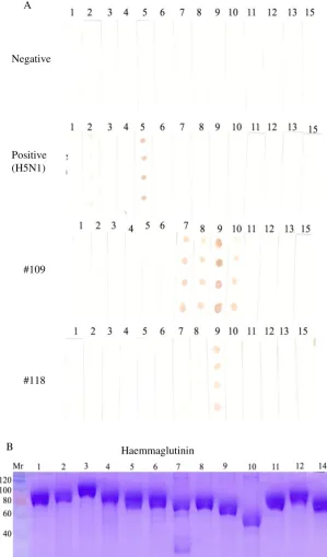
Twenty-one sera (39%) were supposedly positive for AI because they were both positive in cNP ELISA and MM2e-ELISA (MM2e-ELISA’s OD>0.25). When 8 of the 21 sera were tested with iNP ELISA, 7 sera were positive because they had OD, which were higher than that of the negative control serum (Figure 2, red bars). As a matter of fact, the ODs of some of these sera were even higher than that of positive control serum. The majority of the NP-positive sera were also positive for H5 haemagglutinin because 6 of the 8 NP-positive sera were positive in i-H5 ELISA (Figure 2, red bars). Two ducks of this group (#109 and #118), which were negative in the iH5 ELISA had probably been infected by a non-H5 subtype of AI virus.
The assumption that duck no 109 and 118 were not infected by subtype H5 but by other subtype of AI virus was support by the dot blot assay (Figure 3). The reliability of the assay was affirmed by its results on control sera. As expected, the negative control serum did not recognized any of the recombinant haemagglutinin whereas positive control serum which was derived from duck vaccinated and challenged with a H5N1 virus recognised strongly H5 haemagglutinin with some cross reaction with H2 haemagglutinin. In line with the indirect H5 ELISA, sera from duck 109 and 118 did not recognize the H5 haemagglutinin in dot blot assay (Figure 3). Serum from duck 118 only recognised H9 haemagglutinin, whereas serum from duck 109 regonised H7, H8, H9 and H10 haemagglutinins, but the most prominent reaction was with H9 haemagglutinin. The results of this dot blot assay was in line with the indirect ELISA in which all
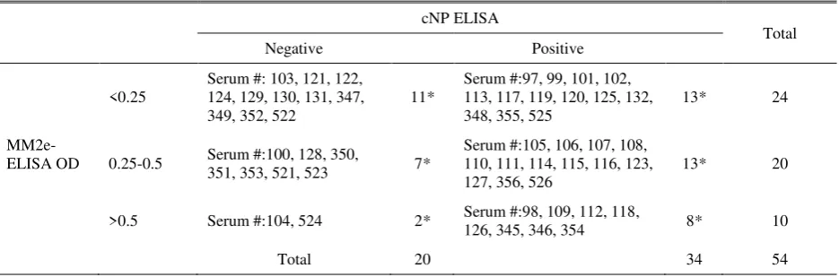
Table 2. Results of MM2e and cNP ELISAs on 54 sera collected from ducks living near commercial layers farms
the recombinant haemagglutinins were used to coat the microtitre plate. The highest ELISA OD in this ELISA was found with the H9 haemagglutinin, followed by H8, H10 and H7 haemagglutinins (Figure 4). All recombinant haemagglutinins used in these immunoblot assay and ELISA had high purity and contained the same protein concentration as indicated by the SDS PAGE analysis (Figure 3B). Therefore, non-specific reaction between contaminated proteins and immunoglobulin contained in the duck sera, and
‘background noise’ due to uneven concentration of
haemagglutinins in the assays could be neglected.
There were nine ducks that were negative in iNP ELISA, seven of which had moderate MM2e-ELISA’s OD (0.25-0.5) and two had high (>0.5) (Table 2). Further analysis with iNP and iH5 ELISAs on some sera of this group, duck 104 (MM2e-ELISA’ OD=2.698), duck 524 (MM2e-ELISA’ OD= 0.627) and duck 351 (MM2e-ELISA’ OD= 0.401), revealed that the all sera were negative in both iNP and iH5 ELISAs
(Figures 2 and 3, blue bars). Because the ducks were
seronegative to NP protein based on cNP and iNP ELISAs, the ducks were likely to be seronegative to AI virus. The results of MM2e ELISA for those ducks, therefore, were considered to be non-specific.
Figure 1. Antibody level, indicated by ELISA OD, in duck sera to Influenza-A nucleoprotein determined by iNP ELISA
Figure 2. Antibody level, indicated by ELISA OD, in duck sera to influenza haemagglutinin H5 determined by iH5 ELISA 3
2.5
2
1.5
1
0.5
0
3
2.5
2
1.5
1
0.5
0 ELISA OD
ELISA OD
Serum Group and Number
(A). Suspension of each recombinant haemagglutinin (3 µl) at 20, 10, 5, and 2.5 µg/ml were spotted onto nitrocellulose membrane strips then reacted with duck sera at 1:400 dilution
(B). Coomassie-blue-stained SDS PAGE of recombinant haemagglutinin to show that all haemagglutinins were pure and had equal concentration
Numbers above the strips and SDS-PAGE gels are the haemagglutinin type; 1, 2, .,... ., 14 denote haemagglutinins H1, H2, H3, ... H14
Figure 3. Determination of haemagglutinin type recognised by duck sera A
Negative
Positive (H5N1)
#109
#118
Figure 4. Haemagglutinin types recognised by serum from duck #109 in indirect ELISA. Confirmation the result of dot blot
(Figure 3). A microtitre plate was coated with each recombinant haemagglutinin (2.5 µg/ml) then reacted with serum from duck #109 at 1:200 dilution
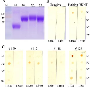
(A). Coomassie-blue-stained SDS PAGE of recombinant neuraminidase N1, N2, N7 and N9 to show that all neuraminidase preparations were pure and in equal concentration. Suspension of each recombinant neuraminidase (3 µl, 20µg/ml) were spotted onto nitrocellulose membrane strips then reacted with duck sera at dilution indicated below the strips (B). Negative and positive control duck sera
(C). Duck serum samples
Figure 5. Neuraminidase types recognised by duck sera
Positive test results in cNP ELISA but negative in MM2e ELISA were found in 13 (24%) sera (Table 3). When five of them were tested in iNP ELISA, only two sera had OD that were higher than that of control
negative serum (Figure 2, green bars). Examination with iH5 ELISA indicated that none of the 5 sera examined was positive for H5 haemagglutinin.
2
1.5
1
0.5
0 ELISA OD
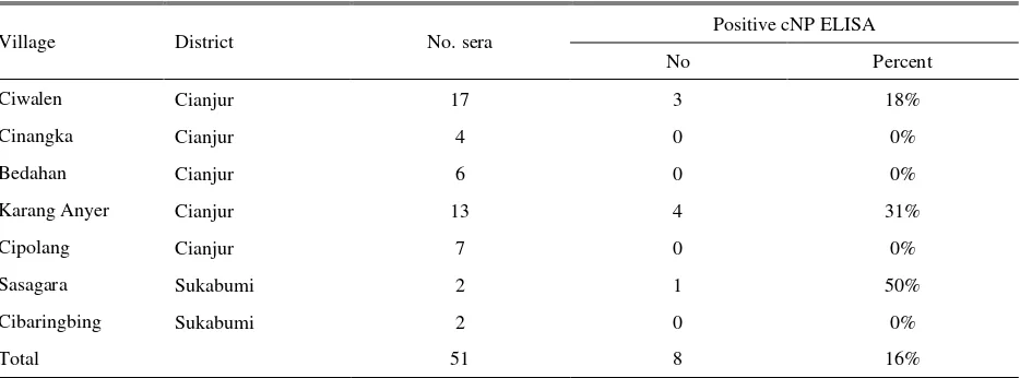
Table 3. Seropositivity in sampled Muscovy duck against Avian Influenza
Village District No. sera Positive cNP ELISA
No Percent
Ciwalen Cianjur 17 3 18%
Cinangka Cianjur 4 0 0%
Bedahan Cianjur 6 0 0%
Karang Anyer Cianjur 13 4 31%
Cipolang Cianjur 7 0 0%
Sasagara Sukabumi 2 1 50%
Cibaringbing Sukabumi 2 0 0%
Total 51 8 16%
Reactivity some of the sera to neuraminidase N1, N2, N7 and N9 is presented in Figure 5. Four ducks tested were all positive in cNP and MM2e ELISAs but had variable OD in iH5 ELISA. Duck 126 had high, duck 112 had moderate and ducks 109 and 118 had low, as low as negative control duck, iH5 ELISA’s OD. Sera from duck 109, 112 and 118 reacted prominently with neuraminidase N2 whereas that from duck 126 withneuraminidase N1. The dot blot assay considered being reliable since the positive and negative sera control reacted as expected. The positive control serum, which was derived from duck that had been vaccinated and challenged with H5N1 virus, reacted only with the N1 protein, and the negative serum reacted with none of the neuraminidases. (Figure 5B). In addition, the recombinant neuraminidases, based on SDS PAGE, had high purity and even concentration (Figure 3B).
Taken together with previous haemagglutinin assay, ducks 109 and 118 might have been infected with a sub-type H9N2 AI virus because their sera recognized recombinant haemagglutinin H9 and neuraminidase N2. Whereas, duck 112 might have been infected with a sub-type H5N2 AI virus because its sera recognized recombinant haemagglutinin H5 and neuraminidase N2.
Muscovy ducks
As compared with ducks, seropositivity for avian influenza in muscovy ducks was lower. Only 8 of 51 (16%) were positive in cNP ELISA (Table 3). The subtype of AI virus responsible for the seropositivity was not assayed as that for ducks. However, on HI test using a clade 2.1.3 isolate of H5N1 virus (A/Ck/WJ/PWT-WIJ/2006) as antigen, four sera were found positive, one serum from Ciwalen village (HI titre 4 log2), and 3 sera from Karang Anyer village (HI titre 5log2, 3log2, 3log2).
Discussion
Since it first reported in 2003 until 2008, H5N1 AI was endemic with high incidence among native chicken and ducks in West Java, especially in the districts of Cianjur and Sukabumi (Yupiana et al. 2010). Since then, the number of cases declined gradually until the outbreak of clade 2.3.2 H5N1 in duck in 2012
(Dharmayanti et al. 2014). At the time of sample
collection, and a couple of years previously, no report of H5N1 outbreak in the Districts of Sukabumi and
Cianjur (Districts’ PDSR, personal communication).
Despite the absence of report on the outbreak of HPAI H5N1, the present study shows that subclinical avian influenza is still endemic and common among ducks and Muscovy ducks living around commercial layer farms in the Sukabumi and Cianjur Districts, West Java. The prevalence of infection in those birds seems to be very high as 63% and 16% of sampled ducks and Muscovy ducks respectively were seropositive for AI. This is in contrast to the situation in AI-vaccinated, commercial layer farms in the area, where no AIV infection was recorded for the last 12 months (S. Tarigan personal observation). Further of interest was, that native chickens which were not vaccinated against AI and scavenging together with the ducks had much lower seroprevalence of AI (Tarigan et al. 2015b).
to NP, commonly used as a marker for AIV infection, was initially detected with cNP ELISA. The cNP ELISA is a competitive ELISA that had been proved to be sensitive and specific for detection of antibody to the NP of type-A influenza viruses in birds and mammals
(Sergeant et al. 2009; Sergeev et al. 2013) and was used
in the surveillance of AI in wild and domesticated birds in Australia (OCVO 2010). The iNP ELISA, although has not been validated previously, has high agreement with the the cNP ELISA. This is not surprising because the indirect ELISA used recombinant nucleoprotein with high purity as the coating antigen.
The MM2e ELISA was shown to be highly specific based on a validation study using chicken serum samples from vaccination and challenge trials (Tarigan
et al. 2015a). This MM2e ELISA has not been validated
for used in ducks. However, based on the present study and previous study, this MM2e ELISA could also be adapted for use in ducks (Lambrecht et al. 2007). Most sera with high OD in MM2e ELISA were positive in cNP ELISA and those with low OD in MM2e ELISA were negative in cNP ELISA. Our previous study in layer chicken revealed that the MM2e ELISA was highly specific for identifying chicken infected with H5N1 virus. In the present study, however, the MM2e ELISA might not be as specific as in layer chicken, as two sera derived from non-infected ducks, based on cNP ELISA, had high OD in MM2e ELISA. This false positive, the cause of which was unknown, suggest that the MM2e ELISA is still need to be adjusted and validated for use in ducks. The finding that some ducks were serologically positive for nucleoprotein but had low MM2e-ELISA’s OD was not unexpected. The same incident has been observed in chicken and ducks in virus subtype (HI titre of <3 log2) in the present study. Since the iH5 ELISA and immunoblot assay indicated the presence antibody to haemagglutinin in some of the sera, the HI test used in this study may not be sensitive enough to detect the presence of the antibody. The lack of sensitivity may be caused by the unmatched antigenicity between the H5N1 subtype used in the HI test and the virus infecting the ducks.
Although the number of serum samples was small, results obtained in this study were important. First, the seroprevalence of AI in ducks in the vicinity of big commercial layer farms was very high. Even though the
virus seems to cause only subclinical infection in those ducks, the AIV may undergo antigenic drift and shift that become pathogenic for chicken. The spill over of LPAI viruses from ducks into poultry and mutate into HPAI viruses has been documented for a number occasions (Swayne 2007). The HPAI H5N1 which was originated from a LPAI underwent mutation in duck in Guangdong province before it spread to chickens (Wan 2012). Secondly, the present study also indicated that AI virus subtype circulating in the duck population was not only H5N1 but also probably subtypes H9N2 and H5N2. Since the later subtypes have never been identified previously in Indonesia, this serological evidence is still inadequate to claim that those subtypes are present in this country. Confirmation of this serological evidence by meticulous effort to isolate the AI virus subtypes from ducks is required because H5N2 and H9N2 are subtypes that cause great economic loss 2003, the H5N1 AI virus subtype has been known to be the only subtype circulating among ducks in Indonesia, and no other AIV subtype has been identified (Henning
et al. 2010). However, since ducks are the natural where H5N1 was also endemic, identified 22 AI viruses consisting 21 samples H6N1 and 1 sample H9N2 disease in ducks, whereas infection with the clade 2.3.2 usually causes severe disease with high mortality in young ducks (Wibawa et al. 2013; Dharmayanti et al.
2014; Wibawa et al. 2014). This later clade caused
223,042 death in ducks at the peak of the outbreak in September - November 2012 (Ditjennak 2013).
Muscovy ducks and geese in Bogor and Sukabumi Districts, West Java found 21 of 460 samples (4.6%) were positive for H5N1, 13 samples (2.8%) for HxN1, 3 samples (0.7%) for H5Nx and 8 samples (1.7%) for HxNx. Several decades previously, (Ronohardjo 1982) studied avian influenza in ducks in Indonesia and reported that H4N6 and H4N2 were the only subtypes found and the subtypes caused clinical disease in ducks characterised by sinusitis, air sacculitis and poor growth in growing ducks. The samples from which the H4N6 cause great economic losses to the poultry industry and found in many countries. Subtype H5N2 is known to to be enzootic in many Asian countries including China
(Zhu et al. 2013; Wang et al. 2014), Korea (Kim et al.
2006; Lee et al. 2011), Pakistan (Cameron et al. 2000),
Iran (Ghaniei et al. 2013) and Israel (Banet-Noach et al. 2007). Although subtype H9N2 AIV is classified as LPAI, the economic losses associated with this subtype are enormous in many countries (Jakhesara et al. 2014;
Shehata et al. 2015).
Since the H9N2 subtype is widely present in Asia, it is not surprising if the subtype also present in Indonesia. The Asian H9N2 which now has adapted to chicken originally derived from ducks because this subtype was only isolated from duck before 1992 (Guo et al. 2000). This means that the H9N2 that apparently still confine to ducks as observed in this study may one day jump to chickens.
The seroprevalence of AI in Muscovy ducks as found in this study was much lower than that in ducks. It is unknown whether the lower seroprevalence in Muscovy ducks is related to its genetically being less susceptible to AI, or else. Different pathological and immunological responses in Muscovy duck and Peking ducks after challenge with an isolate of H5N1 virus have been described previously (Cagle et al. 2011). In this study attempt to identify AI virus subtype reacting to the sera of Muscovy ducks was not made because the difficulty to obtain anti-muscovy-duck conjugate.
CONCLUSION
This study showed that based on serological examinations ducks living near commercial layer farms in Sukabumi and Cianjur, West Java are infected subclinically with AIV with high prevalence. Based on reactivity of the duck sera to recombinant haemagglutinins and neuraminidases in indirect ELISA and dot blot assays, subtypes H5N2 and H9N2, in addition to H5N1, were suspected to be present in the duck population. Further study, however, is required to confirm the presence of H9N2 and H5N2 subtypes in Indonesia by virus isolation. Although at the time of sample collection most of the infection was subclinical and confines only to ducks, the AIV may undergo mutation in ducks to become pathogenic for, and spread to chicken.
ACKNOWLEDGMENT
This work was supported by the Australian Centre for International Agricultural Research under Grant AH/2010/039. The authors thank Mrs Gita Sekarmila, Mr Achpas and the animal caretakers for their excellent technical assistance.
REFERENCES
Abolnik C, Olivier AJ, Grewar J, Gers S, Romito M. 2012. Molecular analysis of the 2011 HPAI H5N2 outbreak in ostriches, South Africa. Avian Dis. 56:865-879. Alexander DJ. 2000. A review of avian influenza in different
bird species. Vet Microbiol. 74:3-13.
Banet-Noach C, Perk S, Simanov L, Grebenyuk N, Rozenblut E, Pokamunski S, Pirak M, Tendler Y, Panshin A. 2007. H9N2 influenza viruses from Israeli poultry: a five-year outbreak. Avian Dis. 51:290-296.
Cagle C, To TL, Nguyen T, Wasilenko J, Adams SC, Cardona CJ, Spackman E, Suarez DL, Pantin-Jackwood MJ. 2011. Pekin and Muscovy ducks respond differently to vaccination with a H5N1 highly pathogenic avian influenza (HPAI) commercial inactivated vaccine. Vaccine. 29:6549-6557.
Cameron KR, Gregory V, Banks J, Brown IH, Alexander DJ, Hay AJ, Lin YP. 2000. H9N2 subtype influenza A viruses in poultry in pakistan are closely related to the H9N2 viruses responsible for human infection in Hong Kong. Virology. 278:36-41.
DGLAHS. 2013. Livestock and animal health statistics 2013. Jakarta (Indones): Direktorat Jenderal Peternakan dan Kesehatan Hewan, Kementerian Pertanian.
Dharmayanti NLPI, Hartawan R, Pudjiatmoko, Wibawa H, Hardiman, Balish A, Donis R, Davis CT, Samaan G. 2014. Genetic characterization of clade 2.3.2.1 avian influenza A(H5N1) viruses, Indonesia, 2012. Emerg Infect Dis. 20:671-674.
[Ditjennak] Direktorat Jenderal Peternakan. 2013. Perkembangan kasus Avian Influenza (AI) pada unggas kondisi s/d 31 Desember 2013. [accessed August 12th 2015]. http://ditjennak.pertanian.go.id.
Ghaniei A, Allymehr M, Moradschendi A. 2013. Seroprevalence of avian influenza (H9N2) in broiler chickens in Northwest of Iran. Asian Pac J Trop 2000. Characterization of the pathogenicity of members of the newly established H9N2 influenza virus lineages in Asia. Virology. 267:279-288.
Hemmatzadeh F, Sumarningsih S, Tarigan S, Indriani R, Dharmayanti NLPI, Ebrahimie E, Igniatovic J. 2013. Recombinant M2e protein-based ELISA: a novel and inexpensive approach for differentiating avian influenza infected chickens from vaccinated ones. PLoS One. 8:e56801.
Henning J, Wibawa H, Morton J, Usman TB, Junaidi A, Meers J. 2010. Scavenging ducks and transmission of highly pathogenic avian influenza, Java, Indonesia. Emerg Infect Dis. 16:1244-1250.
Hotta K, Takakuwa H, Le QM, Phuong SL, Murase T, Ono E, Ito T, Otsuki K, Yamashiro T. 2012. Isolation and characterization of H6N1 and H9N2 avian influenza viruses from Ducks in Hanoi, Vietnam. Virus Res. 2014. Isolation and characterization of H9N2 influenza virus isolates from poultry respiratory disease outbreak. Springerplus. 3:196.
Khatun A, Giasuddin M, Islam KM, Khanom S, Samad MA, Islam MR, Noor M, Bhuiyan JU, Kim WI, Eo SK, markets continuously evolve and cause the severe clinical signs in layers. Vet Microbiol. 118:169-176.
Kim MC, Choi JG, Kwon JS, Kang HM, Paek MR, Jeong OM, Kwon JH, Lee YJ. 2010. Field application of the H9M2e enzyme-linked immunosorbent assay for differentiation of H9N2 avian influenza virus-infected chickens from vaccinated chickens. Clin Vaccine Immunol. 17:1977-1984.
Lambrecht B, Steensels M, Van Borm S, Meulemans G, van den Berg T. 2007. Development of an M2e-specific enzyme-linked immunosorbent assay for differentiating infected from vaccinated animals. Avian Dis. 51:221-226.
Lee CW, Swayne DE, Linares JA, Senne DA, Suarez DL. 2005. H5N2 avian influenza outbreak in Texas in 2004: the first highly pathogenic strain in the United States in 20 years?. J Virol. 79:11412-11421.
Lin, Shaw YPM, Gregory V, Cameron K, Lim W, Klimov A, Subbarao K, Guan Y, Krauss S, Shortridge K, Webster R, Cox N, Hay A. 2000. Avian-to-human transmission of H9N2 subtype influenza A viruses: relationship between H9N2 and H5N1 human isolates. Proc Natl Acad Sci USA.97:9654-9658.
Loth L, Gilbert M, Wu J, Czarnecki C, Hidayat M, Xiao X. 2011. Identifying risk factors of highly pathogenic avian influenza (H5N1 subtype) in Indonesia. Prev Vet Med. 102:50-58.
Martin V, Sims L, Lubroth J, Pfeiffer D, Slingenbergh J, Domenech J. 2006. Epidemiology and ecology of highly pathogenic avian influenza with particular emphasis on South East Asia. Dev Biol. 124:23-36.
Matrosovich MN, Krauss S, Webster RG. 2001. H9N2 influenza A viruses from poultry in Asia have human virus-like receptor specificity. Virology. 281:156-162. Neirynck S, Deroo T, Saelens X, Vanlandschoot P, Jou WM,
Fiers W. 1999. A universal influenza A vaccine based on the extracellular domain of the M2 protein. Nat Med. 5:1157-1163.
Nguyen T, Davis CT, Stembridge W, Shu B, Balish A, Inui K, Do HT, Ngo HT, Wan XF, McCarron M, Lindstrom SE, Cox NJ, Nguyen CV, Klimov AI, Donis RO. 2009. Characterization of a highly pathogenic avian influenza H5N1 virus sublineage in poultry seized at ports of entry into Vietnam. Virology. 387:250-256.
Nomura N, Sakoda, Endo M, Yoshida H, Yamamoto N, Okamatsu M, Sakurai K, Hoang NV, Nguyen LV, Chu HD, Tien TN, Kida H. 2012. Characterization of avian influenza viruses isolated from domestic ducks in Vietnam in 2009 and 2010. Arch Virol. 157:247-257. [OCVO] Office of the Chief Veterinary Officer. 2010.
Officer, Australian Government Department of Agriculture, Fisheries and Forestry.
[OIE] Office International des Epizooties. 2012. Manual of diagnostic tests and vaccines for terrestrial animals. 7th ed. Office International des Epizooties.
Okamatsu M, Saito T, Yamamoto Y, Mase M, Tsuduku S, Nakamura K, Tsukamoto K, Yamaguchi S. 2007. Low pathogenicity H5N2 avian influenza outbreak in Japan during the 2005-2006. Vet Microbiol. 124:35-46. Ronohardjo P. 1982. Virus influenza A itik di Indonesia serta
pengaruhnya pada kesehatan masyarakat dan ekonomi peternakan (Thesis). Bogor (Indones): Institut Pertanian Bogor. Agafonov AP, Kiselev SA, Agranovski IE, Sergeev AA, Shikov AN, Shishkina LN, Safatov AS, Sergeev AN. 2013. Infection of chickens caused by avian influenza virus A/H5N1 delivered by aerosol and other routes. Transbound Emerg Dis. 60:159-165.
Shehata AA, Parvin R, Sultan H, Halami MY, Talaat S, Abd-Elrazek A, Ibrahim M, Heenemann K, Vahlenkamp T. 2015. Isolation and full genome characterization of avian influenza subtype H9N2 from poultry respiratory disease outbreak in Egypt. Virus Genes. 50:389-400. Susanti R, Soejono RD, Mahardika GN, Wibawan I-WT.
2008. Prevalence of Avian Influenza Virus Subtype H5n1 in Waterfowl in West Java Province of Indonesia. 13th International Congress on Infectious Diseases, Kuala Lumpur June 19-22, 2008: e127.
Swayne DE. 2007. Understanding the complex pathobiology of high pathogenicity avian influenza viruses in birds. Avian Dis. 51:242-249.
Tarigan S, Indriani R, Durr PA, Ignjatovic J. 2015a. Characterization of the M2e antibody response following highly pathogenic H5N1 avian influenza virus infection and reliability of M2e ELISA for identifying infected among vaccinated chickens. Avian Pathol. 44:259-268.
Tarigan S, Indriani R, Igniatovic J. 2015b. Circulating H5N1 virus among native chicken living around commercial layer farms. JITV. 20:224-232.
Tiensin T, Nielen M, Songserm T, Kalpravidh W, Chaitaweesub P, Amonsin A, Chotiprasatintara S, Chaisingh A, Damrongwatanapokin S, Wongkasemjit S, Antarasena C, Songkitti V, Chanachai K, Thanapongtham W, Stegeman J A. 2007. Geographic and temporal distribution of highly pathogenic avian influenza A virus (H5N1) in Thailand, 2004-2005: an overview. Avian Dis. 51:182-188.
Villareal CL, Flores AO. 1997. The Mexican avian influenza (H5N2) outbreak. Proc. 4th Int. Symp.Avian Influenza, Athen, Georgia, USA May 28-31, 1997:18-22.
Wan XF. 2012. Lessons from emergence of A/goose/Guangdong/1996-like H5N1 highly pathogenic avian influenza viruses and recent influenza surveillance efforts in southern China. Zoonoses Public Health. 59:32-42.
Wang Q, Ju L, Liu P, Zhou J, Lv X, Li L, Shen H, Su H, Jiang L, Jiang Q. 2014. Serological and Virological Surveillance of Avian Influenza A Virus H9N2 Subtype in Humans and Poultry in Shanghai, China, Between 2008 and 2010. Zoonoses Public Health.
Wibawa H, Bingham J, Nuradji H, Lowther S, Payne J, Harper J, Junaidi A, Middleton D, Meers J. 2014. Experimentally infected domestic ducks show efficient transmission of Indonesian H5N1 highly pathogenic avian influenza virus, but lack persistent viral shedding. PLoS One. 9:e83417.
Wibawa H, Bingham J, Nuradji H, Lowther S, Payne J, Harper J, Wong F, Lunt R, Junaidi A, Middleton D, Meers J. 2013. The pathobiology of two Indonesian H5N1 avian influenza viruses representing different clade 2.1 sublineages in chickens and ducks. Comp Immunol Microbiol Infect Dis. 36:175-191.
Woo JT, Park BK. 2008. Seroprevalence of low pathogenic avian influenza (H9N2) and associated risk factors in the Gyeonggi-do of Korea during 2005-2006. J Vet Sci. 9:161-168.
Yupiana Y, de Vlas SJ, Adnan NM, Richardus JH. 2010. Risk factors of poultry outbreaks and human cases of H5N1 avian influenza virus infection in West Java Province, Indonesia. Int J Infect Dis. 14:e800-805.