Complexes of 2,6-dihydroxybenzoic acid with divalent metal ions:
Synthesis, crystal structure, spectral studies, and biological
activity enhancement
Shella Permatasari Santoso
a, Suryadi Ismadji
b, Artik Elisa Angkawijaya
a,c, Felycia Edi Soetaredjo
b,
Alchris Woo Go
d, Yi Hsu Ju
a,⁎
aChemical Engineering Department, National Taiwan University of Science and Technology, Taipei 106-07, Taiwan b
Department of Chemical Engineering, Widya Mandala Surabaya Catholic University, Kalijudan 37, Surabaya 60114, Indonesia c
Institute of Plant and Microbial Biology, Academia Sinica, Taipei 115-29, Taiwan
dDepartment of Chemical Engineering, University of San Carlos, Talamban Campus, Cebu 6000, Philippines
a b s t r a c t
a r t i c l e
i n f o
Article history: Received 21 January 2016
Received in revised form 25 May 2016 Accepted 5 June 2016
Available online 07 June 2016
2,6-Dihydroxybenzoic acid orγ-resorcylic acid (DHBA) is a phenolic compound which is known to have poor bi-ological performance such as DPPH scavenging activity and microbial growth inhibition. Combining DHBA with divalent metal ion to form complex is expected to improve its biological properties. The complexes of DHBA with divalent Ni/Co were synthesized and characterized in this study. The complex structures were determined by X-ray single crystal analysis and it was found that metal interacted with DHBA through H-bonds. The results on the biological properties indicate that the complexes have remarkable DPPH scavenging activities and microbial growth inhibition abilities. IC0.5value of DPPH is 35.68 mg/L (231.51μM), 9.21 mg/L (18.09μM) and 25.63 mg/
L (50.33μM) for DHBA, NiDHBA and CoDHBA respectively. The microbial growth inhibitory value (%) at a sample concentration of 400 mg/L is 2.9, 6.4 and 96.1 againstEscherichia coliand 4.9, 16.8, 98.2 againstStaphylococcus aureus, for DHBA, NiDHBA and CoDHBA, respectively.
© 2016 Elsevier B.V. All rights reserved. Keywords:
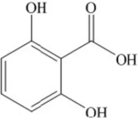
Dihydroxybenzoic acids are aromatic compounds containing two phe-nolic and a carboxylic acid functional groups[1]. 2,6-Dihydroxybenzoic acid or γ-resorcylic acid (DHBA) is one of the six isomerics of
dihydroxybenzoic acids (Fig. 1)[2,3]. DHBA can be synthesized through the Kolbe-Schmitt carboxylation of resorcinol[4,5]. This compound also can be found as one of the phenolic compounds contained in white grape pomace[6]. There are few studies that reported the biological prop-erties of DHBA since this compound possesses poor biological activity. Nishibe[7]reported that DHBA possesses poor DPPH (2,2-diphenyl-1-picrylhydrazyl) radical-scavenging activity whereinb200μM (30 mg/L) of DHBA was required to decrease by 0.200 (IC0.2) in the absorbance
after 30 min reaction with DPPH[7].
The synthesis of organic ligand and metal ion is an interesting re-searchfield since the product complex may enhance the biological properties of the ligand[8–12]. To the best of our knowledge there is
no study utilize combination of DHBA and metal ions in order to en-hance the biological properties of DHBA. In this work, DHBA and diva-lent metal ion (Ni/Co) were combined to form a metal-ligand complex. The structure of the complexes was determined by X-ray sin-gle crystal analysis. Several physical measurements such as SEM, UV– Vis, FTIR, TGA and1H NMR were conducted on the complexes. Biological
activities of the complexes were tested, specifically the DPPH radical-scavenging activity and microbial inhibition growth activity against Escherichia coliandStaphylococcus aureus.
2. Experimental
2.1. Materials
Analytical grade DHBA (C7H6O4, 98% purity),
2,2-diphenyl-1-picrylhydrazyl (DPPH) and metal salts of cobalt nitrate hexahydrate (Co(NO3)2·6H2O, 98% purity) were purchased from Sigma Aldrich
(St. Louis, MO), nickel chloride hexahydrate (NiCl2·6H2O, 98%
puri-ty) was purchased from Alfa Aesar (Lancashire, UK). Ammonium hy-droxide (NH3, 30%) was obtained from Yakuri Pure Chemical (Kyoto,
Japan). ⁎ Corresponding author at: Department of Chemical Engineering, National Taiwan
University of Science and Technology, #43, Sec. 4, Keelung Rd., Taipei 106-07, Taiwan. E-mail address:[email protected](Y.H. Ju).
http://dx.doi.org/10.1016/j.molliq.2016.06.015 0167-7322/© 2016 Elsevier B.V. All rights reserved.
Contents lists available atScienceDirect
Journal of Molecular Liquids
2.2. Physical property measurements
Acid dissociation constants of the ligand DHBA were spectroscopi-cally determined by refining the spectrum as function of pH using HypSpec program[13]. Several measurements were done to character-ize the physical properties of complexes. Surface topography analysis was done by using a JEOL JSM-639 scanning electron microscope at an accelerating voltage of 20 kV and Pt for sample coating. UV–Vis spec-trum analysis was carried out using a JASCO V-550 spectrophotometer equipped with halogen and deuterium lamp. The sample was placed in a standard 10 mm quartz cell. FTIR spectrum analysis was recorded on a Bio-Rad FTS-3500 instrument on KBr disc with a spectra range of 400–4000 cm−1. Thermogravimetric analyses (TGA) under N
2
atmo-sphere and a heating rate of 10 °C/min were done by using a Perkin Elmer Diamond TG/DTA. Elemental analysis on carbon and hydrogen atom was done by using an Elementar Vario EL III.1H NMR spectra
anal-ysis was measured on a Bruker AVIII-600 MHz FT-NMR in D2O solution.
Structure determination was performed by X-ray single crystal analysis using an Oxford Gemini Dual system Single-crystal XRD equipped with Cryojet.
2.3. Spectrophotometric method
The acidity constant of DHBA was determined spectroscopically in the range of 200–400 nm. The spectrum measurements were done using a solution containing DHBA at a concentration of 2 × 10⁻⁴mol/ L. The ligand solution was acidified by using 0.1 mol/L HCl until pHb1.0. Subsequently spectra of the ligand were measured as a
func-tion of pH, where the pHs were sequentially adjusted by using 0.1 mol/L NaOH until pH 13.0. HCl and NaOH were standardized before use. The spectrum data were then used as input to the HypSpec program for determining acidity constant.
2.4. Synthesis of complexes
DHBA (0.39 g, 2.5 mmol) was dissolved in distilled water (15 mL). The solution was heated to 70 °C and a few drops of 2 M ammonia solution were added until DHBA was completely dissolved. Five milliliters of metal salt solution (0.61 g (2.5 mmol) of NiCl2·6H2O or 0.74 g
(2.5 mmol) of Co(NO3)2·6H2O) were added into the DHBA solution.
The pH of the mixture was adjusted to ~4 by adding a few drops of 2 M ammonia solution. The reaction was carried out at 70 °C for 4 h with con-stant stirring, then the solution was cooled to room temperature and left to stand until crystal was formed. The crystal complex was isolated by vacuumfilter, washed several times with distilled water and air-dried.
Small needle green crystals of NiDHBA complex were formed be-tween DHBA and Ni2+. The complex was formed after the solution was
kept overnight with a yield of 46.6%.Anal.Calcd for C14H26NiO16: C,
33.03; H, 5.15. Found: C, 32.66; H, 5.19%.Form. Weight509.06 g/mol.
1H NMR (methanol-d
4, ppm): 7.41, 9.02.
Cubic red crystals (CoDHBA) were formed after DHBA and Co2+
so-lution was kept for 3 days (yield 59.1%).Anal.Calcd for C14H26NiO16: C,
33.02; H, 5.15. Found: C, 32.90; H, 5.27%.Form. Weight509.28 g/mol.1H
NMR (methanol-d4, ppm): 6.18, 6.82.
2.5. Biological property measurement
2.5.1. Radical scavenging activity
Radical scavenging activity of complex was tested against the stable radical DPPH[14]. The tested compound was dissolved in methanol at dif-ferent concentrations. DPPH solution (0.2 mL, 5 × 10⁻⁴mol/L in methanol) was added to the prepared tested compound solution (0.8 mL). The tested complex was incubated at 37 °C for 30 min and the absorbance was mea-sured at 517 nm against methanol as the blank. DPPH (0.2 mL) in metha-nol (0.8 mL) without any tested complex was used as the control. Percent DPPH scavenging activity was calculated as:
%DPPH scavenging activity¼Ac−As Ac 100
whereAcis the absorbance of control andAsis the absorbance of sample at 517 nm. Ascorbic acid was used as the positive control.
2.5.2. Microbial growth inhibitory activity
Broth macro dilution method[15]was used to determine the micro-bial growth inhibitory activity of the complexes against the gram nega-tive microorganismEscherichia coliand gram positive microorganism Staphylococcus aureus. Ampicilin (895.5μg/mL potency) was used as the antibiotic reference. Lysogeny Broth (LB) media prepared from the mixture of tryptone, yeast extract and sodium chloride with ratio 2:1:2 w/w was used as the medium.
The assay was performed in tubes containing different concentra-tions of the tested complex dissolved in LB medium with a total volume of 3 mL for each tube. The prepared bacteria suspension (15μL, 1 × 10⁸cfu/mL) was injected into each test tube. All test tubes were
in-cubated for 24 h at 37 °C. The OD600of each tube was measured after
in-cubation. As the control, 15μL of bacteria suspension was injected into a test tube containing no tested compound. The antimicrobial activity was expressed as %inhibition, calculated as:
%inhibition¼Ic−Is
Ic 100
whereIcis the absorbance of control andIsis the absorbance of sample at 600 nm.
3. Results and discussion
3.1. Acidity constants of the ligand
The acidity constants of DHBA were presented as minus logarithm of stepwise hydrogen dissociation constants, pKa. At neutral form, DHBA possesses three hydrogen atoms at its active donor groups (i.e. carboxyl
group, hydroxyl groups at second and sixth positions of the benzene ring) and can be symbolized as [H3DHBA]. Thefirst dissociation occurs
at the carboxyl group yielding the negative charged [H2DHBA−] as rep-resented by Eq.(1). The second dissociation as well as the third dissoci-ation occur at either the hydroxyl group at second or sixth position, yielding [HDHBA2−] and [DHBA3−] as represented by Eqs.(2) and (3), respectively.
H3DHBA⇄HþþH2DHBA−;Hþ
H2DHBA−
½ ¼Ka1½H3DHBA ð1Þ
2DHBA−⇄HþþHDHBA2−;Hþ
HDHBA2−
h i
¼Ka2½H2DHBA− ð2Þ
HDHBA2−⇄Hþ
þDHBA3−;Hþ
DHBA3−
h i
¼Ka3 HDHBA2−
h i
ð3Þ
The dissociation of an H atom can be observed from the shape-shift of the spectra as function of pH, as shown inFig. 2. The dissociation con-stants of DHBA at 25 °C and an ionic strength of 0.15 mol dm−3NaCl were refined by HypSpec program. The shape-shift of the spectra can be observed more clearly from the insertfigure ofFig. 2. The spectrum shape-shift of DHBA initially occurs at 0.79bpHb2.02 which indicates
thefirst dissociation (pKa1= 0.94 ± 0.07). Afterwards there were no
obvious shifts at 2.02bpHb11.03 indicating that at this pH range
dis-sociation did not occur. The next shape-shifts occur at pHN11.03
which indicate the second and third dissociation (pKa2= 12.07 ± 0.06, and pKa3= 12.66 ± 0.05).
3.2. Physical properties of complexes
Several physical measurements were done to characterize the com-plexes of DHBA with Ni/Co. The measurements include X-ray single crystal, SEM, UV–Vis, FTIR, and thermogravimetric analysis.
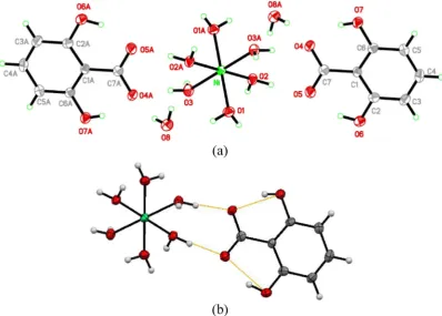
Fig. 3.(a) Molecular structure of NiDHBA, thermal ellipsoid drawn at 50% probability level. (b) H-bonds coordination of complexes. Table 1
Crystal data and structure refinement for the complexes.
Complex NiDHBA CoDHBA
Formula C14H26NiO16 C14H26CoO16
Formula weight 509.06 509.28
T (K) 200(2) 200(2)
Wavelength (Å) 0.71073 0.71073
Crystal system Triclinic Triclinic
Space group P-1 P-1
a (Å) 6.5060(10) 6.5323(10)
b (Å) 7.9239(2) 7.9528(2)
c (Å) 9.8330(2) 9.8628(2)
α(°) 96.2566(14) 96.3144(14)
β(°) 93.5196(13) 93.7198(12)
γ(°) 100.3730(16) 100.4011(12)
V (Å3) 493.930(18) 499.011(18)
Z 1 1
dcalc(Mg/m3) 1.711 1.695
μ(mm−1) 1.066 0.943
F (000) 266 265
Crystal size (mm) 0.300 × 0.300 × 0.150 0.250 × 0.160 × 0.150 Reflections collected 7023 6849
Idependent reflections (Rint) 2263(0.0536) 2275(0.0371) Max. and min. transmission 0.888, 0.752 0.871, 0.830 Data/restraints/parameters 2263/0/149 2275/0/149 Goodness-of-fit onF2 1.048 1.066 R1,wR2((IN2σ(I)) 0.0342, 0.0762 0.0308, 0.0703 R1,wR2(all data) 0.0497, 0.0854 0.0426, 0.0762 Largest diff. peak and hole (e Å−3) 0.381,
−0.756 0.307,−0.558
3.2.1. X-ray crystallographic data
Crystallographic data of the complexes are given inTable 1. It can be seen that the complexes possess similar structure. The complexes were obtained from reacting DHBA with metal salt in water. The complexes were found to be soluble in methanol and ethanol, slightly soluble in water. Since dissociation of the ligand at its carboxyl moiety have a pKa1of 0.94, thus ideally pH ~ 4 was sufficient to provide the
environ-ment for the dissociation of the ligand. The dissociation provides nega-tive charge to the ligand thus allowed it to bind metal ion which has positive charge[16,17]. However, results on crystal structure of the complexes indicate that formation of the complex did not occur through the coordination of dissociated–O donor group of DHBA. The coordina-tion of the complex occurred by hydrogen bonding between the nearby water molecules (which coordinated to the metal ion) with the–O atoms at the carboxylic moiety of DHBA.Fig. 3(b) illustrates the molec-ular structure and H-bonds coordination of NiDHBA.
As can be seen in the result of X-ray single crystal analysis, the pre-dominant synthesized complex is the one which consists of one metal ion and two ligands (ML2complex,Fig. 3(a)), meanwhile the molar
ratio of metal to ligand used for the synthesis was 1:1. One possible
explanation is that while both complexes were formed, ML2complex
has lower solubility than ML and was easier to become saturated and form crystals. Since DHBA has low solubility in water, it is reasonable that complex that consists of more ligand molecules also has lower sol-ubility. X-ray results indicate that the type of counter anion of metal salt (Cl−1for Ni salt and NO
3
−1for Co salt) did not affect the main structure of complex. Counter anions were found not to involve in the formation of complexes.
3.2.2. Surface topography by SEM
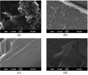
The surface of DHBA powder is depicted inFig. 4(a). It was observed that particles of DHBA powder are not organized. Re-crystallization of Fig. 4.Surface topography of (a) powder DHBA, (b) re-crystallized DHBA, (c) NiDHBA, (d) CoDHBA at magnification of 1000×.
Table 2
Selected FTIR spectra of DHBA, NiDHBA and CoDHBA.
Compound IR spectra (cm −1)
v(O\\H) v(C_O) v(C_C) v(C\\O)
DHBA 34802636 1685 1470 12931036
NiDHBA 3309 1604 1458 1291
1027
CoDHBA 3317 1604 1458 12911027 Fig. 5.DPPH scavenging activities of DHBA and its complexes. *Ascorbic acid was used as the standard reference. , ascorbic acid; , DHBA; , NiDHBA; , CoDHBA.
DHBA in water allowed the particles to solidify and formed more packed structure (Fig. 4(b)). Reaction of DHBA with Ni2+/Co2+resulted in
complexes with different surface topography. As shown inFig. 4(c) and (d), surfaces of the complexes arefiner than the re-crystallized DHBA, especially the Ni2+complex. The change on surface structure
was caused by the interaction of DHBA with metal through hydrogen bonding.
3.2.3. UV-Vis spectra
The spectra of the complexes were measured in water solvent. The complexes possessed two absorption bands. One in UV region repre-sents DHBA and the other in Vis wavelength region indicates thed- or-bital of metal ion. For NiDHBA, DHBA absorption bands occurred at 245 and 306 nm while thed-orbital of Ni was represented by bands at 400 and 700 nm. Since thed-orbital absorption bands have low absor-bance unit[18]thus higher sample concentration was needed in order to observe the bands. Similarly for CoDHBA complex, the absorption bands at the UV wavelength region of 245 and 306 nm represent DHBA while the band at 510 nm representsd-orbital of Co.
3.2.4. FTIR spectra
The IR spectra of the complexes show shifting from that of DHBA (Table 2, Fig. S1). Thev(O\\H) of DHBA attributed to the phenolic and carboxylic group were found at 3480 and 2636 cm−1, respectively. For
the complexes these peaks disappear and are replaced by a broad band centered at 3309 and 3317 cm−1for Ni2+and Co2+complex, re-spectively indicating the presence of water molecules. The disappear-ance of thev(O\\H) bands indicates that the carboxylic group was involved in complex formation. The carbonyl stretching vibration v(C = O) of DHBA was found at 1685 cm−1, for the complexes this peak shifted to 1604 cm−1(
Δv= 81 cm−1). Thev(C = C) stretching of DHBA at 1470 cm−1 shifted to 1458 cm−1for the complexes (Δv= 12 cm−1). The carboxylic stretching vibrationv(C
\\O) of DHBA found at 1293 cm−1shifted slightly to 1291 cm−1for the complexes. The shifting of phenolicv(C\\O) vibration from 1036 cm−1for DHBA to 1027 cm−1(Δv= 9 cm−1) for the complexes confirms the coordina-tion of phenolic oxygen to metal.
3.2.5. Thermal analysis
Thermal analyses for the complexes were carried out at 37 to 900 °C under N2atmosphere. The thermograms are shown in Fig. S2
(Supple-mentary data). The total weight loss of NiDHBA is 81.30% in which ~29% weight loss at 80 to 165 °C is attributed to loss of water molecules, ~19% weight loss at 180 to 240 °C followed by ~28% weight loss at 270 to 530 °C may be attributed to the decomposition of DHBA leaving the metal which is stable at temperaturesN530 °C.
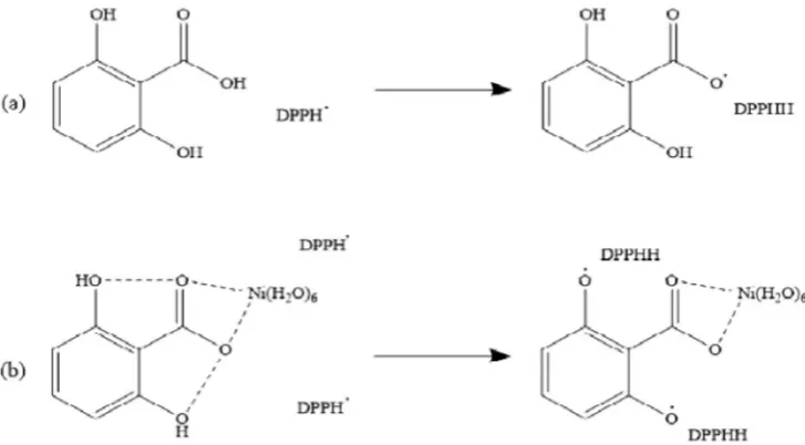
For CoDHBA the total weight loss is 76.33%, ~25% initial weight loss is attributed to water molecules which occurred at 70 to 146 °C, ~23% Fig. 6.Proposed mechanism of DPPH radical scavenging activity by (a) DHBA and (b) NiDHBA complex.
weight loss at 175 to 280 °C and ~7% weight loss at 475 to 550 °C are at-tributed to the decomposition of DHBA leaving the metal residue.
3.3. Biological properties
3.3.1. Radical scavenging activity
The radical scavenging activities of DHBA and its complexes were measured against a DPPH. As shown inFig. 5, the complexes show higher scavenging activity than DHBA, especially the complex NiDHBA. The IC0.5values are 35.68 mg/L (231.51μM), 9.21 mg/L (18.09μM) and
25.63 mg/L (50.33μM) for DHBA, NiDHBA and CoDHBA, respectively. Better scavenging activities of both complexes suggest that metal ion induces the release of more hydrogen atoms of DHBA to stabilize more DPPH radicals. As proposed inFig. 6(a), the free ligand DHBA may only stabilize DPPH through its hydrogen atom at the carboxylic site; meanwhile as shown inFig. 6(b), the complex (containing metal ion) may stabilize more DPPH through hydrogen atoms at the phenolic sites. The release of hydrogen atoms at phenolic sites was anchored by the present of metal ion[19].
3.3.2. Microbial growth inhibitory activity
The microbial inhibitory growth activities of DHBA and its com-plexes were measured againstE. coliandS. aureus. Ampicilin was used as the standard control. The results inFig. 7show that DHBA and its NiDHBA possess weak inhibitory against the two bacteria strains. The most remarkable inhibitory activity against the bacteria was observed in CoDHBA. It was also observed that CoDHBA is more effective against S. aureusthan againstE. coli; for instance at 250 mg/L (491μM), CoDHBA was able to inhibit 57.4% ofS. aureusgrowth while only 38.6% ofE. coli growth was inhibited. The difference may be due to different cell wall structures. As Gram negative bacteria,E. coli possesses additional outer membrane which contains lipid that hinders the complexes to penetrate into the cell[20,21].
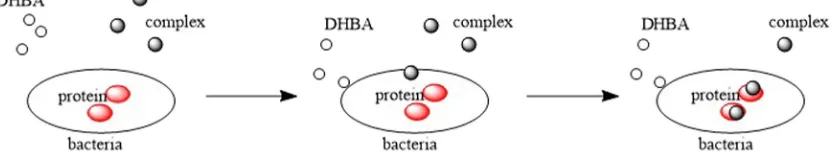
Complex that contains metal ion is more capable to penetrate into bacteria cell than DHBA, thus more effective to inhibit bacteria growth
[22,23]. As proposed inFig. 8, metal-containing complex that penetrates bacteria cell may cause protein disruption and promote bacteria death.
4. Conclusion
The complexes of DHBA with Ni2+/Co2+have been synthesized and
structurally characterized. The coordination of DHBA with metal pro-duced complexes withfiner surface than that of DHBA. Metal ion an-chored the release of more hydrogen atoms of DHBA to neutralized DPPH, therefore complexes possessed better DPPH scavenging activity than that of DHBA. The complexes also showed better microbial growth inhibition againstE. coliandS. aureusthan DHBA. CoDHBA complex was found to possess better microbial growth inhibition than NiDHBA due to that Co ion is more capable to penetrate bacteria cell and cause bacteria death. Since the coordination of DHBA with Ni2+/Co2+only involves
weak hydrogen bond, thus metal can be easily released from the ligand thus the complexes might also be useful in the treatment of metal deficiency.
Acknowledgements
This work was supported by the Ministry of Science and Technology of Taiwan (NSC 104-2221-E-011-146) and National Taiwan University of Science and Technology (102H451403).
Appendix A. Supplementary data
Supplementary data to this article can be found online athttp://dx. doi.org/10.1016/j.molliq.2016.06.015.
References
[1] S. Khadem, R.J. Marles, Monocyclic phenolic acids; hydroxy- and polyhydroxybenzoic acids: occurrence and recent bioactivity studies, Molecules 15 (2010) 7985–8005. [2] D.R. Knapp, Handbook of Analytical Derivatization Reactions, Ed. John Wiley & Sons,
Canada, 1979.
[3] M. Mormann, S. Bashir, P.J. Derrick, D. Kuck, Gas-phase basicities of the isomeric dihydroxybenzoic acids and gas-phase acidities of their radical cations, J. Am. Soc. Mass Spectrom. 11 (2000) 544–552.
[4] Y. Ishii, Y. Narimatsu, Y. Iwasaki, N. Arai, K. Kino, K. Kirimura, Reversible and nonoxidativeγ-resorcylic acid decarboxylase: characterization and gene cloning of a novel enzyme catalyzing carboxylation of resorcinol, 1,3-dihydroxybenzene, from rhizobium radiobacter, Biochem. Biophys. Res. Commun. 324 (2004) 611–620. [5] L. Pesci, S.M. Glueck, P. Gurikov, I. Smirnova, K. Faber, A. Liese, Biocatalytic carboxyl-ation of phenol derivatives: kinetics and thermodynamics of the biological Kolbe-Schmitt synthesis, FEBS J. 282 (2015) 1334–1345.
[6] S.M. Szkudlarz, J. Bajerska, R.Z. Wojtasiak, D. Gorecka, White grape pomace as a source of dietaryfibre and polyphenols and its effect on physical and nutraceutical characteristics of wheat biscuits, J. Sci. Food Agric. 93 (2013) 389–395.
[7] S. Nishibe, Bioactive Phenolic Compounds for Cancer Prevention from Herbal Med-icines, Ed, Japan, Springer Science & Business Media, 1997.
[8] S.B. Bukhari, S. Memon, M.M. Tahir, M.I. Bhanger, Synthesis, characterization and an-tioxidant activity copper-quercetin complex, Spectrochim. Acta A Mol. Biomol. Spectros. 71 (2009) 1901–1906.
[9] A.E. Fazary, E. Hernowo, A.E. Angkawijaya, T.-C. Chou, C.H. Lin, M. Taha, Y.-H. Ju, Complex formation between ferric(III), chromium(III), and cupric(II) metal ions and (O,N) and (O,O) donor ligands with biological relevance in aqueous solution, J. Solut. Chem. 40 (2011) 1965–1986.
[10] C. Abbehausen, T.A. Heinrich, E.P. Abrao, C.M. Costa-Neto, W.R. Lustri, A.L.B. Formiga, P.P. Corbi, Chemical, spectroscopic characterization, DFT studies and initial pharma-cological assays of a silver(I) complex withN-acetyl-L-cysteine, Polyhedron 30 (2011) 579–583.
[11] R.M.S. Pereira, N.E.D. Andrades, N. Paulino, A.C.H.F. Sawaya, M.N. Eberlin, M.C. Marcucci, G.M. Favero, E.M. Novak, S.P. Bydlowski, Synthesis and characterization of a metal complex containing Naringin and Cu, and its antioxidant, antimicrobial, antiinflammatory and tumor cell cytotoxicity, Molecules 12 (2007) 1352–1366. [12]A.F. Santos, D.F. Brotto, L.R.V. Favarin, N.A. Cabez, G.R. Andrade, M. Batistote, A.A.
Cavalheiro, A. Neves, D.C.M. Rodrigues, A.d. Anjos, Study of the antimicrobial activity of metal complexes and their ligands through bioassays applied to plant extracts, Rev. Bras 24 (2014) 309–315.
[13] I.K. Chandra, A.E. Angkawijaya, S.P. Santoso, S. Ismadji, F.E. Soetaredjo, Y.-H. Ju, Solu-tion equilibria studies of complexes of divalent metal ions with 2-aminophenol and 3,4-dihydroxybenzoic acid, Polyhedron 88 (2015) 29–39.
[14]I. Wiegand, K. Hilpert, R.E.W. Hancock, Agar and broth dilution methods to deter-mine the minimal inhibitory concentration (MIC) of antimicrobial substances, Nat. Protoc. 3 (2008) 163–175.
[15]T.P. Popova, R.I. Alexandrova, R. Tudose, E.M. Mosoarca, O. Costisor, Antimicrobial activityin vitroof four nickel complexes, Bulg. J. Agric. Sci. 18 (2012) 446–450. [16] M. Streat, D.J. Malik, B. Saha, Adsorption and Ion Exchange Properties of Engineered
Activated Carbons and Carbonaceous Materials, Ed. Marcel Dekker, New York, 2004. [17] J. Jover, R. Bosque, J. Sales, QSPR prediction of pKafor benzoic acids in different
sol-vents, QSAR Comb. Sci. 27 (2008) 563–581.
[18] V. Jena, M. Satnami, Electronic Spectra of Transitions Metal Complexes, Ed. Lulu.com, United States, 2015.
[19] S.K. Sahoo, R.K. Bera, M. Baral, B.K. Kanungo, Spectroscopic and potentiometric study of 2,3-dihydroxybenzoic acid and its complexation with La(III) ion, Acta Chim. Slov. 55 (2008) 243–247.
[20] S. Atmaca, K. Gul,Ρ. Cicek, The effect of zinc on microbial growth, Tr. J. Med. Sci. 28 (1998) 595–597.
[21] T.J. Silhavy, D. Kahne, S. Walker, The bacterial cell envelope, Cold Spring Harb. Perspect. Biol. 2 (2010) 1–16.
[22] M. Shebl, M.A. Ibrahim, S.M.E. Khalil, S.L. Stefan, H. Habib, Binary and ternary copper(II) complexes of a tridentate ONS ligand derived from
2-aminochromone-3 carboxaldehyde and thiosemicarbazide: synthesis, spectral studies and antimicro-bial activity, Spectrochim. Acta Mol. Biomol. Spectros. 115 (2013) 399–408. [23]M. Rehman, M.K. Baloch, A. Badshah, Synthesis, spectral characterization and