Introduction
Penetrating skull injury (PSI) is one of many kinds of neurosurgical emergencies.1 Most penetrating skull injury cases reported were caused by gunshot wounds.1–3 Other smaller fractions of such injuries were caused by sharp objects such as scissors, knives, nails, forks, and other sharp objects.4–8 Those injuries occur commonly in areas with thin bones, such as orbital surfaces or squamous portion of the temporal bones.4 PSIs are accounted as neurosurgical emergencies due to the risk of neuronal and vascular injuries, not to mention the risk for infections.5,7
Case
A 5-year-old girl was sent to our center with a scissor penetrating her right temple. About 10 hours prior to admission, when she was playing with a pair of scissors in her hand, she was accidentally pushed by her friend and fell down with the scissor puncturing
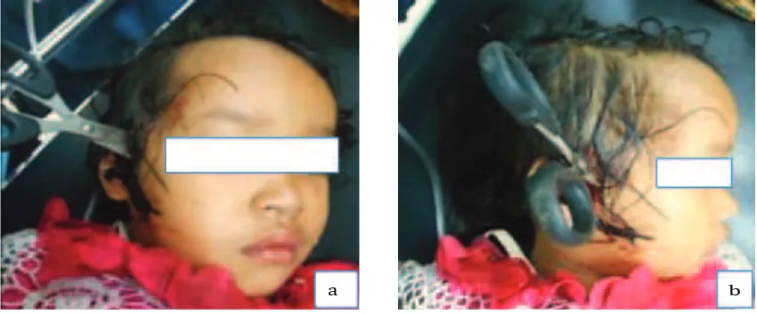
her right temple. The patient was brought to a nearby hospital before transferred to our center. At admission, the patient general and neurological status was within normal limit. At visual inspection, there was a pair of scissors penetrating her right temple (Fig. 1). The scissor was firmly penetrated without any significant bleeding.
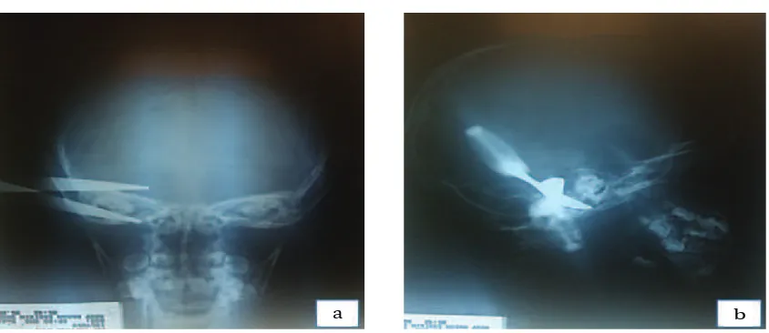
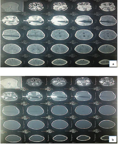
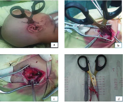
Plain X-ray showed that the scissors penetrated the skull in two points in the temporal region, about 4 cm in depth each (Fig. 2). Head computerized tomography (CT) scan did not show any clear signs of intracranial hemorrhages; the scissors and bone discontinuities were poorly visualized due to metal scattering (Fig. 3). Head magnetic resonance imaging (MRI) was not performed due to the metallic nature of the scissor. After completing the necessary diagnostic tests and preoperative management, the patient was taken to the operating room for foreign object removal and debridement. The patient was put in supine position with her head slightly turned to the left. The original wound was extended into a lazy ‘S’ shape incision and scalp flap was retracted (Fig. 4b). After the flap was raised, the scissor was found penetrating the bone and surrounding tissue in two points. A craniectomy was performed, using two burr holes and bone rongeurs, surrounding the object for better exposure.
The patient was sent home without any neurological deficits or other
complications.
Conclusions: Penetrating skull injuries were interesting due to its mechanism, management and complications. Early diagnosis and appropriate treatment should result in good outcomes.
Keywords:Penetrating skull injury, a sharp metal object
IJIHS. 2015;3(2):79–84
Ahmad Faried, Department of Neurosurgery, Faculty of Medicine, Universitas Padjdajaran-Dr. Hasan Sadikin General Hospital
Jl. Pasteur No. 38, Bandung, Indonesia
During careful removal of the object, dural tears as big as 1x0.1 cm and 1x0.1 cm were found, with minimum cerebral prolapsed (Fig. 4c). There was no significant bleeding found. The post-evacuation wound was irrigated using mixtures of saline and antibiotic. The dural tears were then primarily sutured in water-tight fashion. Postoperative recovery was uneventful. The patient was discharged seven days after the operation without any neurological deficits.
Discussion
Penetrating skull injuries (PSIs) are relatively uncommon wounds, which represent about only 0.4% of all head injuries.1 While having a high number of cases in head injury, our center only had a small number of such cases. Several reports from our center, such as written by Arifin et al.2 and Yudoyono et al.3 had proven the number of such cases. While most PSI in Western countries is caused by gunshot wounds, interestingly, this is quite different in developed and third world countries.8 There is a variety of nonmissile PSI that can occur with weapons ranging from knifes, scissors, pencils, etc.4–8 All of these injuries are defined as wounds that involve a smaller area with relatively lower velocity impact. Most PSIs, regardless of the size of the penetrating bodies, are rarely associated with major neurological symptoms, except in high velocity injuries.4–8
In this case, a large amount of kinetic energy contact with the skull and the force probably acts for a shorter time which causes local deformation resulting in penetration and skull fracture.3,8 Even in the newborn, the kinetic energy contact with the baby’s skull during delivery can caused skull fracture.9
Although quite rare, the incidence of PSI had been reported as early as the 1806. One of the most classical cases was the case of Mr. Phineas Gage, reported on 1848. Mr. Gage was a foreman on a railway construction who was unfortunately engaged in an accidental explosion, resulted in a piece of iron blown and penetrated his head. Mr. Gage survived the initial accident; he was later found to have personality changes and became the first known case of relationship between cerebral function and structural brain lesions. Mr. Gage ultimately succumbed to infection and epileptic state several times later.8 Since the publication of Phineas Gage8 the importance of the frontal lobe as the CEO of the brain is well known. Recently, one case of frontal and cerebellar tumor patient with personality changes and suicidal tendencies has been reported.10
The mechanisms of neuronal and vascular injuries that are caused by these kinds of PSI may differ from those caused by other types of head trauma. Unlike missile injuries, there were no concentric zone of coagulative necrosis that is caused by dissipated energy is present. Unlike motor vehicle accidents, no
Clinical Picture of the Patient from Anterior (a), and Lateral View (b) Fig. 1
diffuse shearing injury to the brain occurs. And unless there were associated hematomas or infarction, the cerebral damage caused by the penetration is largely restricted to the wound tract. A narrow elongated defect on the skull, namely slot fracture, sometimes occurs and is diagnostic when identified. Penetrating injuries to the temporal region are more likely to produce major neurological deficits, because of the thinness of the temporal region, the shorter distance to the brain stem and important vascular structures.4,7
Patients in whom the penetrating object is left in place have a significantly lower mortality than those in whom the objects are inserted and then removed. Frantic, unsuccessful attempts to remove the penetrating objects could make the object dislodged or broken, making it further difficult to remove. Another disastrous complication that was accounted for is uncontrolled hemorrhages from the previously clotted vessels. Other had also reported a case of traumatic aneurysm of intracerebral artery due to such attempts.5,6 Infections can easily complicate PSIs; patients were in risk for developing infections such as meningitis, epidural abscess, subdural empyemas or brain abscess. Prevention and proper infectious complications management can lead to better outcomes. As compound wounds, these injuries require appropriate antibiotic cover and tetanus prophylaxis.4,6,7
It is fundamental to stress that in patients
with PSIs, imaging such as skull X-rays and subsequent CT-scan and possibly cerebral angiography should be requested. Blind removal of the intracranial object may cause neurological and vascular injuries if the trajectory is close to major vessels or to important neural structures. Therefore, an adequate surgical access allowing intracranial direct visualisation of the object should be performed before its withdrawal. Another possibility is to remove the foreign body under CT control to enable prompt detection and management of any complications; in this case, both operating theatre and angiography should be ready. In patients in whom there is no CT evidence of intracranial hematomas or possible damage to vascular structures, the penetrating object can be directly removed before surgical repair under general anaesthesia. Finally, the bone defect caused by PSIs and its operative management, if deemed necessary, could be reconstructed. But early reconstruction was not advocated due to the risk of infections, such as local osteomyelitis and abscess formation.5–7
Postoperative management should cover proper antibiotic and anticonvulsant administration. Serial imaging was advocated to make sure there were no remnant objects or other lesions. As for the patient in this case study, she was neurologically intact without any signs of infections, making the postoperative imaging was deemed unnecessary.
Plain Skull X–ray of the Patient from Anterior (a), and Lateral View (b) Fig. 2
Head Non-contrast CT-scan of the Patient (a) with It’s Bone Window (b) Fig. 3
a
In conclusions, a case of penetrating skull injury in a 5-year-old girl caused by a pair of scissors has been reported. The patient was admitted, operated, and discharged from our center in fine condition and neurologically intact. Penetrating skull injuries are interesting due to its mechanism, management and
complications. Early diagnosis and appropriate treatment should resulted in good outcomes.
Informed consent was obtained from the patient for publication of this case report and any accompaning images. Her family was present at the time.
Intraoperative Findings. The Patient was Put in Supine Position with Her Head Slightly Turned to the Left (a). The Original Wound was Extended into a Lazy ‘S’ Shape Incision (b). We Found Dural Tears in Two Places with Minimal Cerebral Prolapse (c). The Sharp Object, Scissors in Our Case, after Removed from the Temporal Region (d)
Fig. 4
a b
rifle gun pellet: Bandung teaching hospital
experience. Unpublished data 2014.
4. Bozzeto-Ambrosil P, Costa LF, Filho HA. Penetrating screwdriver wound to the head. Arq Neuropsiquiatr. 2008;66(1):93–5.
5. Kim MS, Sim SY. Traumatic aneurysm of the callosomarginal artery-cortical artery junction from penetrating injury by scissors. J Korean Neurosurg Soc. 2014;55(4):222–5.
6. Kumar A, Pandey R, Singh K, Sharma V.
9. Arifin MZ, Gill AS, Anwar AD, Djuwantono T, Faried A. Spontaneous depressed skull fracture during vaginal delivery: a report of two cases and literature review. Indian J Neurotrauma. 2013;10:33–7.