21
O R I G I N A L A R T I C L E
PROGNOSTIC FACTORS OF LEPTOSPIROSIS PATIENTS IN DR.
SARDJITO GENERAL HOSPITAL, YOGYAKARTA, INDONESIA
1 2 2* 2
Kurniaatmaja, E.R , Priambodo D , Humardewayanti R , Loehoeri S 1. Faculty of Medicine, Universitas Gadjah Mada, Yogyakarta
2. Subdivision of Tropical Medicine and Infectious Diseases Department of Internal Medicine Universitas Gadjah Mada / Dr. Sardjito General Hospital, Yogyakarta, Indonesia
*Corresponding Author:rizka_asdie@yahoo.com
ABSTRACT
Background:
. Severe disease can be fatal, although majority of cases are mild and self-limited.
Objective: To determine the prognostic factors for leptospirosis that associated with mortality in patients with leptospirosis in Dr. Sardjito General Hospital, Yogyakarta.
e conducted a retrospective study of data collected in our hospital between Jan 2010 until May 2011, from whom the diagnosis of leptospirosis was confirmed based on pertinent clinical and epidemiological data and positive serology.
Result: Thirty two patients were included in this study, including 29 survivors (90.62%) and 3 non-survivors (9.38%). Of these 32 patients, 26 patients (81.25%) were admitted to the medical ward and 6 patients (18.75 %) were admitted to the ICU. Multivariate logistic regression demonstrated that three factors were independently associated with mortality: higher level of potassium (OR 10.8; CI 1.194-97.728; p<0.01) on admission and neurological dysfunction (altered mentation or seizure) (OR 30; CI 4.367–206.07; p<0.01)
Conclusion: The mortality of leptospirosis remains high despite improvements in patients care. In order to improve the early treatment of high-risk patients, these higher levels of potassium on admission and neurological dysfunction, which are associated with mortality, can be used at the time of admission as prognostic factors.
Leptospirosis, an infectious disease that affects humans and animals, is a common zoonosis with a variety of clinical manifestations. Yogyakarta is one of the cities with a high incidence of leptospirosis. It is important to recognize the clinical features and prognostic factors of this disease
Methods: W
Keywords : Leptospirosis, prognostic factors, mortality
Background
Leptospirosis is the most widespread zoonosis with recent outbreaks in several Asian, 1 Central, and South American countries. Leptospirosis is a zoonotic disease, which is caused by Leptospira and transmitted to human by contact with Leptospira contaminated animal urine or Leptospira contaminated environment. Thirty-two patients with Leptospirosis had admitted in Dr. Sardjito General Hospital from January 2010 until
2
May 2011. In Indonesia, the spread of leptospirosis is in the Province of West Java, Central Java, Yogyakarta, Lampung, South Sumatra, Bengkulu, Riau, West Sumatra, North Sumatra, Bali, NTB, South Sulawesi, North Sulawesi, East Kalimantan and West Kalimantan. The mortality rate of leptospirosis in Indonesia is high, reaching 2.5 to 16.45%. At the age of over 50 years mortality rate can be up to 56%. When Leptospirosis patient is accompanied by a yellow lining of the eye indicating damage to liver tissue, the risk of death will be higher. Several publications reported mortality rates between 3% -54% depending on the infected organ
3 system.
We believe that early evaluation of disease severity at the time of admission might be useful in improving the care of patients with leptopsirosis.
Objective
23 Prognostic Factors of Leptospirosis Patients Volume 1, Number 2, December 2011
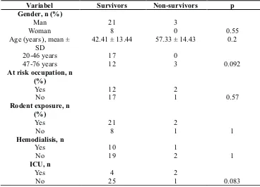
Table 1. The mean hospital stay ± SD for the survivors
Variabel Survivors Non-survivors p
Gender, n (%) Man Woman
21 8
3
0 0.55
Age (years ), mean ± SD
42.41± 13.44 57.33± 14.43 0.2
20-46years 47-76 years
17 12
0
3 0.092
At risk occupation, n (%)
Yes No
12 17
2
1 0.57
Rodent exposure, n (%)
Yes No
21 8
2
1 1
Hemodialisis, n Yes
No
10 19
1
2 1
ICU, n Yes
No
4 25
2
1 0.083
The mean age ± SD for the survivors was 42.41 years ± 13.44 years, and for the non-survivors was 57.33 years ± 14.43 years. In age variable there was no differences between the survivors and non survivors (p = 0.092)
The gender ratio between male and female patients with leptospirosis was 3:1, man 75% and women 25%. There was no differences in gender between the survivors and non survivors (p = 0.55)
Table 2. Clinical sign and symptoms in survivors and non survivors among patients with Leptospirosis
Sign and symptoms
Survivors(n =29) Non survivors(n =3) pvalue
Fever
2 0 1.000
Dyspnea 15 2 1.000
Jaundice 19 3 0.534
Cardiovascular collapse
16 1 0.589
Conjunctival Suffusion
21 2 1.000
Oliguria 9 3 0.044*
Respiratory symptoms
7 2 0.184
Neurological dysfunction
0 2 0.006*
Hemorragic 1 1 0.181
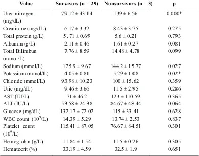
Table 3. Result of univariate analysis of laboratory values between survivors and non-survivors among patients with leptospirosis.
Value Survivors (n = 29) Nonsurvivors (n = 3) p
Urea nitrogen (mg/dL)
79.12 ±43.14 139 ±6.56 0.000*
Creatinine (mg/dL) 6.17 ±3.32 8.43 ±3.75 0.275
Total protein (g/L) 5. 71 ±0.69 5.6 ±0.21 0.793
Albumin (g/L) 2.11 ±0.46 1.61 ±0.27 0.081
Total Bilirubun (mmol/L)
7.76 ± 8.59 14.48 ± 4.78 0.099
Sodium (mmol/L) 125.9 ±9.67 144.2 ±15.77 0.027
Potassium (mmol/L) 4.05 ±0.81 5.29 ±1.08 0.02*
Chloride (mmol/L) 93.98 ±10.23 100 ±15.62 0.359
Uric (mg/dL) 9.46 ±3.66 11.5 ±2.95 0.286
AST (IU/L) 71 ±46.2 123 ±110.59 0.365
ALT (IU/L) 53.58 ±24.38 84.67 ±48.44 0.064
Glucose (mg/dL) 132.17 ±72.02 115 ±33.41 0.628
WBC count (109/L) 14.39 ±5.29 13.74 ±2.53 0.837
Platelet count (109/L)
115.41 ±87.05 76.67 ±84.51 0.301
Hemoglobin (g/L) 11.84 ±1.54 11.5 ±0.26 0.305
Hematocrit (%) 33.19 ± 4.59 32.5 ± 1.9 0.651
*statistically significant
Table 4. Result of multivariate stepwise logistic regression analysis of risk factors for patients with Leptospirosis.
Risk Factor OR 95% CI P value
Oliguria 0.75 0.541 –1.04 0.018
Urea nitrogen (mg/dL)
0.813 0.642 -1.028 0.025
Potassium (mmol/L)
10.8 1.194 –97.728 0.009
Neurological symptoms
30 4.367 –206.07 0.000
Discussion
In this study, several poor prognostic factors in leptospirosis have been identified, including higher admission serum potassium and neurological dysfunction (altered mentation or seizures) were associated with high in-hospital
8
fatality. In another retrospective study, advanced age, oliguria, cardiac arrhythmia, dyspnea, and
Mortality in leptospirosis ranges from 1% 1, 10, 11
to 20 %. In South India, a mortality of 5.32% was reported from Kolenchery and 3.5% from Madras. The study showed that there was no difference in mortality when patients were grouped by age, gender, at-risk occupation, or rodent exposure.
25 Prognostic Factors of Leptospirosis Patients Volume 1, Number 2, December 2011
10 with a worse prognosis for oliguric renal failure. Ninety-seven percent of the patients had renal failure and all patients who died were oliguric. In our study, oliguria was a very sensitive criterion of the severity of the disease. Similarly, Seguro et al noted that the mortality rate for oliguric patients with acute renal failure were higher than that for patients with persistent dieresis. Again, 90% of leptospirosis is anicteric with a lower mortality compared with icteric forms. In our group, 68% of patients were icteric and there was no difference in mortality.
Neurologic dysfunction (altered mentation or seizures) was the most significant predictor of mortality; most patients with neurologic dysfunction also had significant renal and hepatic disease contributing to encephalopathy. Altered mental status was the strongest independent predictor of death in urban leptospirosis in Brazil; other poor prognostic factors were oliguria, advanced age, renal and respiratory insufficiency.In this study, there was an association between altered
12 mental status and mortality.
Conclusions
The mortality of leptospirosis remains high despite improvements in patients care. In order to improve the early treatment of high-risk patients, these two clinically and two laboratory criteria, independently associated with mortality, could be used at the time of admission.
References
1. Vinetz JM. Leptospirosis. Curr Opin Infect Dis. 2001; 14:527–538.
2. Tunissea A. Anlisis Spasial Faktor Risiko Lingkungan pada Kejadian Leptospirosis di Kota Semarang. 2008; 1-4.
3. Murtiningsih B, Budiharta S, Supardi S. Faktor Risiko Leptospirosis di Provinsi Yogyakarta dan sekitarnya. 2005; BKM; 21:1-8.
4. Dupont H, Perdrizet DD, Perie JL, et al. Leptospirosis: Prognostic Factors Associated with Mortality. Clinical Infectious Diseases 1997; 25:720–4.
5. Unnikrishnan D, Pisharody R, Vijayalakshmy N. Prognostic Factors in Leptospirosis. A study from Kerala, India. Infectious Diseases in Clinical Practice 2005; 13:104-107.
6. Riwidikno H. Uji Beda. In Statistik Kesehatan : Belajar mudah teknik analisis data dalam Penelitian Kesehatan. Jogjakarta: Mitra Cendikia Press. 2007; 4-70.
7. Dahlan, M. Sopiyudin. Uji Hipotesis. In statistika untuk Kedokteran dan Kesehatan. Jakarta : PT. Arkans. 2006; 1-135.
8. Lopes AA, Costa E, Costa YA, et al. The association between serum potassium at hospital admission and the case-fatality rate of leptospirosis in men. Rev Inst Med Trop Sao Paulo. 2001; 43:217–220.
9. Daher E, Zanetta DM, Cavalcante MB, et al. Risk factors for death and changing patterns in leptospirosis acute renal failure. Am J Trop Med Hyg. 1999; 61:630–634
10. Levett PN. Leptospirosis. Clin Microbiol Rev. 2001;14:296–326
11. Muthusethupathi MA, Shivakumar S, Vijayakumar R, et al. Renal involvement in leptospirosis—our experience in Madras City. J Postgrad Med. 1994; 40:127–131.