* Corresponding author. Tel/Fax : +62-281-642840
ANTICANCER ACTIVITY OF CALANONE ON HeLa CELL LINE
Heny Ekowati
1.*, Indwiani Astuti
2, and Mustofa
2 1Department of Pharmacy, Faculty of Medicine and Health Sciences, Jenderal Soedirman University, Karangwangkal, Purwokerto
2
Department of Pharmacology and Toxicology, Faculty of Medicine, Universitas Gadjah Mada, Jl. Sekip Utara, Yogyakarta
Received November 24, 2009; Accepted June 28, 2010
ABSTRACT
Calanone (coumarin derivate compound), isolated from Calophyllum sp. had been shown to have cytotoxic activity on leukemia L1210 cell line with IC50= 59.40g/mL. Calanone presumed have anticancer activity on HeLa
cervical carcinoma cell. This study was conducted to investigate the cytotoxic and apoptotic activity of calanone and its effect to p53 and p21 expression on HeLa cervical carcinoma cell. Cytotoxic assay of calanone was performed on HeLa cell line, using MTT assay. Apoptotic assay was performed on HeLa cell line incubated with calanone for 24 h, by immunofluororescence method, using fluorochromes ethidium bromide and acridine orange. Expression of p53 was examined on HeLa cell line, by PCR with p53 wild-type primer. Expression of p21 was examined on HeLa cell line, by immunohistochemistry method. 5-fluorourasil was used as positive control in cytotoxic, apoptotic assay, and p53 expression. The result showed that calanone has cytotoxic activity on HeLa cell line, with IC50= 22.887g/mL,
caused cytotoxicity through apoptotic mechanism, increase p53 tumor suppressor gene expression, while the p21 expression test showed a negative result.
Keywords:Calanone, cytotoxic, HeLa cell line
INTRODUCTION
Neoplasm/tumor indicated increased level in the pattern of diseases causing general death in the world. World Health Organization (WHO) described that 12% of all death in the world was caused by neoplasm [1]. In America, 2004, proximally 10,500 new cases of invasive cervix cancer occurred, and >50,000 cases of carcinoma in situ were detected. There were 3,900 deaths from the disease, and of those patients, proximally 85% had never had a Pap smear [2]. In 2005, cervical cancer in hospitalized patients in Indonesia was in 2ndrank [1].
The random screening of natural products for their direct use as drugs was successful, introducing compounds such as anthracyclines, Vinca alkaloids, epipodophyllotoxins, and taxanes into clinical use [3]. Coumarins have attracted intense interest in recent years because of their diverse pharmacological properties. Among these properties, their cytotoxic effects were most extensively examined [4].
Calanone, coumarin derivate compound, isolated fromCalophyllum sp.had been shown to have cytotoxic activity on leukemia L1210 cell line with IC50 are 59.09 g/mL [5]. Development of calanone was done by Iswanto et al. [6], that succeed synthesized derivate calanone, a recommendation from analysis QSAR, 2,4-dinitrophenilhidrazone calanone. Several coumarins had been shown caused apoptosis on cancer cell line and one of them, i.e. 2-(8-Hydroxy-6-methoxy-1-oxo-1H -2-benzopyran-3-yl) Propionic Acid (NM-3). The exposure
to NM-3 is associated with increases in expression of the p53 tumor suppressor. In human MCF-7 and ZR-75-1 breast cancer cells, NM-3 induces the p21 cyclin-dependent kinase inhibitor, cell cycle arrest at G1-S phase, and necrotic cell death [7].
Cervical carcinoma is one of the most common cancers in women. High-risk human papilloma viruses (HPV) such as HPV18 play an important role in the development of essentially all cases of cervical carcinoma [8]. Chemotherapy uses drugs of several different classes to destroy cells in the S, M, or initial G stages of the cell cycle [9]. The p53 gene appears to trigger programmed cell death (apoptosis) as a way of regulating uncontrolled cellular proliferation in the setting of aberrant growth signals. Mutations in the p53 gene result in loss of the ability of the gene product to bind to DNA, thereby removing its suppressive effect [10]. The E6 protein of HPV-18 has sequence homology to the SV40 large T antigen and has the capacity to bind and inactivate the tumor-suppressor gene p53 [2]. p53 is a transcription factor that activates the transcription of a large number of cellular genes including p21WAF1 (here after referred to as p21). The induction of the cyclin-dependent kinase inhibitor p21 is one of the principal effectors of growth arrest [11-12].
EXPERIMENTAL SECTION
Materials
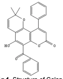
Calanone were kindly gift from organic chemistry laboratory, Jenderal Soedirman University (Indonesia), the chemical structure is shown in Fig. 1. 5-fluorouracil (5-FU), were obtained from parasite laboratory, Faculty of Medicine, Universitas Gadjah Mada. HeLa cervical cancer cells were obtained from parasite laboratory, Faculty of Medicine, Universitas Gadjah Mada, HeLa cells were grown in RPMI 1640 medium (Sigma) supplemented with 10% Fetal Bovine Serum (FBS) (Sigma-Aldrich, USA) at 37 °C in a 5% CO2atmosphere,
3% penicillin- streptomycin and 1% fungison. Subcultures were obtained after treatment with 0.05% trypsin (Gibco, Auckland) in phosphate buffer saline. To examine the effects of calanone the cells were treated with 100, 50, 25, 12.5, 6.25g/mL. And to examine the effects of 5-FU the cells were treated with 300, 150, 75, 37.5, 18.75g/mL, for 24 h, respectively.
Procedure
MTT assay
HeLa cervical cancer cells were cultured in a 96-well plate (Nalge Nunc International, Denmark) at a density of 2 × 10⁴ cells per well, incubated 24 h. The cells were then treated with varying concentrations of calanone and 5-FU. After 24 h, the cells were washed and treated with 0.01 mL MTT per well. Plates were incubated at 37 °C in a 5% CO2atmosphere for 4 h, and
0.1 mL of the extraction buffer (10% sodium dodecyl sulfate in 0.01% HCl) was added. After an overnight incubation at 37 °C, the absorbance was measured at 595 nm using an ELISA reader (Bio-Rad) and was compared with the control cultures without compound. To determine cell viability, percent viability was calculated as [(absorbance of drug-treated) sample/(control absorbance)] × 100.
Determination of Apoptosis
HeLa cells were grown on glass coverslips in tissue culture dishes (Falcon) and were allowed to attach for 24 h prior to the addition of drug. After the cells were incubated with drug for 24 h, the coverslips were washed once in phosphate-buffered saline and fixed in object glass. Treated cells were stained with acridine orange and ethidium bromide 5 L and visualized by fluorescence microscopy. Viable (normal, green nuclei), early apoptotic (condensed, green nuclei) and late apoptotic (condensed, red nuclei) cells were counted.
Fig 1. Structure of Calanone
Determination of p53
For DNA isolation, cells were lysed in 35L proteinase K and 700 L buffer lyses (50 mM Tris.Cl pH 8, 100 mM NaCl, 100 mM EDTA, 1% SDS), incubated at 55 °C for 1 h. Fenol was added and shake for 5 min. Then alcohol absolute 300 L was added, shakes gently, then take the DNA and add with TE 1x. For DNA amplification, mixed 15 L PCR mix, 11 L aquadest sterile, 2 L primer p53 wild type (forward and reverse @ 1 L), and 2 l DNA. And then processed in PCR machine with warm up at 95 °C for 5 min, then denaturizing (94 °C for 45 sec), annealing (63 °C for 1 min), and extension (72 °C for 4 min), repeated for 35 cycles. PCR product then analyzed with electrophoresis on a 2% agarose gel (with ethidium bromide 2.5L/100mL aquadest). Identified the band then photographed under UV illumination.
Determination of p21
HeLa cervical cancer cells were cultured in a 24-well plate (Nalge Nunc International, Denmark) at a density of 2 × 10⁵ cells per well, incubated 24 h. The cells were then treated with 2x IC50, 1x IC50, 0.5x IC50
concentrations of calanon and 5-FU. After 24 h, cells were plated in poly-L-Lisin slide. Cells were fixed with methanol (pro analysis) for 5 min, permeabilized for 5 min in PBS containing 0.2% Triton X-100, blocked in 2% BSA for 1h, and stained with the monoclonal antibody p21 (1:400) for 1 h. Then washed with PBS 3x 5 min, stained withbionylated secondary antibodyfor 1 h. Incubation in HRP-streptavidin for 10 min, added DAB for 5 min, washed with aquadest,Counterstained
with Harry’s hematoxylinfor 20 sec, mounted on glass slides. The cells were examined using a confocal microscope.
RESULT AND DISCUSSION
Calanone had cytotoxic activity on HeLa Cell Line
Fig 2.Inhibition rate on HeLa cell line with calanone and 5-fluorourasil. HeLa cells were treated with various doses of calanone and 5-fluorourasil, incubated for 24 h at 37 °C in humidified 5% CO2 atmosphere. Cell viability
was determined by MTT assay, absorbance was read at 595 nm. Inhibition rate (%) was defined as: ((live cell in the control – live cell in the test group)/live cell in the control) x 100. Standard curve: y = 0.00005x + 0.3405 (R2= 0.9963). Results are average of three independent experiments (mean ± SD)
Fig 3. IC50 of calanone and 5-fluorourasil on HeLa
cancer cells. Concentrations inhibiting 50% of the cell were determined by probit analysis using SPSS software
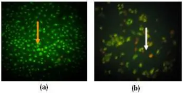
Fig 4. Cells were treated with acridine orange and ethidium bromide. Viable cell (orange arrow) (a), apoptotic cell (white arrow) (b). HeLa cells treated with calanone underwent the classical morphologic changes indicate of apoptosis
Fig 5. The effects of calanone (a), marker DNA (b), untreated cell (c) and 5-fluorourasil (d) on p53 expression. HeLa cells growing at the exponential growth phase were harvested by trypsinization. Cells were carried to plates (2x10⁴ cells /well), then treated with calanone, and 5- fluorourasil (2x IC50, 1x IC50, and
0.5x IC50) and incubated for 24 h at 37 °C, 5% CO2in
RPMI medium. At the end of the incubation period we collected the DNA samples from treated cells and equal amounts of DNA samples were electrophoresed on 2% agarose gel in TAE buffer. The gel was stained with 1 mg/mL ethidium bromide for 30 min, and then photographed under UV illumination
have attracted intense interest in recent years because of their diverse pharmacological properties. Among these properties, their cytotoxic effects were most extensively examined. These cytotoxic coumarins represent an exploitable source of new anticancer agents, which might also help addressing side-toxicity and resistance phenomena [4].
Calanone was derivative compound of coumarin, isolated fromCalophyllum sp., and had cytotoxic activity on leukemia L1210 cell line, with IC50 59.04 µg/mL [5]. In 2007, derivative calanone, 2,4-dinitrofenilhidrazone calanone was synthesized [6] and had cytotoxic activity on leukemia L1210 cell line, with IC50 47.09 µg/mL. In Fig. 2 and 3, we conclude that calanone have cytotoxic activity on HeLa cancer cell line, with IC50 22.88 µg/mL. In over 90% of cervical carcinoma cancer, the p53 tumor suppressor pathway is disrupted by the human papilloma virus (HPV). The HPV E6 protein promotes
accelerated ubiquitin-mediated degradation of p53. HeLa cells express E6 protein from integrated HPV18 DNA and this causes an aberrant checkpoint control in these cells [8].
Calanone induced apoptosis and increase p53 tumor suppressor gene expression
The present studies demonstrate that calanone had cytotoxic activity on HeLa cervical carcinoma cell, with IC50 22.88 µg/mL. Calanone had cytotoxicity on
Fig 6. Expression of p21 on HeLa cancer cells. Untreated cell (a), calanone (b), by immunohistochemistry method. Treated cells were plated on poly-L-lysine and stained with a p21-specific mAb. Blue nucleus showed negative p21 expression results (black arrow)
ovarian carcinoma and HeLa cervical carcinoma cells. Our study found that IC50 of 5-fluorourasil was
129.29 µg/mL. In 2002, Okca & Ozes found that IC50 of
5-fluorourasil was 1.74 µg/mL. The result indicate that in our study, Hela cell was resistant to 5-fluorourasil in vitro. Nicolantonio et al. [13] reported the increased TS levels in ECF and 5-FU exposed cells are consistent with a number of previous reports. Gene amplification of TS with consequent increases in TS mRNA and protein has been observed in cell lines that are resistant to 5-FU and fluorodeoxyuridine (FUDR). Treatment with 5-FU has been shown to acutely induce TS expression in cell lines, animal models and human tumors [14-15].
The effects observed with calanone can be explained, at least in part, by activation of the p53 tumor suppressor. Rapid induction of p53 activity occurs by mechanisms involving posttranslational stabilization in the response of cells to various types of stress [16-17]. The present results demonstrate that calanone treatment is associated with dose-dependent increases in p53 levels. HeLa cervical carcinoma cell responded to calanone and 2,4-dinitrophenilhidrazone calanone with activation of p53 and the induction of apoptosis.
Calanone did not induced p21 expression
Calanone increase the expression of p53 tumor suppressor on HeLa cervical carcinoma cell but were not induced p21 (Fig. 6). Yin et al. [7] reported, 2-(8-Hydroxy-6-methoxy-1-oxo-1H-2-benzopyran-3-yl)
propionic acid (NM-3) is an isocoumarin derivative, increases in expression of the p53 tumor suppressor on human PA-1 ovarian carcinoma and HeLa cervical carcinoma cells. In human MCF-7 and ZR-75-1 breast cancer cells, NM-3 induces the p21 cyclin-dependent kinase inhibitor, cell cycle arrest at G1-S-phase, and necrotic cell death. Since p53 requires some coactivators, like CBP/p300, and it is not known whether p53 activates transcription of all of its target genes by using identical transcriptional complex, it is very possible that transcription of each p53 target gene may require
CONCLUSION
Calanone has cytotoxic activity on HeLa cell line, with IC50= 22.887 g/mL, caused cytotoxicity through
apoptotic mechanism, increase p53 tumor suppressor gene expression, while the p21 expression test showed a negative result.
ACKNOWLEDGEMENT
Support from organic chemistry laboratory Jenderal Soedirman University and Chemistry Research Center of LIPI for obtaining calanone is gratefully acknowledged.
REFERENCES
1. Indonesia Ministry of Health, 2007, Indonesia Health Profile 2005, Jakarta.
2. Young, R.C., 2004, Gynecologic Malignancies in Harrison’s Principles of Internal Medicine, 16th ed., McGraw-Hill, New York.
3. Denny, W.A., 2002, The Contribution of Synthetic Organic Chemistry to Anticancer Drug Development in Anticancer Drug Development. Oxford-United Kingdom.
4. Kostova, I., 2005,Curr. Med. Chem.,5, 1, 29–46. 5. Chasani, M., 2002, Synthesis of calanone
derivates and biology activity test, Thesis, University of Indonesia, Jakarta
6. Iswanto, P., Chasani, M., and Vaulina, E., 2007,
Synthesis of calanone derivatives as antileukemia compound with QSAR approach (Quantitative structure-activity relationship), Research report for research institute, Jenderal Soedirman University. 7. Yin, L., Ohno, T., Weichselbaum, R., Kharbanda,
S., and Kufe, D., 2001, Mol. Cancer Ther., 1, 1, 43–48
8. Akca, H., and Özefi, O.D., 2002, Turk J. Biol.,26, 145-150.
9. Corwin, E.J., 2008, Handbook of Pathophysiology, 3rded., Lippincott Williams & Wilkins, Philadelphia. 10. Rugo, H.S., 2006, Cancer in Current Medical
Diagnosis and Treatment, 45th Ed., Eds. Tierney, L., McPhee, S.J., and Papadakis, M., McGraw-Hill-New York.
11. Kubbutat, M.H.G., Ludwig, R.L., Ashcroft, M., and Vousden, K.H., 1998, Moll. Cell. Biol., 18, 10, 5690–5698.
12. Szymanska, K., and Hainaut, P., 2003, Acta Biochim. Pol.; 50, 1, 231–238.
P., Somers, S.S., Toh, S., Higgins, B., Lamont, A., Gulliford, T., Hurren, J., Yiangou, C., and Cree, I.A., 2005,BMC Cancer,5, 78.
14. Swain, S.M., Lippman, M.E., Egan, E.F., Drake, J.C., Steinberg, S.M., and Allegra, C.J., 1989, J. Clin. Oncol.,7, 7, 890-899.
15. Chu, E., Koeller, D.M., Johnston, P.G., Zinn, S., and Allegra, C.J., 1993, Mol. Pharmacol., 43, 4, 527–533.
16. Oren, M., and Rotter, V., 1999,Cell. Mol. Life Sci.,
55, 1, 9–11.
17. Vogelstein, B., Lane, D., and Levine, A., 2000,