CHAPTER OUTLINE
N
o tissue is more important than blood:
it delivers oxygen and nutrients, it
removes carbon dioxide, and it plays a key
role in fighting infections. At first glance,
blood looks like a simple tissue: essentially a
bunch of different cells floating in salty water,
more or less. That means it should be pretty
easy to make artificial blood. But it’s not.
Many factors account for the interest in
artificial blood. The military recognizes blood
loss as a primary cause of death, owing to
both pressure loss as blood volume
de-creases and decreased oxygen delivery with
fewer red blood cells present. Blood is
typi-cally in short supply, and it cannot be frozen
forever. Blood transfusions can carry viral
dis-eases, and even in an emergency, it’s
neces-sary to check the patient’s blood type before
an infusion.
The most common way to deal with blood
loss during emergencies is to infuse saline
so-lution. This procedure maintains blood
vol-ume but does not replace the essential
oxy-gen-carrying function. The major goal of
artificial blood is to carry oxygen, since
oxy-gen starvation can destroy brain cells in four
minutes. Scientists have tried to use
hemo-globin, the oxygen-carrying molecule in the
red blood cell, but unless it is “packaged”
in-side a cell, it can damage the kidney.
At present, several experimental versions
of oxygen-carrying artificial blood are being
tested. But blood is a complex and subtle
tis-sue, and so artificial blood is likely to serve as
a supplement, not a replacement, for the
nat-ural stuff.
92
Tissues
4
■
Cells Are the Building Blocks of
Tissues
p. 000
■
Organization increases with
Organs, Organ Systems, and the
Organism
p. 000
tissue is a group of similar cells and
ex-tracellular substances that have
com-bined to perform a single function. The
human body has four tissue types.
Ep-ithelial
tissue covers the body, lines all cavities, and
com-poses the glands. Connective tissue literally connects
the structures of the body, providing structural support
and holding organs together. Stretchy and strong,
con-nective
tissue maintains the body’s integrity.
Muscular
tissue provides movement and heat.
Nervous
tissue
re-sponds to the environment by detecting, processing,
and coordinating information.
EPITHELIAL TISSUE IS AT
THE SURFACE
Epithelial tissue (singular: epithelium) is composed of
cells laid together in sheets (
Figure 4.1
). One side
of these cells is oriented toward the body cavity or
ex-94
CHAPTER 4
Tissues
Cells Are the Building Blocks
of Tissues
L
EARNINGO
BJECTIVESListthe four tissue types in the human body.
Describethe function of each tissue type, explaining its unique characteristics.
ternal environment and may
have cilia or microvilli. The
other surface is joined to
deeper connective tissue at
the basement membrane. This
basement layer, an acellular
membrane, is composed of a collection of
polysaccha-rides and proteins that help to cement the epithelial
tis-sue to the underlying structures.
Epithelium is little more than cells tightly
con-nected, one to the next. It has neither blood vessels nor
any extracellular substances between the cells. The
classes of epithelium are identified by both the number
of cell layers and the shape of the cells in the upper
layer.
Simple
epithelium has one layer of cells and
usually functions as a diffusion or absorption
mem-brane. The lining of your blood vessels and the
respira-tory membranes of your lungs are simple epithelium.
Stratified
epithelia have many layers of cells and are
de-signed for protection. Examples are found in the outer
layer of your skin and the ducts of your salivary glands.
Epithelial cells can be flattened, cube-like, or
columnar. Each shape mirrors the function of the
tis-sue. Flattened cells, reminiscent of fried eggs, are called
squamous
cells. Squamous epithelium is thin enough
to form a diffusion membrane.
Cuboidal
and
columnar
epithelia are plumper and usually compose mucous
membranes, where the epithelial cells secrete mucus
and other compounds (see
Figure 4.2
).
A
Epithelial cells
Figure 4.1
These epithelial cells have been removed from the body; however, their strong cell-to-cell attachments help to keep them together.
Cell shape
Squamous Cuboidal Columnar
Basement membrane Arrangement
of layers
Simple Stratified
Basement membrane
Microvilli
Folded parts of the
cell membrane that
increase the cell’s
surface area.
Cell shapes and arrangements
Figure 4.2
Cell shapes and arrangement of layers are the basis for classifying epithelial tissues. The shape of the topmost layer of cells is used to determine the name of stratified epithelium because this layer is not deformed by those above it. Layers of flattened cells that look like “piles of tiles” would be classified as stratified squamous epithelium, whereas the image here shows layers of plump cells, classified as stratified cuboidal epithelium.
Outlinethe various types of epithelial, connective, and muscular tissue.
Cells Are the Building Blocks of Tissues 95
Glands are composed of
epithelial tissue (
Figure 4.3
)
and classified by how their
secre-tions are released. Glands that
se-crete into ducts are
exocrine
glands. Salivary glands and sweat
glands are exocrine glands. Each
one secretes its products into a
duct that directs the secretion to
the surface of the gland.
Endocrine
glands have no
ducts. Instead, they secrete directly
into the extracellular fluid
sur-rounding the gland. Endocrine
glands secrete hormones that are
then picked up by the blood
stream and carried throughout
the body. The adrenal, thyroid,
and pituitar y glands are all
en-docrine glands.
Glands
Figure 4.3
Glands are composed of epithelium, with characteristics like other sheets of epithelium and the ability to secrete specialized compounds for metabolic or regulatory purposes.PINEAL GLAND
PARATHYROID GLANDS (behind thyroid glands)
HYPOTHALAMUS
PITUITARY GLAND SALIVARY GLANDS THYROID GLAND
THYMUS
Cells Are the Building Blocks of Tissues 97
96
CHAPTER 4
Tissues
CONNECTIVE TISSUE KEEPS
IT TOGETHER
As the name implies, connective tissue connects bodily
structures. It binds, supports, and anchors the body, and
is the most abundant type of tissue in the body.
Connec-tive tissue is composed of cells suspended in a
noncellu-lar
matrix
. The matrix, or “ground substance,” is
se-creted by the connective-tissue cells, and it determines
the characteristics of the connective tissue. The matrix
can be liquid, gel-like, or solid, depending on the cells.
The ground substance of all connective tissue contains
fibers of
collagen
(for strength) and
elastin
(for
flexibil-ity, stretch, and recoil). Collagen is one of the main
com-ponents of all connective tissue and consequently is the
most abundant protein in the animal kingdom.
The nature of the ground substance leads us to
classify connective tissue as
soft connective tissue
or as
specialized connective tissue
(
Figure 4.4
). Cartilage,
bone, blood, and lymph are types of connective tissues.
CARTILAGE JOINS AND CUSHIONS
Cartilage is a unique
connec-tive tissue because it is
avascu-lar
(other types of connective
tissue all have rich blood
sup-plies). (
Figures 4.5 and
4.6
, page 00)
Chondrocytes
,
the cartilaginous cells, secrete a gel-like matrix that
even-tually surrounds and imprisons them, segregating them
from direct contact with one another or any nutrient
supply. Cartilage heals slowly because nutrients must
dif-fuse through the matrix to the chondrocytes; nutrients
cannot reach the cells directly via the blood stream. Each
chondrocyte resides in a small “lake” within the matrix
called a lacuna. The fluid bathing the cell in this lacuna
diffuses through the matrix to and from the blood
sup-ply. This indirect route is far slower than bringing the
fluid directly to the cells and is the reason cartilage is so
slow to repair itself. Arthritis is a serious disease of the
joints, targeting the cartilage found within them. It is
dif-ficult to treat, in part because the cartilage is avascular
and therefore does not respond quickly to medications
(see the “I Wonder . . .” feature on arthritis on page 00).
BONE IS SIMILAR TO
STEEL-REINFORCED CONCRETE
Bone is a hard mineralized tissue found in the skeleton,
which is a defining characteristic of vertebrates (
Fig-ure 4.7
, page 99). Bone cells secrete an
osteoid
ground substance that
eventu-ally hardens and surrounds
the cells in an
ossified
matrix.
The osteoid ground substance
includes proteins, water,
cal-cium, and phosphorous salts.
Once the matrix ossifies, the cells remain in contact
with one another through small channels called
canali-culi
. Like other connective tissues, bone has collagen
fibers in the matrix for flexible support. Young bone
has a higher percentage of collagen fibers than older
bone, accounting for the greater flexibility of bones in
infants and young people. Where an adult’s bone would
snap under excessive force, a young child’s bone will
bend. The convex surface may fray, like a bent green
stick, but the bone does not break.
BLOOD AND LYMPH COMMUNICATE
WITH THE ENTIRE BODY
Blood and lymph are considered fluid connective
tis-sues because their matrix is not a solid. Blood is
com-posed of specialized cells that
are carried in the fluid matrix,
or
plasma
. The main function
of blood is to transport
nutri-ents, gases, hormones, and
wastes. Chapter 11 devotes an
entire section to blood.
Lymph
is another
fluid connective tissue. It is
de-rived from the
interstitial
fluid
that bathes the cells and
is collected in the lymphatic
vessels. Like blood, lymph
in-cludes cells as well as proteins and other compounds in
its fluid matrix. Chapter 10 deals with lymph in greater
detail.
Soft connective tissues: loose and dense
Figure 4.4
Soft connective tissue has a matrix composed of a semifluid ground substance, fibroblaststhat secrete fibers, and white blood cells that fight infection. The fibers of the matrix can be either loosely arranged, or densely packed together. Loose connective tissue is sometimes called areolarconnective tissue. Dense connective tissue includes the dense irregulartissue of the dermis of the skin, where the collagen fibers are arranged in a network, and the dense regulartissue of tendons, where the collagen fibers are aligned to resist tearing. Elastic connective tissue is made up of freely branching elastic fibers, with fibroblasts in the spaces between fibers.
Avascular
Without blood
vessels.
Osteoid
Bone matrix before
it is calcified.
Plasma
The clear, yellowish
fluid portion of
blood.
Interstitial fluid
Fluid that fills the
spaces between
cells of tissues.
LM LM
LM
Aorta
Heart 435x
Sectional view of elastic connective tissue of aorta
Elastic connective tissue
300x
Sectional view of subcutaneous areolar connective tissue
Areolar connective tissue Subcutaneous
layer
Collagen fiber Mast cell
Elastic fiber Reticular fiber Skin
Dense regular connective tissue Sectional view of dense regular connective
tissue of a tendon
250x
Skeletal muscle Tendon
Collagen fiber Nucleus of fibroblast
Elastic lamellae (sheets of elastic material) Nucleus of fibroblast
Macrophage
EVEN FAT HAS A JOB TO DO
Adipose tissue contains fat cells—cells that are
special-ized for lipid storage. Unlike other connective tissues,
adipose tissue does not have an extensive extracellular
matrix (see
Figure 4.8
). Its matrix is a soft network
of fibers holding the cell together and binding it to
sur-rounding tissues.
Cellulite
“bumps” on the skin indicate
where the adipose matrix is connected to the skin. The
Cells Are the Building Blocks of Tissues 99
LM 450x
Perichondrium
Nucleus of chondrocyte Lacuna containing chondrocyte
Ground substance Fetus
Skeleton
Sectional view of hyaline cartilage of a developing fetal bone
Hyaline cartilage
LM LM
Lacuna containing chondrocyte Patella
(knee cap) Tendon of quadriceps femoris muscle
Chondrocyte
Collagen fibers in ground substance
1100x
Sectional view of fibrocartilage of tendon
Portion of right lower limb
Fibrocartilage Lacuna
containing chondrocyte Ear
Chondrocyte
Elastic fiber in ground substance
420x
Sectional view of elastic cartilage of ear Elastic cartilage
Elastic cartilage and Fibrocartilage
Figure 4.6
a
Elastic cartilage contains many elastic fibers in the matrix. Elastic cartilage allows the outer ear to bend, then return to its original shape. The epiglottisthat prevents food and liquid from entering your respiratory tract also contains elastic cartilage. When you swallow, the epiglottis bends to cover the opening of the trachea. Afterward, the epiglottis snaps back to its original position, allowing air to flow through the windpipe.b
The matrix of fibrocartilage is packed with collagen fibers, so it is found where extra strength is needed. Cushions in your knee joints and the disks between the vertebrae are made of fibrocartilage.Epiglottis
Large,
leaf-shaped piece of
cartilage lying over
top of the larynx.
LM
Lamellae Lacuna Central (haversian) canal Canaliculi
Calcified extracellular matrix
Lacuna Osteocyte
Canaliculi
550x
Sectional view of an osteon (haversian system) of femur (thigh bone)
Femur
Details of an osteocyte
Compact bone
Figure 4.7
Bone consists of a hard matrix surrounding living cells. Bone has both a blood supply and a nerve supply running through it. The matrix of compact bone is found in long cylinders called osteons or Haversian systems. Lighter spongy bone has less structure, forming in struts and supports rather than a solid mass.
LM
Blood vessel Fat-storage area of adipocyte Nucleus of adipocyte
Plasma membrane Cytoplasm
Fat
Sectional view of adipose tissue showing adipocytes of white fat
Adipose tissue Heart
300x
Adipose
Figure 4.8
The nucleus and cytoplasm in adipocyte play second fiddle to the main action: the huge droplet of stored lipid.
Trachea
The main trunk
of the respiratory
tree.
Hyaline cartilage
Figure 4.5
The most common type of cartilage is hyaline cartilage. The matrix of hyaline cartilage contains many collagen fibers and looks crystal blue in living tissue. Hyaline cartilage covers the ends of bones, allowing them to slide against one another without damage. It is also found in your nose and
trachea. During development, your entire skeleton was modeled in hyaline cartilage, which then ossified—that is, turned to bone.
8
8
8
8
0
0
0
0
%
%
%
%
8
8
8
8
0
0
0
0
%
%
%
%
100
CHAPTER 4
Tissues
Cells Are the Building Blocks of Tissues 101
Nucleus of smooth muscle fiber
Artery
Longitudinal section of smooth muscle tissue
350x
Smooth muscle fiber (cell)
Smooth muscle fiber Smooth
muscle
LM
Nucleus Striations Skeletal muscle fiber (cell) Skeletal
muscle
Longitudinal section of skeletal muscle tissue Skeletal muscle fiber
400x LM
Striations Nucleus
Heart
Cardiac muscle fiber (cell)
Longitudinal section of cardiac muscle tissue
600x
Intercalated disc
Cardiac muscle fibers
LM
adipose cells within the fibrous matrix can expand with
the swelling of the fat droplets they contain, while the
matrix fibers cannot stretch as far. The different
stretch-ing capacities of these two components of adipose
tis-sue form dimples on the skin. Cellulite is a normal
function of fat deposition and storage. It is not an
in-herently evil tissue that must be removed from the
body, despite what you may have read in the
supermar-ket tabloids. Even newborns have cellulite!
MUSCULAR TISSUE MOVES US
The function of muscular tissue is to contract. The cells
get shorter, generating force and often movement. The
three types of muscular tissue are:
skeletal muscle,
smooth muscle
, and
cardiac muscle
Skeletal muscle
tis-sue is highly organized, with the cells lying parallel to
each other, much like a cable. When stimulated, groups
of muscle cells contract in unison. (
Figure 4.9
).
www.wiley.com/ college/ireland
Smooth muscle lines hollow organs such as the blood vessels and the digestive tract. Smooth muscle cells are short, cylindrical cells that taper at both ends and have only one nucleus. They are not striated, and are not under voluntary control. This is helpful. Wouldn’t it be nerve-wracking to consciously manage the diameter of your blood vessels to maintain blood pressure, or to consciously create the rhythmic constrictions that the digestive tract uses to move food during digestion?
Cardiac (heart) muscle has short, branched, and striated cells, with one nucleus at the center of each cell. Specialized communication junctions called intercalated discs facilitate the heartbeat by spreading the signal to contract. Intercalated discs are gap junctions found within areas of closely knit cell membranes, helping to spread the impulse while also binding the cells together. Cardiac muscle will be described in more detail in Chapter 11.
Comparison of three types of muscle tissue
Figure 4.9
Skeletal muscle is the tissue that makes up the muscles that move your limbs and stabilize your trunk, muscles such as your biceps brachiiand rectus abdominus. This tissue is composed of long, multinucleate cells with visible striations. The cells of skeletal muscle extend the length of the muscle and are arranged in parallel groups called fascicles. Skeletal muscle is described in full detail in Chapter 5. Because you consciously control their contractions, skeletal muscle is called voluntary muscle.
Biceps brachii
The anterior muscle
of the upper arm.
Rectus
abdominus
“Six-pack” muscles
that stabilize the
trunk.
Striations
102
CHAPTER 4
Tissues
Cells Are the Building Blocks of Tissues 103
I W
ONDER .
.
.
What is arthritis?
A
rthritis is a general term for degradation of the joints, for a variety of causes. Rheumatoid arthritis results from an au-toimmune attack on the joint. Osteoarthritis results from various sorts of wear and tear. Only one type of arthritis, the one that results from an infection, can be cured (using antibiotics). Others forms must be managed to reduce pain and improve quality of life.Osteoarthritis affects 21 million Americans. The dis-ease damages cartilage that normally forms a smooth bear-ing surface inside joints. Pain begins as bones rub on bones. Osteoarthritis tends to be worst when it affects the spine, hips and knees.
Rheumatoid arthritis is an inflammation of the syn-ovium, which lines the joints. The disease can seriously de-form the hands, but it often affects joints throughout the body. Rheumatoid arthritis is two or three times as common among women, indicating that females are genetically more susceptible to this type of autoimmune attack. About 2 mil-lion Americans have the disease.
Common symptoms of arthritis include stiffness, espe-cially in the morning, swelling, warmth or redness in the joint, and constant or recurring pain. Many patients reduce their movement to avoid pain, but inactivity harms muscles, ligaments, and the cardiovascular system. A proper diagno-sis must precede treatment, as doctors want to rule out other diseases that can affect the joints, such as lupus and
fibromyalgia. After a good patient history, diagnostic tools may include:
• X-rays, to detect abnormal growth or erosion in the joints.
• CT (computed tomography) or MRI (magnetic resonance imaging) scans, to view soft tissue, such as cartilage. • An arthroscopic (tube-mounted) camera, to determine
the extent of damage inside a joint.
• Blood analysis, to test for proteins and antibodies that accompany an immune attack, a hallmark of rheumatic arthritis.
Treatment options include various kinds of anti-inflam-matory drugs, rehabilitative therapy, pain management, and, in extreme cases, joint replacement. Both heat and cold can improve symptoms by reducing muscle spasms and pain. Exercises can improve flexibility, endurance, and strength, helping restore mobility and quality of life. A vari-ety of new medicines called biological response modifiers may limit inflammation in rheumatoid arthritis by interfer-ing with an immune protein called tumor necrosis factor. Surgeons may fuse bones to prevent movement at the af-fected joint or replace the joint with a metal joint.
Arthritis research continues. Scientists want to under-stand the role of genetics or a prior infection in triggering joint damage. What exactly is going wrong with the im-mune system and cells in the joint? Because joint damage can be permanent, researchers want to intervene to stop the damage at an early stage. That explains the interest in “biological markers”—unique compounds or proteins that are associated with arthritic processes. As the population ages, we can expect more interest in arthritis, and as we learn more about the disease process, we can expect more progress as well.
www.wiley.com/ college/ireland
AXON
Axon hillock Mitochondrion
Cytoplasm
Nucleus
Nissl bodies Nucleus of
Schwann cell
Node of Ranvier
Axon terminal Synaptic end bulb Axon:
Myelin sheath
DENDRITES CELL BODY
Axon collateral
Neuron
Figure 4.10
Neurons are the cells that carry electrical impulses. They can be extremely short, like those found within the spinal cord and brain, or they can be longer than 125 centimeters, like those that extend from the spinal cord to the end of the great toe. The cell body of a neuron has long, slender projections.
NERVOUS TISSUE IS THE BODY’S
PHONE AND COMPUTER SYSTEM
Ner vous tissue, the final type of tissue in the human
body, is “irritable,” which means it responds to changes
in the environment. Ner vous tissue contains two
categories of cells:
neurons
(
Figure 4.10
) and
neuroglia
.
Neuroglia
are the supporting cells of nervous
tissue (“glia” means “glue”). It was once thought that
these cells merely held the neurons together. Now we
know that the various neuroglial cells have specific
sup-porting roles. Neuroglia do not send or receive
electri-cal impulses. Instead, they improve nutrient flow to the
neurons, provide physical support, remove debris, and
provide electrical insulation.
104
CHAPTER 4
Tissues
Cells Are the Building Blocks of Tissues 105
Health, W
ellness, and Disease
Are we making progress in the struggle against cancer?
C
ancer is one of the scariest of all diagnoses. In 2004, cancer passed heart disease to become the largest cause of death in the United States, claiming about 560,000 lives a year. Lung cancer will kill an estimated 162,000 Americans in 2006; other major killers include colorectal (55,000), breast (41,000), pancreas (32,000), and prostate (27,000) cancer.After decades of research effort, the five-year cancer survival rate has been inching up, from 50 percent in the mid-1970s to 65 percent in 2000. The most famous sur-vivor is bike racer Lance Armstrong. After nearly dying of testicular cancer, Armstrong went on to win the Tour de France an unprecedented seven times. Testicular cancer is one of the most curable cancers, with a five-year vival rate of 96 percent. For breast cancer, five-year sur-vival is up to 88 percent. To some extent, cancer sursur-vival is affected by the nature of the organ where it originates. Organs with large blood supply, like the lung (five-year survival: 15 percent) and liver, tend to have lower survival rates.
Cancer is tenacious: tumor cells are hard to kill. In essence, cancer is uncontrolled cell replication. Normally, all cells are kept under tight regulation, dividing only when necessary. Cancer results from some sort of “insult”
to this regulatory apparatus. The insult may be caused by a chemical or by radiation, or it may arise when DNA makes a mistake while a cell is replicating. This genetic de-fect can promote cell growth by producing hormones or other sig-naling molecules that encourage cells to divide. Or it may produce receptors that become hypersensi-tive to a signal to divide. Defects can add up if other mutations im-pair genes that normally restrain cell division or the natural DNA re-pair process. When a cancer cell di-vides, the two new cells carry the same defects, and cancer is under way.
A large number of carcinogens can cause cancer. The best known is tobacco smoke, which contains compounds that cause lung, bronchial, and throat cancer. The ultraviolet portion of sunlight can cause skin cancer. A variety of organic chem-icals can cause the kind of mutation that will eventually become cancer. Alcohol and estrogen can both promote cancer by stimulating cell division which in turn raises the risk of defective copying of DNA. Viruses are another ma-jor cause of cancers, probably because they must take command of a cell’s genetic machinery in order to copy themselves.
At some point, cancer cells may leave their origin (the “primary” site) and move through the blood or lymph to a distant site. These metastatic tumors may continue to grow even if the primary tumor is removed or killed. Breast cancer often metastasizes to the bones or lungs, and lung cancer to the brain.
Cancer treatment has long focused on killing or re-moving the primary tumor and then attacking any metastatic tumors. Surgical removal of cancerous tumors began even before the discovery of anesthetics. After the discovery of X-rays in 1895, radiation began to be em-ployed, as it is particularly deadly to cells that are divid-ing. The third leg of the cancer-fighting triad is
chemotherapy: compounds that kill fast-growing cells. Though helpful, each kind of therapy has drawbacks.
Surgery and radiation do not effectively treat metastatic tumors. Because radiation and chemotherapy kill dividing cells, they are particularly toxic to cells that normally di-vide rapidly. These occur in the scalp (causing the typical hair loss of chemotherapy) and, more importantly, in the gastrointestinal tract, causing bleeding and nausea.
Advanced diagnostic techniques are often used to monitor the success of cancer treatments. For example, PET (positron emission tomography) scans can measure glucose usage from outside the body. Cancer cells show up because they tend to be heavy users of this simple sugar.
Newer forms of cancer therapy reflect the fact that cancer, at its root, is many different diseases, caused by many problems with cells and the intercellular signaling system inherent in tissues. Gradually, the traditional ap-proach to treatment—killing tumor cells—is giving way to more targeted therapies that can either identify cancer cells more precisely or block the signals that cause their growth.
A good example of this approach involves human epidermal growth factor. This natural compound attaches to receptors on cell membranes and signals them to grow. In about 25 percent of breast cancers, a genetic de-fect causes extra copies of this receptor to form on the cancer cells, causing them to overreact to this normal growth signal and grow uncontrollably. In 1998, a genetic engineering firm began selling a protein called Herceptin that blocks this pathway. The compound was an early sign of how better science can work to defeat cancer, but it is only effective in cancers that involve one specific defect.
A second “intelligent” approach is even more experi-mental: using the body’s immune system to fight cancer with a vaccine. Cancer vaccines may be designed to fight emerging cancers or to kill existing tumors. The vaccine might consist of an inactivated portion of the tumor cell membrane that is not found on normal cells. Vaccination would prime the immune system to make antibodies that would attach only to tumor cells and then direct an im-mune attack on the cell. The concept is promising, but progress has been slow.
Inevitably, however, the many specific diseases we group together as “cancer” will succumb to some form of this more targeted attack. In medicine, as in so many fields, knowledge is power.
CONCEPT CHECK
What
is the difference between simple and stratified epithelium?List
and briefly describe three types of cartilage.Which
types of muscular tissue are involuntary?What
are the two main types of cell in nervous tissue?Nerves
are clusters of neurons and their
projec-tions, sheathed in connective tissue. Because nerves
ex-ist in the body’s periphery, they are part of the
periph-eral nervous system
. Sensory nerves conduct sensory
messages from the body’s
sensory organs
to the spinal
cord, which route the information to the brain. Motor
nerves carry impulses that cause muscular movement
or glandular secretion from the spinal cord to the
mus-cles and glands. The brain and spinal cord contain
neurons that receive and integrate information, and
stimulate motor neurons to fire. These
information-processing neurons occur in the central axis of the
body, so they comprise the
central nervous system
. The
breakdown of the nervous system and the histology of
nervous tissue are covered extensively in Chapter 7.
7
4
5 6
2 CHEMICAL LEVEL
1 ATOMIC LEVEL
Atoms
3 CELLULAR LEVEL
Molecule (DNA)
ORGANISM LEVEL
ORGAN SYSTEM LEVEL
ORGAN LEVEL TISSUE LEVEL
106
CHAPTER 4
Tissues
Organization Increases with Organs, Organ Systems, and the Organism 107
Organization Increases with
Organs,
Organ Systems, and the Organism
L
EARNINGO
BJECTIVESExplorehow organisms display the hierarchy of life.
ecall that one characteristic of life is a
high degree of organization (
Figure
4.11
). This layering of organization,
or hierarchy, is visible in all life forms:
While each organ system has one specialized
function, the continuation of life requires that these
systems be integrated into a whole, cohesive unit. The
ultimate level of organization, then, is the
organism
. In
human biology, the human being is the pinnacle of
or-ganization. You are composed of cells cooperating in
tissues, which are in turn positioned together to
effi-ciently carr y out an organ’s processes. Organs then
work together to perform a larger function such
cleans-ing the blood, compriscleans-ing an organ system. There are
11 organ systems in the human body, as follows:
R
skeletal (support), muscular (movement and heat pro-
Integumentar y (protecting and covering),
duction), nervous (sensing and responding),
cardiovas-cular (transporting fluids and oxygen), respiratory (gas
exchange), urinary (fluid balance), endocrine
(regulat-ing sequential growth and development), digestive
(ob-taining nutrients), lymphatic (immunity) and
reprodu-tive systems (continuation of species). All 11 organ
systems, integrated and working together, maintain life
as you know it. When something goes wrong with an
or-gan, the system as well as the entire organism suffers.
Replacement organs are usually in short supply,
neces-sitating the creation of new medical solutions. (See the
Ethics and Issues feature “Should we, could we, grow
new organs?” on page 00.)
Outlinethe role organ systems play in maintaining homeostasis.
Atom
Molecule
Organelle
Cell
Tissue
Organ
Organ system
Organism
Levels of structural organization
Figure 4.11
The goal of the organism is to maintain
home-ostasis, keeping the internal environment stable despite
constant internal and external changes. You put food
into the digestive tract, requiring water and energy to
digest it into nutrients, which are consumed during
movement and metabolic activity. You lose fluids
through sweating, breathing, and urinating. You alter
your gas concentrations with every breath. Every
mus-cular contraction changes your blood chemistry and
in-ternal temperature. Each subtle change in body
chem-istr y must be corrected in order to maintain
homeostasis. Alterations in one system affect the
func-tioning of all other systems; metabolism in the muscles
requires oxygen, which is delivered through the
respi-ratory and cardiovascular systems. You are a finely
bal-anced machine, and ever y mechanical action, ever y
chemical reaction, requires that balance be restored.
Negative feedback loops keep your vital statistics in
ac-ceptable ranges despite the myriad changes you put
your body through every day.
108
CHAPTER 4
Tissues
Organization Increases with Organs, Organ Systems, and the Organism 109
Ethics and
Issues
Should We, Could We, Grow New Organs?
Dr. Anthony Atala and his team of researchers have been able to grow new, functional urinary bladders for seven young patients ranging from toddlers to adolescents. How did they do this? The process seems deceptively simple on the surface. The doctors take samples of healthy bladder cells and muscle tissue from the patient and grow these in an incubator under sterile conditions. When these tissues are growing well and appear to be healthy, the doctors move them from the original sample dishes to a sterile mold of a bladder. By layering these new tissues over the mold, they give the tissues a “pattern” to follow. Within a few weeks, the patient’s own tissues have grown over the blad-der mold, creating a brand new bladblad-der. The newly created bladder is smaller than the patient’s original bladder, but once placed inside the patient’s pelvic cavity, the new bladder grows to reach adult size. Even more amazingly, when these newly created bladders were properly posi-tioned in the patients’ bodies, all seven worked just as they should.
I
t sounds like a Doctor Seuss rhyme, and growing human or-gans in the lab sounds like the plot of the next-generation Frankenstein story. But there is a basis of truth to it. Of course, the need is there. We have many more organ re-quests than donated organs every year. According to the National Women’s Health center, every day 63 people in America receive organ transplants, but another 16 die for lack of appropriate organs. Growing the needed organ from just a few of the patient’s own cells would solve this prob-lem, giving everyone a seemingly limitless supply of re-placement organs. It might be the perfect solution to our current transplant woes—but is it feasible?Amazingly enough, researchers at Wake Forest Univer-sity have recently been able to do just that. Urinary blad-ders are not on the list of organs that are usually donated, but there is a growing need for replacement bladders. Pa-tients with bladder cancer, spina bifida, and other debilitating nervous and urinary diseases often need re-placement bladders. In Wake Forest University laboratories,
CONCEPT CHECK
Order
the following terms by increasing complexity: cell, molecule, organism, organelle, organ, tissue, organ system.Define
organ system.Give
three examples of natural processes in your body that are balanced via homeostaticmechanisms. Although this seems very promising, there are
con-cerns. The urinary bladder is a simple organ, basically com-posed of a layer of transitional epithelium covering smooth muscle and connective tissue. Its main function is to store urine, without absorbing the contents back into the blood stream, until the bladder can be emptied. More complicated organs, such as the heart and lungs might prove more diffi-cult to diffi-culture. News of preliminary successes like these seven bladders may be causing unfounded hope in those still on organ waiting lists.
Another concern that arises from this exciting news is the possibility of “organ farms,” where healthy clients pro-vide cells and tissues, perhaps for monetary reward, and or-gans are created before the need arises. Can you imagine a competitive market for human organs? Where there is cur-rently a small black market for kidneys and hearts, with the success of technologies such as this, would we see a new “biological market” filled with organs grown to specifica-tions? In many ways this is a brave new world. Let’s hope we handle it wisely.
Sectional view of transitional epithelium of urinary bladder in relaxed state
350x
Nucleus of transitional cell
Connective tissue Transitional epithelium
LM
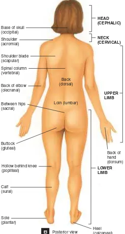
A
Posterior viewHEAD (CEPHALIC) NECK (CERVICAL)
Back of elbow (olecranal) Between hips (sacral)
Buttock (gluteal)
Sole (plantar) Calf (sural) Base of skull (occipital) Shoulder (acromial)
Spinal column (vertebral) Shoulder blade (scapular)
Back of hand (dorsum)
UPPER LIMB
Back (dorsal)
Loin (lumbar)
Heel (calcaneal)
LOWER LIMB
Hollow behind knee (popliteal)
B
Anatomical position with directional terms
Figure 4.12
In the “anatomical position,” the bones of the forearm lie straight instead of crossing over one another as they do when our hands rest by our sides.
A
Anterior view Chest (thoracic)TR
UNK
Pelvis Abdomen
NECK (CERVICAL)
Anterior surface of knee (patellar)
Foot (pedal)
Toes (digital or phalangeal) Ankle (tarsal)
Leg (crural) Thigh (femoral)
Top of foot (dorsum) Navel (umbilical)
Front of elbow (antecubital) Hip (coxal)
Groin (inguinal) Wrist (carpal)
Palm (palmar) Forearm (antebrachial) Breast (mammary) Breastbone (sternal) Armpit (avillary) Arm (brachial) Chin (mental) Mouth (oral) Nose (nasal) Cheek (buccal) Ear (otic) Eye (orbital) Forehead (frontal) Lateral
Proximal
Distal
Lateral
Superior
Inferior Medial
Skull (cranial) Face (facial)
HEAD (CEPHALIC)
Fingers (digital or phalangeal)
Pubis (pubic) Hand (manual)
L
EARNINGO
BJECTIVESLearnto use anatomical directional terms.
Scientists Use a Road Map
to the Human Body
tudying human biology—human
anatomy and physiology—is a daunting
task because we are concerned not only
with the location of organs and organ
systems, but also their interconnection. To discuss these
complicated matters clearly, we need a system to
pre-cisely name the structures of the body. Whenever we talk
about an organ’s placement, or the appearance of a
por-S
tion of the body, we assume we
have placed the body in the
anatomical position
(see
Fig-ure 4.12
). Using this
posi-tion as a standard allows us to
make sense of directional
terms such as proximal
and
distal,
superior
and inferior.
The body has natural
boundaries that we exploit for
describing position in human
biology. The body has two
large cavities, the ventral and
dorsal cavities (see
Figure
4.13
). The
ventral cavity
comprises the entire ventral
(or belly) aspect of your torso.
The ventral portion of the body contains distinct
sec-tions. The
thoracic cavity
includes the chest area and
houses the heart, lungs, vessels, and lymphatic system
of the mediastinum. The “guts” are found within the
abdominal cavity
, which is lined with
peritoneum
. The
organs of the urinary system and the reproductive
sys-tem are located in the
pelvic cavity
.
Identifythe body cavities and the organs that each contains.
110
CHAPTER 4
Tissues
Scientists Use a Road Map to the Human Body 111
Proximal/distal
Opposite terms
meaning near the
core of the body
versus farther from
the core.
Superior/inferior
Opposite terms
meaning above and
below.
Mediastinum
[image:10.1440.801.1047.317.777.2]112
CHAPTER 4
Tissues
B
C
A
RIGHT
HYPOCHONDRIAC REGION
EPIGASTRIC REGION
RIGHT LUMBAR REGION
LEFT LUMBAR REGION UMBILICAL
REGION
RIGHT INGUINAL (ILIAC) REGION
LEFT INGUINAL (ILIAC) REGION
HYPOGAS-TRIC (PUBIC) REGION
RIGHT
HYPOCHONDRIAC REGION
EPIGASTRIC REGION
RIGHT LUMBAR REGION
UMBILICAL REGION
RIGHT INGUINAL (ILIAC) REGION
HYPOGAS-TRIC (PUBIC) REGION
Anterior view showing location of abdominopelvic regions
Anterior view showing abdominopelvic regions
Anterior view showing location of abdominopelvic quadrants LEFT LOWER QUADRANT (LLQ) LEFT UPPER QUADRANT (LUQ) RIGHT LOWER
QUADRANT (RLQ) RIGHT UPPER QUADRANT (RUQ)
LEFT
HYPOCHONDRIAC REGION
LEFT LUMBAR REGION
LEFT INGUINAL (ILIAC) REGION Subcostal line Right
Clavicles
Clavicles
Left Midclavicular lines
Right Left Midclavicular lines
Transtubercular line
LEFT
HYPOCHONDRIAC REGION
Body regions and cavities
Figure 4.14
CONCEPT CHECK
Identify
a body part that is proximal to the hand.What
are the divisions of the ventral cavity?Scientists Use a Road Map to the Human Body 113
A
Right lateral viewB
Anterior viewCranial cavity Vertebral cavity Thoracic cavity
Abdominopelvic cavity:
Abdominal cavity Diaphragm
Pelvic cavity
Formed by cranial bones and contains brain. Formed by vertebral column and contains spinal cord and the beginnings of spinal nerves. Chest cavity; contains pleural and pericardial cavities and mediastinum.
Each surrounds a lung; the serous membrane of the pleural cavities is the pleura.
Surrounds the heart; the serous membrane of the pericardial cavity is the pericardium.
Central portion of thoracic cavity between the lungs; extends from sternum to vertebral column and from neck to diaphragm; contains heart, thymus, esophagus, trachea, and several large blood vessels.
Subdivided into abdominal and pelvic cavities. Contains stomach, spleen, liver, gallbladder, small intestine, and most of large intestine; the serous membrane of the abdominal cavity is the peritoneum.
Contains urinary bladder, portions of large intestine, and internal organs of reproduction.
Cranial cavity Vertebral cavity
Thoracic cavity*
Pleural cavity
Pericardial cavity
Mediastinum
Abdominopelvic cavity
Abdominal cavity
Pelvic cavity
CAVITY COMMENTS
Body cavities
Figure 4.13
The
dorsal body cavity
includes the
cranial
cavity
hous-ing the brain, and the
vertebral
cavity in which lies the spinal
cord. The meninges
line these
two continuous cavities.
Medical specialists
of-ten refer to the
abdominopelvic
regions and
quadrants
of the body when diagnosing pain. There are nine
re-gions, and four quadrants, each of which describes a
particular area housing just a few of the organs of the
abdominal cavity (
Figure 4.14
).
As we study human biology, we will refer to
these regions, quadrants, and cavities as landmarks for
identifying the position of organs and the relationships
between them. They also provide a common language
to facilitate communication about location or organ
function. In this age of digital surgery and online
med-ical diagnoses, having a common language becomes
even more important. Digital clinical assistance, or even
distance education in this field, would be impossible
without these conventions.
Knowing the organization of the chemicals,
cells, and tissues that make up the human body is a
pre-requisite for understanding how humans function in
the environment. Armed with this basic knowledge, an
in-depth look at humans and their environment
be-comes much more interesting. The remainder of this
text will explore the relationship between human
physi-ology and the environment in which humans live.
Meninges
Three protective
membranes
covering the brain
and spinal cord.
1
Cells Are the Building Blocks
of Tissues
The human body has four tissue types: epithelial, connective, nervous, and muscu-lar tissue. Epithelium covers and lines all body cavities and is classified based on cell shape (squamous, cuboidal, or columnar) and number of cell layers (simple or strati-fied). Connective tissue can be soft and
loose or dense. Cartilage, bone, blood, and lymph are all examples of connective tissue. Muscular tissue can contract, and it comes in three varieties: smooth, skeletal, and car-diac muscle. Nervous tissue includes the im-pulse-carrying neurons and the neuroglia, which provide support for neurons.
spinal cavity, surrounding the spinal cord. The ventral cavity includes the tho-racic cavity, the abdominal cavity, and the pelvic cavity. The ventral cavity can be subdivided into quadrants for specifi-cally pinpointing the location of an or-gan, a structure, or a physiological event in the body.
3
Scientists Use a Road Map
to the Human Body
When discussing the placement of human anatomical structures, we assume the body is in anatomical position. This is a face-forward position, with the palms of the hands forward. The two main body cavities are the dorsal cavity and the ven-tral cavity. The dorsal cavity includes the cranial cavity, holding the brain, and the
2
Organization Increases with
Organs, Organ Systems, and
the Organism
Tissues are grouped to-gether in organs. Organs per-forming a similar function come
together in organ sys-tems. A group of organ
systems comprises an organism. The 11 organ systems of the human
are the: skeletal (bones providing
support and pro-tection),
muscu-lar (muscles for movement and heat genera-tion), nervous (sensing and re-sponding to the environment), cutaneous (skin serving as a protective and sensitive layer), lymphatic (providing specific immunity), car-diovascular (transporting oxygen and nutri-ents to cells), respiratory (obtaining oxygen and removing carbon dioxide), digestive (obtaining nutrients), urinary (maintaining fluid balance), reproductive (producing new individuals), and endocrine (regulating se-quential growth and development).
1.As a research assistant in a cytology lab, you are handed a
stack of photographs from an electron microscope. Each repre-sents a different type of cell. You are asked to identify photos of animal cells that secrete large amounts of protein and include a mechanism for moving their secretions along their surface. What organelles would be required by this cell? In which tissues might these cells be found?
2.The digestive tract has two surfaces; an inner surface that lines
the gut and allows food to pass, and an outer surface that sepa-rates the gut from the rest of the abdominal organs. What spe-cific tissue would you expect to find on each of these surfaces? Would the inner surface have the same lining as the outer? Why or why not?
3.There are many types of connective tissue in the body, from
adipose to bone to blood. What is it that makes these tissues different? More immportantly, what are the unifying character-istics found in all connective tissues?
4.You are given the opportunity to create artificial skin in a
labo-ratory to help burn patients. Remember that the skin must be protective, relatively water-tight, and yet have some sensory function. What tissues will you need for this organ? Which type of epithelium will you use for the outer layer? What tissue will you need to house the blood vessels and the nerves? Will you need muscular tissue? nervous tissue?
5.Physicians often use the quadrants and regions of the body to
diagnose pathologies. If a patient complained of stabbing pain in the abdominal cavity, which organs might be involved? Look at Figure 4.13 to help with your diagnosis. How would you de-scribe the location of the urinary bladder using the nine ab-dominopelvic regions given in Figure 4.14?
CRITICAL THINKING QUESTIONS
CHAPTER SUMMARY
■ avascular 000
■ biceps Brachii 000
■ epiglottis 000
■ interstitial Fluid 000
■ mediastinum 000
■ meninges 000
■ microvilli 000
■ osteoid 000
■ plasma 000
■ proximal/Distal 000
■ rectus Abdominus 000
■ striations 000
■ superior/Inferior 000
■ trachea 000
KEY TERMS
11.Identify the tissue illustrated
a. Hyaline cartilage b. Skeletal muscle c. Cardiac muscle d. Smooth muscle
17.Which term correctly describes the relationship indicated as C?
a. Superior b. Inferior
c. Proximal d. Distal
18.The __________houses the heart, lungs, vessels and lymphat-ics of the mediastinum.
a. Ventral cavity b. Abdominal cavity
c. Cranial cavity d. Thoracic cavity
19.What membrane lines the indicated cavity?
a. Pericardial membrane b. Meninges
c. Peritoneum d. Mediastinum
20.Which label indicates the quadrant in which the majority
of the liver lies? b. B
c. C d. D
1.The four major tissue types that comprise the human body
in-clude all of the following EXCEPT:
a. Epithelial tissue b. Muscular tissue c. Areolar tissue d. Nervous tissue e. Connective tissue.
2.The tissue that can be found covering and lining openings in
the body is
a. Epithelial tissue b. Muscular tissue c. Areolar tissue d. Nervous tissue e. Connective tissue
3.The tissues that do not have a blood supply include
a. Epithelial tissue only
b. Epithelial and connective tissue c. Some types of connective tissue only
d. Epithelial and some types of connective tissue.
4.Identify the tissue type pictured a. Stratified epithelium b. Cuboidal epithelium c. Simple epithelium d. Columnar epithelium
7.The structure labeled A in this diagram is
a. Fibroblasts b. Collagen fibers c. Matrix
d. White blood cell
SELF TEST
116
CHAPTER 4
Tissues
Self Test 117
A
D B
C A
6.The specific type of cell that comprises most diffusion mem-branes is a
a. Squamous epithelial cell b. Cuboidal epithelial cell c. Columnar epithelial cell d. Exocrine cell
10.The connective tissue whose functions include lipid storage
and organ protection is
a. Blood b. Lymph c. Adipose d. Bone
C
D
A
B
8.The connective tissue that is composed of regular, linear arrangements of collagen fibers packed tightly into the matrix is called
a. Areolar connective tissue b. Loose connective tissue
c. Dense irregular connective tissue d. Dense regular connective tissue
9.Identify the type of connective tissue illustrated below. a. Bone
b. Hyaline cartilage c. Elastic cartilage d. Lymph
e. Fibrocartilage
5.The function of the pictured tissue is most likely a. A diffusion membrane
b. A protective membrane c. A contractile organ d. A connective support
12.Which type of muscle tissue can be described as involuntary, striated and connected via intercalated discs?
a. Skeletal muscle b. Cardiac muscle c. Smooth muscle
d. Two of these have the listed characteristics
13.Identify the structure indicated as “A” on this image a. Neuroglia
b. Dendrites c. Axon d. Neuron body
14.The functions of neuroglia include
a. Improving nutrient flow to neurons b. Supporting neurons
c. Sending and receiving electrical impulses d. Two of the above are correct
e. All of the above are correct
15.The correct order from least to most complex is:
a. Organ, organ system, organelle, organism b. Cell, tissue, organism, organ system c. Tissue, organ, organ system, organism d. cell, organelle, tissue, organ
16.Which term correctly describes the relationship indicated as “A” on
this figure?