MEDIA MEDI KA
I NDONESI ANA
Hak Cipta©2009 oleh Fakultas Kedokteran Universitas Diponegoro dan Ikatan Dokter Indonesia Wilayah Jawa Tengah
Risk Factors for Mortality in Dengue Shock Syndrome (DSS)
Catharina Suharti1, Tatty E Setiati2, Eric CM Van Gorp3, Robert J Djokomoeljanto1, Moeljono S Trastotenojo2, Jos WM van der Meer4, Wil MV Dolmans4
ABSTRACT
Background:Dengue shock syndrome (DSS) is the most severe form of dengue hemorrhagic fever (DHF) and has a high mortality. There are two major pathological changes in DHF determining the severity of disease, plasma leakage and bleeding. Cytokines released dur ing the immune response to dengue virus have been thought to be mediators of the process.
Methods: The study involved 50 children with DSS, of whom 13 (26%) died. We investigated which clinical signs and laboratory findings are r elated to mor tality.
Results:We found that gastrointestinal bleeding and bilater al pleural effusion were significantly more frequent in non-survivors than in survivors (p< 0.02 and p= 0.0006, respectively). Also, mean admission levels of thrombin-antithrombin complexes (TATc) and plasminogen activator inhibitor type 1 (PAI-1), activation markers of coagulation and fibrinolysis, respectively, were significantly higher in non-survivors (p= 0.004 and p= 0.0006, respectively). In regression analysis, bilateral pleural effusion and admission levels of TATc were significantly associated with mortality (p= 0.007 and p= 0.048, respectively).
Conclusions: Our data provide evidence for a relationship of mortality with pleural effusion, a marker of plasma leakage, and coagulation activation, both characteristic pathological changes in dengue shock syndrome.
Keywords: Dengue shock syndrome, mortality, risk factor.
ABSTRAK
Faktor risiko kematian pada demam berdarah dengue dengan sindroma syok (DSS)
Latar belakang: DSS merupakan bentuk klinik yang paling berat dari demam berdarah dengue (DBD) dan mempunyai angka kematian yang tinggi. Terdapat dua kelainan patologik utama pada DBD yang menentukan beratnya penyakit, yakni kebocor an plasma dan perdarahan. Sitokine yang dilepas sewaktu terjadi respon imun virus dengue diduga merupakan mediator proses ini.
Metode: Studi dilaksanakan pada 50 penderita DSS anak, dimana 13 (26%) diantaranya meninggal. Investigasi dilakukan untuk mencari temuan klinik dan laboratorik yang berhubungan dengan kematian.
Hasil: Perdarahan gastrointestinal dan efusi pleura bilateral secara bermakna lebih banyak ditemukan pada penderita yang meninggal dibandingkan dengan penderita yang hidup (berturut-tur ut p< 0,02 dan p= 0,0006). Rerata kadar thrombin-antithrombin complexes (TATc) sewaktu masuk rumah sakit dan kadar plasminogen activator inhibitor type 1 (PAI-1) juga merupakan petanda aktivasi koagulasi dan fibrinolisis, secara bermakna (berturut-tur ut p= 0,004 dan p= 0,0006) lebih tinggi pada penderita yang meninggal. Pada analisis regresi didapatkan bahwa efusi pleura bilater al dan kadar TATc sewaktu masuk rumah sakit berhubungan bermakna dengan kematian (ber turut-turut p= 0,007 dan p= 0,048).
Simpulan:Data dari studi ini membuktikan adanya hubungan antar a kematian dan efusi pleura bilateral (suatu petanda kebocoran plasma) dan aktivasi koagulasi dimana keduanya merupakan kelainan patologik khas untuk DSS.
1
Department of Internal Medicine, School of Medicine, Diponegoro University and Dr. Kariadi Hospital, Semarang, Indonesia, Jl. Dr. Sutomo 18 Semarang
2
Department of Paediatrics, School of Medicine, Diponegoro University and Dr. Kariadi Hospital, Semarang, Indonesia
3
Department of Internal Medicine Slotervaart Hospital, Amsterdam, The Netherlands
4
BACKGROUND
Dengue virus infection is an arthropod-borne disease, caused by any of four closely related serotypes of dengue virus, belonging to the genusFlavivirus, family
Flaviviridae: DEN-1, DEN-2, DEN-3 and DEN-4. In-fection confers life-long homotypic, but not heterotypic, immunity. Clinical manifestation can be asymptomatic infection, dengue fever (DF) and dengue hemorrhagic fever (DHF). The severity of DHF can vary from DHF I to DHF IV. The severe forms (DHF III and IV) are also referred to as dengue shock syndrome (DSS), cha-racterized by shock due to leakage of plasma from blood vessels.1
The pathogenesis of DSS is controversial. Epidemiolo-gical studies indicate that pre-existing dengue virus anti-bodies may predispose an individual to DHF on sub-sequent infection with another dengue virus serotype.2-4 Other studies have demonstrated that 2 and DEN-3 are more virulent than DEN-1 and DEN-4, but all four serotypes can cause severe or fatal disease.5-9 Epidemio-logical observations have provided evidence that age and race may be risk factors for DHF. DSS has been almost confined to children10 and the age group most severely affected was 5-9 years.11 Blacks are less susceptible to dengue shock syndrome than whites and Asians.3
There are two major pathological changes in DHF determining the severity of disease, plasma leakage and bleeding. The mechanism responsible for the increased permeability of vascular endothelial cells and bleeding in DHF has not been elucidated, although cytokines released during the immune response to dengue virus have been thought to be mediators of the process.12-14 The mortality rate of DHF is high, ranging from 2 to 10.9%,15-19 reaching 47% in patients with profound shock.20
We conducted a prospective clinical study, to investi-gate which clinical signs and laboratory parameters are associated with mortality in DSS.
METHODS
This prospective study was performed in Dr. Kariadi Hospital, the university hospital of Diponegoro Univer-sity, Semarang, Indonesia. The research protocol was approved by the Review Board of the Dr. Kariadi Hospital. Written informed consent was obtained from children’s parents or legal guardians.
Between June and November 1996, during an outbreak of dengue in Indonesia, 50 consecutive children with the clinical diagnosis of DSS, who were admitted to the Paediatric Intensive Care Unit were enrolled in the study. Only children with age 3 years were included.
The clinical diagnosis of DSS (DHF grade III and grade IV) was finally based on the 1997 WHO criteria.(1) Patients presenting all criteria for DHF: (i) fever or history of acute fever 2-7 days; (ii) bleeding (mild to severe); (iii) thrombocytopenia 100.000/mm3or less (iv) evidence of plasma leakage plus evidence of circulatory failure manifested by (a) rapid and weak pulse; (b) narrow pulse pressure (20 mmHg); (c) hypotension and (d) cold, clammy skin and restlessness, were classified DHF III. If profound shock with undetectable blood pressure and pulse was present, patients were classified as DHF IV. Decreased consciousness was based on the Glasgow Coma Scale.21
Through clinical assessment was performed daily du-ring hospitalization, using a medical record form. Labo-ratory tests to support clinical management included blood cell counts, tests for hemostasis (prothrombin time and activated partial thromboplastin time) as well as biochemical tests for kidney and liver functions and electrolyte status. Thrombin-antithrombin complexes (TATc) and plasminogen activator inhibitor type 1 (PAI-1), activation markers for coagulation and fibri-nolysis, respectively, were also assayed. Chest x-ray and ECG were done routinely.
Blood specimens were collected in vacutainer tubes (Becton Dickinson, Rutherford, NJ 07417). Two ml of blood for serological assays was collected on day of admission and at discharge. Blood was centrifuged 1000-1500 rpm for 10 minutes, after which serum was transferred to screw cap Eppendorf tubes and stored at -800C, until assayed.
electrolytes, were measured with Auto Analyzer, Hitachi, 7050. Normal value for serum protein were 5.9-8.6 g/dl, and for serum albumin were 3.5-5.6 g/dl. TATc and PAI-1 measurements were done at the Slotervaart Hospital, Amsterdam, The Netherlands. For the trans-port from Indonesia to The Netherlands (which last longer than 15 hours) the samples for coagulation, fibrinolysis, and serological assays were kept on dry ice. The diagnosis of dengue virus infection was confirmed by serological assays. A capture and indirect enzyme-linked immunosorbent assay (ELISA) detected dengue specific IgM and IgG antibodies in serum samples, according to a previously described procedure.23 Continuous data were described in mean (SD) and no-minal data in number (%). Nono-minal variables between survivors and non-survivors were compared using x2 Test. Fisher's Exact test was performed if the number of cells with expected frequency less than 5 were more than 20%. Independent t-tests were done to compare numeric variables between survivors and non-survivors. The assumption of normality of the data was checked before the t-test. Mann-Whitney U tests were done when the data were not normally distributed. The cut-of-point of significance was p=0.05 with 95% confi-dence interval. Regression analysis was done to deter-mine factors associated with mortality. Survival time was calculated with Kaplan-Meier curves. Log-rank test was used for curve comparison. All statistical analyses were performed with SPSS for Windows version 9.0.
RESULTS
Between June and November 1996, 50 children with a clinical diagnosis of DSS, were enrolled in the study. Thirteen patients (26%) died during follow up in the Intensive Care Unit. The clinical diagnosis of DSS was confirmed by serological assays in all patients, either by an IgM response or a fourfold rise in IgG titers. All antibody profiles were typical for secondary dengue infection.
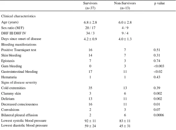
As shown in table 1, most patients were young children who presented after about 4 days of illness. Relatively more female patients died than males. Of the non-survivors 4 of 13 (30.7%) were classified as DHF IV, as compared with 3 of 37 (8.1%) survivors. The proportion of patients with gastrointestinal and gum bleeding was significantly higher in non-survivors than in survivors. A clammy skin, delirium, decrease of consciousness and bilateral pleural effusion were significantly more frequent in non-survivors than in survivors.
Table 2 shows that the prothrombin time (a measure for both the extrinsic and the common coagulant pathway) was significantly longer in non-survivors than in
survivors. Also the levels of thrombin-antithrombin complexes (TATc) and plasminogen activator inhibitor type 1 (PAI-1), activation markers of coagulation and fibrinolysis, respectively, were significantly higher in non-survivors than in survivors. The levels of serum protein were significantly lower in non-survivors than in survivors.
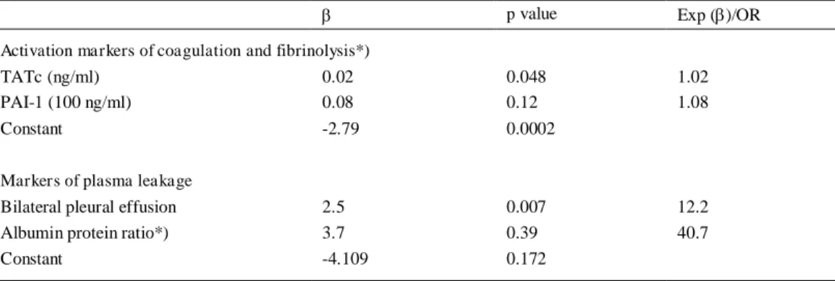
In the logistic regression analysis for mortality of day of admission levels of TATc and PAI-1, activation markers of coagulation and fibrinolysis, respectively, we found a significant association of TATc with mortality (p=0.048; regression coefficient: 0.023). Such asso-ciation was not found for PAI-1 (Table 3).
In the logistic regression analysis of albumin/protein ratio on day of admission and bilateral pleural effusion for mortality, a significant association was found for bilateral pleural effusion but not for albumin/protein ratio. The odds ratio for bilateral pleural effusion was 12.2 and p-value 0.007, implying that patients with bilateral pleural effusion had a 12.2 times increased risk of death (Table 3). This effect is also shown in the cumulative survival curve of patients with and without bilateral pleural effusion (Figure).
DISCUSSION
In this prospective study of 50 children with DSS, we found that bilateral pleural effusion and elevated TATc concentrations were significantly associated with mor-tality in logistic regression analysis. Bilateral pleural effusion indicates severe vascular leakage whereas high TATc concentrations reflect markedly activated coa-gulation. Our finding that bilateral pleural effusion is associated with poor prognosis corroborates the data of a study from Jakarta of 30 fatal cases of virologically confirmed dengue virus infection. In that study plasma leakage appeared to be the main cause of death, because 19 of the 30 cases (63.3%) had profound shock with hemoconcentration.24 In an autopsy study from Thailand of 100 children with fatal DSS plasma leakage was also likely the main cause of death, as evidenced by >4g/dl protein content of serous effusions in the pleural, abdominal and pericardial cavities. Also, the mucosa, submucosa and serosa of the gastrointestinal tract of these children showed oedema and hemorrhage, es-pecially in children with prolonged shock.25,26
Table 1. Clinical characteristics of survivors and non-survivors in 50 patients with dengue shock syndrome
Survivors (n=37)
Non-Survivors (n=13)
p value
Clinical characteristics
Age (years) 6.8 2.8 6.0 2.8
Sex ratio (M/F) 20 / 17 4 / 9
DHF III/DHF IV 34 / 3 9 / 4
Days since onset of disease 4.2 0.9 4.0 1.3
Bleeding manifestations
Positive Tourniquet test 16 7 0.51
Skin bleeding 14 7 0.31
Epistaxis 7 3 0.74
Gum bleeding 0 3 <0.003
Gastrointestinal bleeding 17 11 <0.02
Hematuria 1 1 0.43
Signs of disease severity
Cold extremities 35 13 0.39
Clammy skin 3 6 0.002
Delirium 13 11 0.002
Decreased consciousness 16 11 0.01
Convulsions 2 3 0.07
Bilateral pleural effusion 2 6 0.0006
Lowest systolic blood pressure Lowest diastolic blood pressure
92 11 59 24
83 11 45 31
Values indicated are mean SD and number
Table 2. Laboratory tests for hemostasis and markers of plasma leakage on day of admission in survivors and non-survivors in 50 patients with dengue shock syndrome
Survivors (n=37)
Non-Survivors (n=13)
p value *)
Tests for hemostasis
APTT (sec) 56.6 24.1 107.0 102.2 0.06
PT (sec) 15.3 4.7 24.2 12.0 0.001
PAI-1 (ng/ml) 285.1 491.8 1126.4 1314.6 0.0006
TATc (ng/ml) 32.2 27.3 67.5 43.3 0.004
Platelets (x109/l) 63.8 35.7 48.6 12.9 0.15
Markers of plasma leakage p value (95% CI)
Serum protein (g/l) 5.2 1.0 4.5 0.9 0.035 (0.052 - 1.335)
Serum albumin (g/l) 3.5 0.7 3.2 0.6 0.22 (-0.164 - 0.682)
Albumin/protein ratio 0.67 0.08 0.71 0.07 0.10 (-0.103 - 0.010)
Plasma sodium (mmol/l) 134.9 6.2 133.8 8.0 0.63 (-3.639 - 5.872)
Hematocrit (%) 38.3 6.4 36.4 7.4 0.39 (-2.519 - 6.227)
Table 3. Association of markers of coagulation and fibrinolysis and of plasma leakage with mortality in 50 patients with dengue shock syndrome
p value Exp ( )/OR
Activation markers of coagulation and fibrinolysis*)
TATc (ng/ml) 0.02 0.048 1.02
PAI-1 (100 ng/ml) 0.08 0.12 1.08
Constant -2.79 0.0002
Markers of plasma leakage
Bilateral pleural effusion 2.5 0.007 12.2
Albumin protein ratio*) 3.7 0.39 40.7
Constant -4.109 0.172
: regression coefficient; Exp. : exponent ; OR: odds ratio; TATc: thrombin-antithrombin complex; PAI-1: plasminogen activator inhibitor type-1; *) day of admission levels
Days since admission
16 14 12 10 8 6 4 2 0 -2
%
of patien
ts surv
ivin
g 100
80
60
40
20
0
Legend to figure.
Figure. Kaplan Meier survival curves of patients with bilateral pleural effusion compared with those without bilateral pleural effusion (p=0.002). The median survival time of patients with bilateral pleural effusion was 4 days (95% CI, 1.32 - 6.68).
fatal cases demonstrated evidence of severe bleeding followed by shock24, foci of hemorrhage in the brain28, histological evidence of intravascular coagulation,26,29 sero-hemorrhagic effusions29 and renal impairment.30 We also found that prothrombin time was significantly longer in non-survivors than in survivors. This suggests that the extrinsic (tissue factor/factor VIIa) pathway may be the main route in the activation of coagulation in DSS.
A number of clinical signs were more frequently seen in non-survivors than in survivors although these signs did not qualify in the logistic regression analysis, because these are not specific for DSS. Clammy skin was significantly more frequent (p=0.0002) and the systolic and diastolic blood pressures were lower in non-survivors (Table 1). In addition, we found that delirium and decreased consciousness were significantly more frequent in non-survivors than in survivors (p=0.002
No bilateral pleural effusion
Bilateral pleural effusion
% o
f
p
ati
ent
s s
ur
vi
vi
n
g
and p=0.010, respectively). Dengue virus subtypes 2 and 3 have been isolated from cerebrospinal fluid and have known to be neurovirulent.31 Histological studies from fatal DHF cases with signs of encephalopathy have provided evidence that the brain cell is also a target cell for dengue virus.32-36 In addition, loss of integrity of the cerebral vascular endothelium leading to cerebral oedema and cerebral bleeding has been observed.28,37 Conclusion our data provide evidence for a relationship of mortality with both, plasma leakage and coagulation activation in dengue shock syndrome. In the absence of interventions to stop vascular leakage, close monitoring and the use of plasma expanders to increase intravas-cular oncotic pressure is mandatory, especially in children with pleural effusion. In addition strategies to control coagulation activation would be an important area of development.
ACKNOWLEDGEMENTS
Participants in this project, besides the authors, were Dr. Herawati Juslam from the Department of Paediatrics, Diponegoro University and Dr. Kariadi Hospital, Semarang, Indonesia. We thank Dr. Hardian and Dr. Henry Setiawan from Clinical Epidemiology Unit, Diponegoro University, Semarang, for statistical support, Y.T. van der Heide from Clinical Chemistry and Hematology Laboratory Slotervaart Hospital, Amsterdam for managing the blood samples. We thank also Dr. J. Groen and Prof. A. Osterhaus from Institute of Virology Erasmus Medical Center Rotterdam for performing the diagnostic assays. Also, we thank Royal Netherlands Academy of Arts and Sciences (KNAW) for the financial support.
REFERENCES
1. World Health Organization. Dengue haemorrhagic fever: diagnosis, treatment, prevention and control. 2nd ed. Geneva, 1997;12-47.
2. Sangkawibha N, Rojansuphot S, Ahandrik S, Viriyapongse S, Jatanasen S, Salitul V, et al. Risk factors in dengue shock syndrome: a prospective epidemiologic study in Rayong, Thailand. Am J Epidemiol. 1984;20: 653-69.
3. Guzman MG, Kouri GP, Bravo J, Soler M, Vasquez S, Morier L. Dengue hemorrhagic fever in Cuba, 1981: a retrospective seroepidemiologic study. Am J Trop Med Hyg. 1990;42:179-84.
4. Burke DS, Nisalak A, Johnson DE, Scott RM. A prospective study of dengue infections in Bangkok. Am J Trop Med Hyg. 1988;38:172-80.
5. Rosen L. Comments on the epidemiology, pathogenesis and control of dengue. Med Trop. 1999;59:495-98.
6. Guzman MG, Kouri G, Valdes L, Bravo J, Alvarez M, Vazques S, et al. Epidemiologic studies on dengue in Santiago de Cuba, 1997. Am J Epidemiol. 2000;152:793-99.
7. Graham RR, Juffrie M, Tan R, Hayes CG, Laksono I, Ma’roef C, et al. A prospective seroepidemiologic study on dengue in children four to nine years of age in Yogyakarta, Indonesia. Studies in 1995-1996. Am J Trop Med Hyg. 1999;61:412-19.
8. Kobayashi N, Thayan R, Sugimoto C, Oda K, Saat Z, Vijayamalar B, et al. Type-3 dengue viruses responsible for the dengue epidemic in Malaysia during 1993-1994. Am J Trop Med Hyg. 1999;60:904-9.
9. Pandey BD, Morita K, Hasebe F, Parquet MC, Igarashi A. Molecular evolution, distribution and genetic relationship among the dengue 2 viruses isolated from different clinical severity. Southeast Asian J Trop Med Public Health. 2000;31:266-72.
10. Kouri GP, Guzman MG, Bravo JR, Triana C. Dengue haemorrhagic fever/dengue shock syndrome lessons from the Cuban Epidemic. Bull WHO. 1989;67:375-380.
11. Chareonsook O, Foy HM, Teeraratkul A, Silarug N. Changing epidemiology of dengue hemorrhagic fever in Thailand. Epidemiol Infect. 1999;122:161-66.
12. Kurane I, Ennis FA. Immunopathogenesis of dengue virus infections. In: Gubler DJ, Kuno G, editors. Dengue and dengue hemorrhagic fever. Wallingford, UK: Cab International, 1997; p.273-90.
13. Iyngkaran N, Yadav M, Sinniah M. Augmented inflammatory cytokines in primary dengue infection progressing to shock. Singapore Med J. 1995;36:218-21. 14. Anderson R, Wang S, Osiowy C, Issekutz AC. Activation of endothelial cells via antibody-enhanced dengue virus infection of peripheral blood monocytes. J Virol. 1997;71:4226-32.
15. Tripathi BK, Gupta B, Sinsha RS, Prasad S, Sharma DK. Experience in adult population in dengue outbreak in Dehli. J Assoc Physicians India.1988;46:273-76. 16. Aggarwal A, Chandra J, Aneja S, Patwari AK, Dutta
AK. An epidemic of dengue hemorrhagic fever and dengue shock syndrome in children in Dehli. Indian Pediatr. 1998;35:727-32.
17. Anuradha S, Singh NP, Rizvi SN, Agarwal SK, Gur R, Mathur MD. The 1996 outbreak of dengue hemorrhagic fever in Delhi, India. Southeast Asian J Trop Med Public Health. 1998;29:503-06.
18. Kabra SK, Jain Y, Pandey RM, Madhulika, Singhal T, Tripathi P, et al. Dengue hemorrhagic fever in children in the 1996 Delhi epidemic. Trans R Soc Trop Med Hyg. 1999; 93:294-98.
19. Wali JP, Biswas A, Handa R, Aggarwal P, Wig N, Dwivedi SN. Dengue hemorrhagic fever in adults: a prospective study of 110 cases. Trop Doct. 1999; 29:27-30.
21. Jarvis C. Physical examination and health assessment. Philadelphia: WB Saunders; 1993.
22. De Boer JP, Creasy AA, Chang A, Roem D, Brouwer MC, Eerenberg AJ, et al. Activation patterns of coagulation and fibrinolysis in baboons following infusion with lethal or sublethal dose of escherichia coli. Circ Shock. 1993:39:59-67.
23. Velzing J, Groen J, Drouet MT, van Amerongen G, Copra C, Osterhaus AD, et al. Induction of protective immunity against Dengue virus type 2: comparison of candidate live attenuated and recombinant vaccines. Vaccine. 1999;17:1312-20.
24. Sumarmo, Wulur H, Jahja E, Gubler DJ, Suharyono W, Sorensen K. Clinical observations on virologically confirmed fatal dengue infections in Jakarta, Indonesia. Bull World Health Organ. 1983;61:693-701.
25. Bhamarapravati N, Tuchinda P, Boonyapaknavik V. Pathology of Thailand haemorrhagic fever: a study of 100 autopsy cases. Ann Trop Med Parasitol. 1967;61: 500-10.
26. Bhamarapravati N. Pathology of dengue infections. In: Gubler DJ, Kuno G, editors. Dengue and dengue hemorrhagic fever. Wallingford, UK: Cab International, 1997; p.115-132.
27. van Gorp ECM, Suharti C, Tatty E Setiati, Mairuhu ATA, ten Cate H, Dolmans WMV, et al. Impaired fibrinolysis in the pathogenesis of lethal dengue hemorrhagic fever (DHF). Submitted for publication. 28. Chimelli L, Hahn MD, Netto MB, Ramos RG, Dias M,
Gray F. Dengue: neuropathological findings in 5 fatal cases from Brazil. Clin Neuropathol. 1990;9:157-62.
29. Strobel M, Jattiot F, Boulard F, Lamaury I, Salin J, Jarrige B, et al. Emergence of dengue hemorrhagic fever in French Antilles. Three initial fatal cases in Guadeloupe. Presse Med. 1998;27:1376-78.
30. George R, Liam CK, Chua CT, Lam SK, Pang T,
Geethan R, et al. Unusual clinical manifestations of dengue virus infection. Southeast Asian J Trop Med Public Health. 1988;19:585-90.
31. Lum C, Lam SK, Choy YS, George R, Harun F. Dengue encephalitis: a true entity? Am J Trop Med Hyg. 1996: 54:256-59.
32. Miagostovich MP, Ramos RG, Nicol AF, Nogueira RM, Cuzzi-Maya T, Oliveira AV, et al. Retrospective study on dengue fatal cases. Clin Neuropathol. 1997;16:204-08.
33. Desprès P, Frenkiel MP, Ceccaldi PE, Dos Santos CD, Deubel V. Apoptosis in the mouse central nervous system in response to infection with mouse-neurovirulent dengue viruses. J Virol. 1998;72:823-29.
34. Ramos C, Sanchez G, Pando RH, Baquera J, Hernandez D, Mota J, et al. Dengue virus in the brain of fatal case of hemorrhagic dengue fever. J Neurovirol. 1998; 4:465-68.
35. Hommel D, Talarmin A, Deubel V, Reynes JM, Drouet MT, Sarthou JL, et al. Dengue encephalitis in French Guiana. Res Virol. 1998;149:235-38.
36. Guzman MG, Alvarez M, Rodriquez R, Rosario D, Vazquez S, Vald SL, et al. Fatal dengue haemorrhagic fever in Cuba, 1997. Int J Infect Dis. 1999;3:130-35.