www.elsevier.com / locate / bres
Short communication
Chronic-intermittent hypoxia induces immediate early gene expression
in the midline thalamus and epithalamus
a ,
*
a a b,cAnthony L. Sica
, Harly E. Greenberg , Steven M. Scharf , David A. Ruggiero
a
Department of Medicine, Pulmonary and Critical Care Division, Long Island Jewish Medical Center, Long Island Campus of the Albert Einstein College of Medicine, New Hyde Park, New York, NY 11040, USA
b
Departments of Psychiatry and Anatomy and Cell Biology, Columbia University College of Physicians and Surgeons, New York, NY 10032, USA
c
Neurological Research Institute of Lubec, Lubec, ME 04652, USA
Accepted 2 August 2000
Abstract
Chronic-intermittent hypoxia (CIH) was postulated to activate thalamic regions that are synaptically related to autonomic-related areas of the cerebral cortex. Animals exposed to CIH for 30 days exhibited c-fos labeling in paraventricular thalamic and lateral habenular nuclei. Our findings strongly suggest activation of a diencephalic network that participates in behavioral responses to chronic stress. 2000 Elsevier Science B.V. All rights reserved.
Theme: Endocrine and autonomic regulation
Topic: Cardiovascular regulation
Keywords: Chronic-intermittent hypoxia; Rat; c-fos; Paraventricular thalamic nucleus; Habenular nucleus
Diurnal systemic hypertension is a prominent feature of from the paraventricular thalamus [2,4,10,18], which is many patients with obstructive sleep apnea (OSA) and is known to be activated by various stressors, and has been presumably related to the intermittent hypoxemia ex- described as the thalamic relay for stress-related infor-perienced nightly. Systemic hypertension is a likely conse- mation targeted for viscerolimbic regions of cortex [4,13]. quence of a hypoxia-related increase in vasoconstrictive Hence, the paraventricular thalamus may be the principal sympathetic nerve activity as demonstrated clinically in relay for activation of the medial prefrontal and insular humans with OSA [5,11,22,24], and experimentally in cortices during CIH [21]. Thus, in the present inves-rodents exposed to chronic-intermittent hypoxia (CIH) for tigation, we sought to determine whether the paraventricu-30–35 days [1,6–8]. Such long-term hypoxic exposure is lar thalamus was activated by chronic-intermittent hypoxia also associated with robust expression of Fos (the protein by comparing expression of Fos in CIH-exposed rats to product of the protooncogene, c-fos) by sympathetic that of control rats.
regulatory regions of the brainstem, nucleus of the solitary Six male Sprague–Dawley rats (200–250 g) were tract, rostral ventrolateral medulla [9], and of the cerebral randomly assigned to either a control or experimental cortex, especially the medial prefrontal and insular cortices group. Both experimental and control animals were placed [21]. A role for those cortical regions in blood pressure in adjoining, identical plastic chambers that were main-modulation has been demonstrated in a number of studies: tained at room temperature and were large enough to allow the medial prefrontal area has been shown to modulate the unimpeded movement. The fraction of inspired oxygen baroreflex, whereas the insular cortex, especially the right (FiO ) in each chamber was regulated by microprocessor-2
side, exerts tonic sympathoinhibitory influence on mean controlled miniature solenoids attached to each chamber by arterial pressure [23]. Both cortical areas receive inputs high-pressure tubing. For the CIH protocol, the chamber was flushed with 100% nitrogen for 30 s followed by 30 s of 100% O at a low flow rate. The time course of change
*Corresponding author. Tel.: 11-718-470-7058; fax: 11-718-470- 2
1035. in FiO2 for the experimental chamber was determined
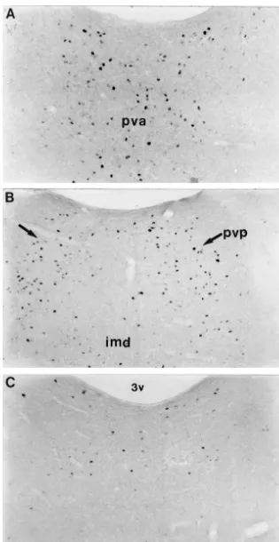
using an oxygen analyzer (Sensormedics, Model OM-11): the paratenial nucleus, which abuts the rostral division of FiO was noted to decline to a nadir of 6.5–7%, remaining2 the paraventricular thalamic nucleus, was devoid of FLI at that level for 5–7 s, and then gradually increased to 21% supported the specificity of the thalamic response pattern. over the next 30 s during which oxygen flowed into the Labeled neurons extended into the caudal division of the chamber. This cycle was repeated each minute over 8 paraventricular thalamic nucleus (Fig. 1B) and, at the daytime hours (8–9 A.M. to 4–5 P.M.) for 30 days. mesodiencephalic junction, were contiguous with those in Control rats were exposed to the same sequence of events the adjoining central gray region. Although sparse and as experimental rats, with the exception that room air was lightly labeled, cells containing FLI were detected in the used instead of nitrogen and oxygen. In this manner, centromedial and paracentral thalamic nuclei in rats ex-control rats experienced the same environmental cues and posed to CIH but not in controls. The ventral tier nuclei degree of handling as CIH rats. At the conclusion of daily were devoid of FLI.
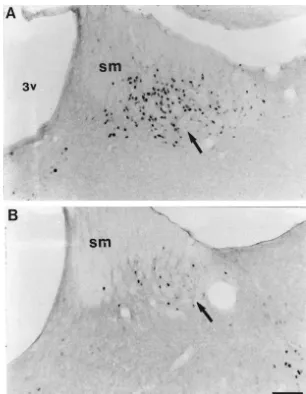
exposure protocols, animals were transferred from plastic Striking evidence for c-fos gene induction was identified chambers to home cages where access to food and water in the epithalamus, where the nuclear reaction product was was freely available. All animals were maintained on a restricted to neurons of the lateral habenular nucleus. By 12:12 light:dark cycle. Brief Summary of CIH exposure contrast, few neurons in the epithalamus expressed FLI on
methods and physiological effects. sections from control rats that breathed room air over the
Control rats breathed room air but were otherwise same time course as experimental animals (Fig. 2A and B). exposed to identical environmental stressors. Two h fol- Our results are the first to demonstrate that the midline lowing the last hypoxic challenge, an experimental and a thalamus and habenular nucleus are activated in response control animal of each pair were anesthetized with pen- to chronic-intermittent hypoxia. Given the known projec-tobarbital (100 mg / kg, i.p.) and perfused with heparinized tions of the paraventricular nucleus to the medial prefrontal normal saline followed by a buffered 4% paraformal- and insular cortices [2,4,10,18], our findings also support dehyde (pH 7.4). Forebrains were blocked from the the idea that the paraventricular nucleus is a relay of stress midcollicular level to the frontal pole, removed and frozen- related information destined for cortex [4,13]. The midline sectioned, transversely on a sledge microtome at 35 mm. thalamus was likely activated by arterial chemoreceptors Serially sectioned tissues were processed immuno- and, perhaps, by more complex behaviorally-conditioned cytochemically to label c-fos proteins or Nissl-stained [9]. cues associated with the stress of intermittent hypoxia. The All histological and immunocytochemical processing possibility that signals conveyed by ascending chemosen-methods were identical to those applied in our previous sory pathways contributed to the thalamic responses was studies to examine responses of the medulla oblongata [9] advanced by evidence demonstrating parallel projections to and cerebral cortex [21] to CIH. In brief, tissues from the the paraventricular nucleus from caudomedial regions of forebrain of a stimulated rat exposed to CIH and from a the nucleus of the solitary tract and rostral ventral medul-control rat breathing room air were placed in individual lary reticular formation [17], areas that were activated by spot test wells filled with the same solutions in order to CIH and that coincided with constituents of the arterial minimize technical variability. Tissues were incubated in a chemoreflex arc [9].
commercially characterized antibody raised in rabbit The epithalamic response to CIH is another new ob-against Fos (Oncogene Science, Manhasset, NY) and servation. Our evidence for a direct habenular projection to processed concurrently by using the avidin–biotin complex the paraventricular thalamic nucleus [17] predicts that the (Vectastain Elite kit) immunohistochemical labeling pro- epithalamus and its afferent networks may modulate cedure. The immunoreaction product was demonstrated by midline thalamic response to chronic hypoxia and perhaps the TMB chromagen reaction. Thalamic nuclei were other stressors, as well. The habenular nucleus modulates examined for Fos-like immunoreactivity (FLI) and their states of arousal and chronic responses to stress [16], nuclear boundaries differentiated under darkfield optics conceivably by serving as a convergence center for striatal and on tissues stained with thionin. Nuclei containing FLI and limbic conduction channels [19] and the retina [20]. were photographed on Kodak TMAX ASA100 film. It is conceivable that neocortical [21] and diencephalic
Fig. 2. Induction of c-fos, in the lateral habenular nucleus in an animal exposed to chronic-intermittent hypoxia (A). Note that chronic-intermittent hypoxia independently of other environmental factors produced increases in Fos expression in the habenular nucleus as compared to the control animal (B). Bar5125mm. sm, stria terminalis; 3v, third ventricle.
sustained, robust induction patterns that we observed in the References
brainstem [9] and cerebral cortex [21], and now in the
[1] G. Bao, P.M. Randhawa, E.C. Fletcher, Acute blood pressure
thalamus and epithalamus are suggestive of chronic
activa-elevation during repetitive hypocapnic and eucapnic hypoxia in rats,
tion of cortical and subcortical regions responsive to
J. Appl. Physiol. 82 (1997) 1071–1078.
sustained stress. Such widespread activation patterns may [2] H.W. Berendse, H.J. Groenewegen, Restricted termination fields of underlie the development of hypertension in chronic the midline and intralaminar thalamic nuclei of the rat, Neuroscience
hypoxic exposures. 42 (1991) 73–102.
[3] A.W. Briujnzeel, R. Stam, J.C. Compaan, G. Croiset, L.M. Akker-mans, B. Olivier, V.M. Wiegant, Long-term sensitization of Fos-responsivity in the rat central nervous system after a single stressful
Acknowledgements experience, Brain Res. 819 (1999) 15–22.
[4] M. Busber, A.Y. Deutch, Stress induces Fos expression in neurons of the thalamic paraventricular nucleus that innervate limbic structures,
We wish to thank M. Anwar and D. Muller for their
Synapse 32 (1999) 13–22.
excellent technical assistance. This work was supported by
[5] E.C. Fletcher, J. Miller, W. Schaaf, J. Fletcher, Urinary
catechol-a Long Islcatechol-and Jewish Mediccatechol-al Center Fcatechol-aculty Awcatechol-ard amines before and after tracheostomy in patients with obstructive (ALS) and National Institutes of Health grant: NS-36363 sleep apnea and hypertension, Sleep 10 (1987) 35–44.
episodic hypoxia causes diurnal elevations of systemic blood [16] C.A. Murphy, A.M. DiCamillo, F. Haun, M. Murray, Lesion of the pressure in rats, Hypertension 19 (1992) 555–561. habenular efferent pathway produces anxiety and locomotor hy-[7] E.C. Fletcher, J. Lesske, J. Culman, C.C. Miller, T. Unger, Sympa- peractivity in rats: a comparison of the effects of neonatal and adult
thetic denervation blocks blood pressure elevation in episodic lesions, Behav. Brain Res. 81 (1996) 43–52.
hypoxia, Hypertension 20 (1992) 612–619. [17] K. Otake, D.A. Ruggiero, Monoamines and nitric oxide are em-[8] H.E. Greenberg, A. Sica, D. Batson, S.M. Scharf, Chronic intermit- ployed by afferents engaged in midline thalamic regulation, J.
tent hypoxia increases sympathetic responsiveness to hypoxia and Neurosci. 15 (1995) 1891–1911.
hypercapnia, J. Appl. Physiol. 86 (1999a) 298–305. [18] K. Otake, Y. Nakamura, Single midline thalamic neurons projecting [9] H.E. Greenberg, A.L. Sica, S.M. Scharf, D.A. Ruggiero, Expression to both the ventral striatum and the prefrontal cortex in the rat,
of c-fos in the rat brainstem after chronic intermittent hypoxia, Brain Neuroscience 86 (1998) 635–649.
Res. 816 (1999 b) 638–645. [19] A. Parent, S. Gravel, R. Boucher, The origin of forebrain afferents [10] H.J. Groenwegen, H.W. Berendse, The specificity of the ‘non- to the habenula in rat, cat and monkey, Brain Res. Bull. 6 (1981)
specific’ midline and intralaminar thalamic nuclei, Trends Neurosci. 23–38.
17 (1994) 52–57. [20] T. Qu, K. Dong, K. Sugioka, T. Yamadori, Demonstration of direct [11] K. Hla, T. Young, M. Bidwell, M. Palta, J. Skatrud, J. Dempsey, input from the retina to the lateral habenular nucleus in the albino
Sleep apnea and hypertension. A population-based study, Ann. rat, Brain Res. 709 (1996) 251–258.
Intern. Med. 120 (1994) 382–388. [21] A.L. Sica, H.E. Greenberg, S.M. Scharf, D.A. Ruggiero, Immediate-[12] A. Keilmann, T. Herdegen, The c-Fos transcription factor in the early gene expression in cerebral cortex following exposure to
auditory pathway of the juvenile rat: effects of acoustic deprivation chronic-intermittent hypoxia, Brain Res. 870 (2000) 204–210. and repetitive stimulation, Brain Res. 753 (1997) 291–298. [22] V.K. Sommers, M.E. Dyken, M. Clary, F.M. Abboud, Sympathetic [13] P. Kent, H. Anisman, Z. Merali, Are bombesin-like peptides neural mechanisms in obstructive sleep apnea, J. Clin. Invest. 96
involved in the mediation of stress response?, Life Sci. 62 (1997) (1995) 1897–1904.
103–114. [23] A.J.M. Verberne, N.C. Owens, Cortical modulation of the car-[14] M. Lanteri-Minet, P. Isnardon, J. de Pommery, D. Menetrey, Spinal diovascular system, Progr. Neurobiol. 54 (1997) 149–168.
and hindbrain structures involved in visceroception and vis- [24] C.J. Worsnop, M.T. Naughton, C.E. Barter, T.O. Morgan, A.I. ceronociception as revealed by the expression of Fos, Jun and Anderson, R.J. Pierce, The prevalence of obstructive sleep apnea in Krox-24 proteins, Neuroscience 55 (1993) 737–753. hypertensives, Am. J. Respir. Crit. Care Med. 157 (1998) 111–115. [15] G.D. Mower, Differences in the induction of Fos protein in cat