The measurement of the alveolar bone crest in aggressive
periodontitis using Cone Beam Computed Tomography imaging
Astia Dwiputri Lestari*, Azhari*, Sri Wendari**
*Department of Dentomaxilofacial Radiology Faculty of Dentistry Universitas Padjadjaran **Department of Periodontics Faculty of Dentistry Universitas Padjadjaran
ABSTRACT
The normal alveolar bone crest located at a distance of 1 to 2 mm from CEJ towards the apical. If there is a bone loss, the alveolar bone crest located at 2 mm more to the apical from CEJ. The alveolar bone loss is one characteristic of aggressive periodontitis and the onset of the disease at the age of puberty. The purpose of this research was to know and to assess the height of alveolar bone crest using Cone Beam Computed Tomography (CBCT) in patients with aggressive periodontitis. The type of this research was descriptive. A total of 317 sample CBCT imagery from 6 aggressive periodontitis patients. The result of this research showed that average height of alveolar bone crest second premolar distal,
the irst molar mesial, irst molar distal and second molar mesial on the irst region were 3 mm, 4.2 mm, 6.1 mm, and 4.8 mm respectively. On the second region were 3.2 mm, 3.6 mm, 3.6 mm, and 3.4 mm respectively, and in the third region were 2.4 mm, 2.8 mm, 2.5 mm dan 1.4 mm consecutively. While in the fourth region were 2.9 mm, 3 mm, 2.8 mm, and 2.3 mm respectively. The average height of alveolar bone crest of aggressive periodontitis was 3.8 mm. Slicing CBCT imagery on coronal view for the anterior
region and sagittal view for the posterior region, showed the characteristic of height alveolar bone crest
was arch-shaped which showed the diferent height of alveolar bone crest of the second premolar distal
until second molar mesial.
Keywords: Alveolar bone crest, bone loss, aggressive periodontitis, Cone Beam Computed Tomography (CBCT)
Corresponding author: Azhari, Department of Dentomaxilofacial Radiology Dentistry Faculty of Dentistry Universitas Padjadjaran, Jl. Sekeloa Selatan No.1, Bandung, Indonesia, 40132. Ph./Fax.:+6222-2504985/2532805
INTRODUCTION
Dental problems were placed on the sixth of the highest diseases as reported by Indonesian
National Household Health Survey. Two of the high
prevalence diseases were caries and periodontal disease. Periodontal disease is the second major problems which considered as a problem in society.
Periodontal disease is an inlammation
process of periodontal tissue which starts from an
invasion of microorganisms and its products into
the gingiva. Inlammation from the supporting
structures of the teeth caused by microorganisms resulted in progressive damage in periodontal ligament and alveolar bone by forming a pocket, gingival recession, or combination of both which called periodontitis.2
American Academy of Periodontology (AAP)8
in 1999 described classiications of periodontal
chronic periodontitis, aggressive periodontitis, periodontitis as a manifestation of systemic disease, necrotizing periodontal disease, abscesses of the periodontium, periodontitis associated with endodontic lesions, and developmental or acquired deformities and conditions.2 One of the periodontal diseases classiication is
aggressive periodontitis. Aggressive periodontitis usually happened in younger patients with lower prevalence compared to chronic periodontitis. Aggressive periodontitis epidemiology data in Indonesia has not existed, but there are several experiments about this prevalence.
One of the experiments that have done in
the Periodontics Clinics of Faculty of Dentistry Universitas Airlangga discovered that there was an escalation from 9% in 1991 to 13% in 2002. In 2010, experiment regarding this disease was
done by Nariratih in Student’s Clinic of Faculty
of Dentistry Universitas Padjadjaran from May-July 2010 showed that aggressive periodontitis prevalence was as much as 3,13%.2,3
Aggressive periodontitis is a continuation of
gingival inlammation into a deeper periodontal
tissue structure and alveolar bone crest. CEJ measurement of the alveolar bone crest was >2
mm (3-4mm) (Hodges, 1997). CEJ measurement of alveolar bone crest could diferentiate stages
of periodontal diseases. This could help when diagnosing periodontal diseases. The correct diagnosis is important for treatment planning. Diagnosis determined by a careful analysis of the case, clinical examination, and other examinations (radiography, blood test).2,4,5
Radiography has an important role in making a diagnosis of the periodontal disease.
Radiography gives speciic information about the
condition of periodontal tissue and records about bone condition during the course of the disease, but conventional radiography has limits, one of them is it only showed the two-dimensional image in three-dimensional condition.6
Cone Beam Computed Tomography (CBCT) is the newest radiography equipment represents the maxillofacial area. CBCT shows an accurate image
of the jaw, diferent from the panoramic picture.
Because of the distortion in panoramic, it cannot relied on taking a measurement. Also, CBCT could deliver cross-sectional image (buccolingual), axial, coronal, sagittal, and panoramic. Meanwhile
the conventional panoramic can only produce one-dimensional image, which was mesiodistal or anteroposterior and there is also a superimpose image.7
There has not been any research performed in Faculty of Dentistry Universitas Padjadjaran observing the alveolar bone crest in aggressive periodontitis patient using CBCT imaging. Therefore, the purpose of this research was to discover and assess the alveolar bone crest in aggressive periodontitis patients using Cone Beam Computed Tomography (CBCT) Imaging.
METHODS
The type of this research was descriptive and using a secondary data which was recorded in CBCT Radiography in Faculty of Dentistry Universitas Padjadjaran from May-July 2010 from the patient diagnosed clinically and radiographically as aggressive periodontitis.
Sampling was done by total sampling from 6 subject aggressive periodontitis’s CBCT radiograph
data, consisted of 321 images of a mesial-distal radiograph of alveolar bone crest CBCT imaging
from aggressive periodontitis patient’s alveolar
bone fragments in each teeth interdental which were recorded in Faculty of Dentistry Universitas Padjadjaran from May-July 2010.
The variable of this research was the alveolar
bone crest. The alveolar bone crest deined as the
distance between cementoenamel junction (CEJ) to alveolar bone crest. The measurement was
done by drawing a ictitious horizontal line from
the alveolar bone crest to the mesial-distal of
adjacent teeth, then a ictitious vertical line from
CEJ to the horizontal of the alveolar bone crest. Tools used in this research are Cone Beam Computed Tomography (CBCT) Radiograph and
a computer. The computer used was an ASUS Notebook series A42J with Generic PnP Monitor, with software included the Microsoft® Oice Word
2007, Microsoft® Excel 2007, and the Capture
Screen Program.
The procedure of this research was after obtaining the secondary data of aggressive periodontitis patients with 3D CBCT in the Radiology Installation of Faculty of Dentistry Universitas Padjadjaran Dental Hospital; the EazyDent®
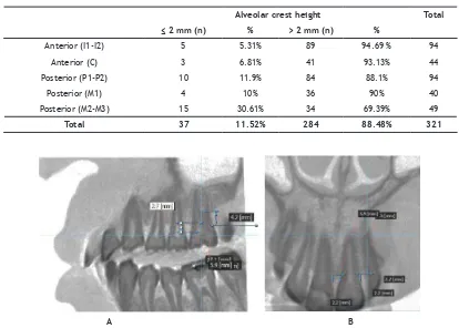
Table 1. Percentage of mesial and distal tooth surface with the alveolar bone crest height of ≤2mm and >2mm
Alveolar crest height Total
≤ 2 mm (n) % > 2 mm (n) %
Anterior (I1-I2) 5 5.31% 89 94.69 % 94
Anterior (C) 3 6.81% 41 93.13% 44
Posterior (P1-P2) 10 11.9% 84 88.1% 94
Posterior (M1) 4 10% 36 90% 40
Posterior (M2-M3) 15 30.61% 34 69.39% 49
Total 37 11.52% 284 88.48% 321
chosen and opened with the EzImplant® program. After having the image in the monitor, the contrast was set by selecting windowing menu. The central axis pointer was moved to the interproximal teeth which the alveolar bone was seen, then zoomed. After having the right position, the attention was focused on the coronal and sagittal view. After
the alveolar bone crest visible, a ictitious line
was drawn using a ruler to get the alveolar bone
crest’s data, from the alveolar bone crest to the
mesial-distal of adjacent teeth, then from CEJ to the horizontal of the alveolar bone crest. The data was then collected and presented using a table to be counted for the average value.
RESULTS
This research was conducted in May 2011 in Faculty of Dentistry Universitas Padjadjaran Dental Hospital. From 6 3D Cone Beam Computed Tomography (CBCT) radiograph archives of
aggressive periodontitis patient, 4 of it consisted
of CBCT radiograph archives of localized aggressive periodontitis and 2 archives of generalized
aggressive periodontitis taken during May-July 2010, so the mesial-distal radiograph of alveolar bone crest in each tooth interdental from CBCT images obtained were 321 images.
The image results of the alveolar bone crest of the aggressive periodontitis patient that viewed from the coronal direction to see the anterior teeth and from the sagittal direction to see the posterior teeth were presented in Table 1-Table 3. Table 1 described the CBCT radiograph imaging of 37 teeth surface with the alveolar bone crest of
≤2 mm (11.52%), and 284 teeth surface with the alveolar bone crest of >2mm (88.48%).
Figure 1 showed the alveolar bone crest height of aggressive periodontitis using CBCT imaging in anterior region (I1) and posterior region (M1). It can be seen that there were damage on the anterior region (I1) and posterior region (M1). Table 2 showed the average alveolar bone
crest height in anterior region was 4.08 mm, while in posterior region was 3.38 mm. This table also
showed the average alveolar bone crest height of
all aggressive periodontitis patient was 3.8 mm.
The lowest alveolar bone crest height was found
Figure 1. (A) Alveolar bone crest height of aggressive periodontitis patient in the anterior region (I1) using CBCT Imaging, (B). Alveolar bone crest height of aggressive periodontitis patient in the posterior region (M1) using CBCT Imaging.
in the anterior region (I1-I2), which was 4.39 mm and in the posterior region (M1) was 4.07 mm.
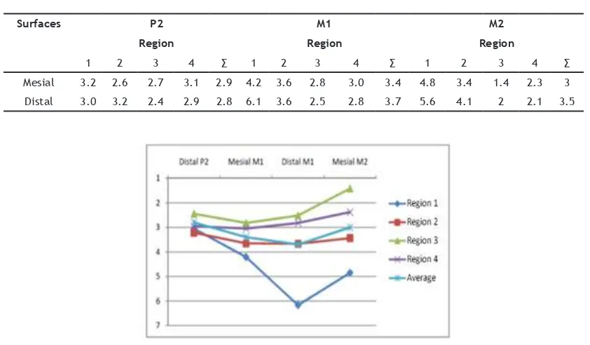
Table 3 showed the diference between the
mesial-distal second premolar (P2) height and the
second molar (M2). This diference will make an
arch-shaped pattern of the bone damage that can be seen in Figure 2. The arch-shaped pattern was visible in the distal second premolar (P2) to the mesial second molar (M2) located in all four regions.
Figure 3 showed the arch-shaped pattern
generated from the height diference of the
average alveolar bone crest height between the
distal second premolar, mesial irst molar, distal irst molar, and the mesial second molar in the all four region.
DISCUSSION
Aggressive periodontitis is an infectious disease in the periodontal tissue with a
two-dimensional radiographical image of alveolar bone
resorption that usually happened in the irst molar
and incisivus tooth.8 The higher distance from
alveolar bone crest to CEJ was a proof of the bone loss. The higher the distance from CEJ to alveolar bone crest, the higher the alveolar bone loss, thus
the lower alveolar bone crest. With bone loss,
the height of the alveolar bone was 2mm closer towards the apical from CEJ (Langland, 2002).
Such circumstances it the results of CBCT imaging
in 6 aggressive periodontitis patients with the lowest alveolar bone crest in the anterior region
which was 4.39 mm (I1-I2). The localized state of
periodontal tissue damage in molar and incisivus
caused by molar and incisivus are the irst erupted
teeth.2
Results from the sample of CBCT imaging, aggressive periodontitis patients which having the
alveolar bone crest of ≤2 mm was on the 37 teeth surface (11.52%), and as much as 284 teeth surface
Table 2 The average alveolar bone crest height (in mm) in the anterior region, posterior region, maxilla, and mandible
Anterior
(I1-I2)
Anterior
(C)
Posterior
(P1-P2)
Posterior
(M1)
Posterior
(M2-M3)
Average
Maxilla 4.65 3.75 3.21 4.31 4.52 4.08
Mandible 4.18 3.52 3.85 2.76 3.59 3.38
Average 4.39 3.72 3.05 4.07 3.78
Average 4.05 3.63 3.8
Table 3. The average alveolar bone crest height (in mm) in the mesial and distal aspects
Surfaces P2 M1 M2
Region Region Region
1 2 3 4 ∑ 1 2 3 4 ∑ 1 2 3 4 ∑
Mesial 3.2 2.6 2.7 3.1 2.9 4.2 3.6 2.8 3.0 3.4 4.8 3.4 1.4 2.3 3
Distal 3.0 3.2 2.4 2.9 2.8 6.1 3.6 2.5 2.8 3.7 5.6 4.1 2 2.1 3.5
were having the alveolar bone crest of >2 mm
(88.48%). This result was consistent with research
conducted by Hodges in 1997, which stated that in the early periodontitis stage (in 1999 the term was changed into aggressive periodontitis), the distance from CEJ to the alveolar bone crest was >2 mm. This condition happened due to a gingiva
inlammatory progressivity towards the alveolar
bone crest with deeper periodontal structure.4 The term “juvenile periodontitis” or “early periodontitis,” “prepubertal periodontitis” dan “rapidly progressive periodontitis” now are
classiied as aggressive periodontitis.9 Early periodontitis described as gingiva inlammatory
progressivity towards the alveolar bone crest with deeper periodontal structure, with a little loss of attachment and alveolar bone. The distance from
CEJ to alveolar bone crest was 3-4 mm.4 This
circumstance matches with the result of CBCT imaging from 6 aggressive periodontitis patients that showed the average height of the aggressive
periodontitis bone crest was 3.8 mm. The result of
this study also showed that the maxillary alveolar bone crest height was lower than the mandible. The maxillary alveolar bone crest height was
4.08 mm, while the mandible was 3.38 mm. This
condition can be caused by plaque and calculus accumulation, and more commonly occurred in the maxillary than the mandible. Another reason could be due to the thinner and less dense cortical in the maxillary than the mandible. That is why the maxillary alveolar bone was easier to come through the resorption that the mandible.10
The alveolar bone crest height of aggressive
periodontitis was diferent in the anterior region
and the posterior. The height in the anterior region
was 4.05 mm while in the posterior region was 3.63
mm. This condition happened due to the thickness
diference between the anterior and posterior
region.11 The defect pattern of the alveolar bone
in aggressive periodontitis is arch-shaped.2 This shape was seen from the result showed that the alveolar bone crest from distal second premolar
to mesial second molar has a diferent height that
will form an arch-shaped pattern.
CONCLUSION
The slicing of the CBCT imagery on coronal view for the anterior region and sagittal view for
the posterior region, showed the characteristic of height alveolar bone crest was arch-shaped which
showed the diferent height of alveolar bone crest
of the second premolar distal until second molar mesial.
REFERENCES
1. Tampubolon NS. Dampak Karies Gigi dan
Penyakit Periodontal Terhadap Kualitas Hidup [dissertation]. Medan: USU; 2005.
2. Newman MG, Takei HH, Carranza FA. Carranza’s Clinical Periodontology. 10th ed. Philadelphia:
Saunders-Elsevier; 2006. p. 452-66, 506-12, 561-80.
3. Nariratih D, Rusyanti Y, Susanto A. Prevalence and characteristics of Aggressive Periodontitis.
Padjadjaran J Dent. 2011;23(2):97-104. 4. Hodges K. Concepts in Nonsurgical Periodontal
Therapy. 1st ed. New York: Delmar-Thomson
Learning Inc.; 1998. p. 128-34.
5. Lindhe J, Lang NP, Karring T. Clinical
Periodontology and Implant Dentistry. 2nd ed. Hoboken: Blackwell Publishing; 1990.
6. White SC, Pharoah MJ. Oral Radiology:
Principles and Interpretation. 5th ed. St. Louis: Mosby-Elsevier; 2004. p. 314-24.
7. Oral Health Group [homepage on internet].
Toronto: Perschbacher S. 2009. Making The Most
of Cone Beam CT Responsibly. [cited 2011 Apr]; [about 2 screens]. Available from: https:// www.oralhealthgroup.com/features/making-the-most-of-cone-beam-ct-responsibly/
8. Grant DA, Stern IB, Listgarten MA. Early Onset Periodontitis (juvenile periodontitis/ aggressive periodontitis). In: Eley BM, Soory M, Manson JD. Periodontics. 6th ed. St. Louis:
Mosby-Elsevier; 2010. p. 377-97.
9. Armitage GC. Development of a classiication system for periodontal diseases and conditions.
Ann Periodontol. 1999 Dec;4(1):1-6.
10. Torkzaban P, Arabi B, Torkan R, Khoshhal M. Assessment of cementoenamel junction-alveolar bone crest distance in interproximal areas of intact primary molars in healthy 7-9 year old boys in Hamadan. Avicenna J Dent
Res. 2009;2(1):25-30.
11. Borges MS, Mucha JN. Bone density assessment for mini-implants positioning. Dental Press J