Protective role of
Nigella sativa
oil
against reproductive toxicity,
hormonal alterations, and oxidative
damage induced by chlorpyrifos in
male rats
Rachid Mosbah
1, Mokhtar Ibrahim Yousef
2,
Francesca Maranghi
3and Alberto Mantovani
3Abstract
This study is aimed at elucidating the possible protective effects of Nigella sativa oil (NSO) in alleviating the toxicity of chlorpyrifos (CPF) on reproductive performance in male rats. Animals were orally administered with NSO (1 ml/kg/day), CPF (20 mg/kg/day), and NSOþCPF every day for 4 weeks. Results showed that CPF
decreased spermatid number, sperm count, daily sperm production, and sperm motility while increased dead sperm and abnormal sperm compared with the control. Also the levels of testosterone, thyroxine levels, steroidogenic enzyme 17-ketosteroid reductase, body weight, food intake, and relative weight of reproductive organs were decreased. Thiobarbituric acid reactive substances were increased, while glutathione (GSH) and antioxidant enzymes were decreased in plasma and testes of rats treated with CPF. Histopathological exam-ination of testes showed a decrease in the number of seminiferous tubules, form shrinkage, enlargement of the connective tissue and gametogenic changes in germ cells of rats treated with CPF. NSO alone increased tes-tosterone, semen characteristics, GSH, and antioxidant enzymes and decreased the levels of free radicals. Furthermore, the presence of NSO with CPF alleviates its toxic effects. Our results indicated that NSO can improve semen picture and moderate CPF-induced reproductive toxicity.
Keywords
Reproductive toxicity, chlorpyrifos,Nigella sativa oil, free radicals, hormones, histopathological examination
Introduction
Many pesticides are known to impair reproductive competence of males in laboratory, field, clinical, or occupational settings (Figa-Talamanca et al., 2001; Giwercman and Giwercman, 2011; Mantovani, 2006; Mantovani and Maranghi, 2005). Organophosphates (OPs) are among the most commonly used pesticides/ insecticides in developing countries including Egypt and Algeria.
Chlorpyrifos (CPF) is one of the most widely used OP insecticides throughout the world in both domestic and agricultural applications (Asperlin, 1994). CPF affects the central and peripheral nervous system by inhibiting the acetylcholinesterase (AChE), which breaks down the neurotransmitter acetylcholine (Ach) in the cholinergic synapses and at neuromuscular junc-tions. The neurotoxic effects of CPF are related to the
ability of its metabolite (CPF-oxon) to bind and irrever-sibly inhibit AChE. The resulting accumulation of ACh in the synaptic cleft causes overstimulation of the neu-ronal and muscular cells and thereby disrupting
1Department of Biology, Faculty of Sciences, University of
Boumerdes, Algeria
2Department of Environmental Studies, Institute of Graduate
Studies and Research, Alexandria University, Alexandria, Egypt
3Department of Veterinary Public Health and Food Safety,
IstitutoSuperiore di Sanita`, Rome, Italy
Corresponding author:
Mokhtar Ibrahim Yousef, Department of Environmental Studies, Institute of Graduate Studies and Research, Alexandria University, 163 Horreya Avenue, Chatby 21526, P.O. Box 832, Alexandria, Egypt.
Email: [email protected]
Toxicology and Industrial Health 2016, Vol. 32(7) 1266–1277
The Author(s) 2014 Reprints and permissions:
cholinergic function, which leads to neurotoxicity, paralysis, and eventually death (Eaton et al., 2008).
Previous studies showed that exposure to CPF caused serious health impacts (Ambali et al., 2011; El Mazoudy et al., 2011; Verma et al., 2007). Gultekin et al. (2001) found that CPF increased oxidative stress in different organs, as evidenced by enhanced levels of thiobarbitu-ric acid reactive substances (TBARS), accompanied by concomitant decrease in the activities of superoxide dis-mutase (SOD), catalase (CAT), and glutathione perox-idase (GPx).
The oxidative stress is known to be a key factor in several diseases. Recent findings indicated that the toxic manifestations induced by CPF may be associ-ated with the enhanced production of reactive oxygen species (ROS), which cause damage to the various membrane components of the cell, especially the accumulation of lipid peroxidation products in differ-ent tissues and organs such as liver, kidney, brain, tes-tis, and in fetus (Kalender et al., 2012; Verma et al., 2007; Saulsbury et al., 2009).
ROS can elicit detrimental effects on sperm, indu-cing DNA damage being associated with risk of male infertility (Ji et al., 2012). Antioxidants can protect against the effects of oxygen species on sperm quality and reproduction (Kalender et al., 2012; Yousef, 2004, 2010; Yousef and Salama, 2010; Yousef et al., 2003a, 2003b, 2004, 2005, 2006; 2007, 2010). During the past few years, estimation of free radical generation and antioxidants defense has become an important aspect of investigation in mammals. Previous studies were carried out to evaluate the potential role of antioxidants (some medicinal plants as well as many synthetic and nat-ural antioxidants) for the protection of cells against oxidative damage and reproductive toxicity due to environmental toxins (Kalender et al., 2012; You-sef, 2004, 2010; Yousef and Salama, 2010; Yousef et al., 2003a, 2003b, 2004, 2005, 2006, 2007, 2010). These substances have shown their effec-tiveness to attenuate the oxidative damage, lipid peroxidation, and toxic effects produced in a wide array of systems, organs, and tissues.
Herbalism is an alternative or folk medicine prac-tice, which refers to the use of a plant’s part or its extracts and oil for medicinal purposes. It has increased recently and attracted a tremendous atten-tion of many researchers all over the world. It is note-worthy that in some Asian and African countries, 80%
of the population depends on traditional medicine for primary health care (WHO, 2008).
Among the promising plants that are believed to
have medicinal and healing properties is Nigella
sativa (NS). It is one of the most commonly used plants, the seeds or oil of which is used to fight dis-eases and promote health in the Middle East, Asia, and Africa (Sharma et al., 2009).
N. sativais a spice plant commonly known as black seed, black cumin, or ‘‘El Habat el-Sawda’’; it belongs to the botanical family of Ranunculaceae that was used from prehistoric times as a flavoring agent
in bread and pickles. N. sativa oil (NSO) has been
found to contain over 100 bioactive molecules among
which thymoquinone (TQ; 30–48%), p-cymene
(7–15%), carvacrol (6–12%), 4-terpineol (2–7%),
t-anethol (1–4%), sesquiterpene longifolene (1–8%),
thymohydroquinone, dithymoquinone, and -pinene
are some of the predominant compounds (Burits and Bucar, 2000). The seeds and essential oil ofN. sativa
have been subjected to a range of pharmacological investigations in recent years. Studies have shown a wide spectrum of therapeutic properties due to their antioxidant, digestive, appetite stimulant, laxative, galactogogue, emmenagogue, diuretic, diaphoretic, carminative, analgesic, hepatoprotective, renal pro-tective, insecticide, bronchodilator, immunomodula-tive, hypotensive, choleretic, antibacterial, antifungal, anthelmintic, inflammatory, antispasmodic, anti-pyretic, antitumoral, antidiabetic, anti-astmatic, and anti-ulcerogenic activities. Most of these beneficial activities are attributed to the presence of TQ, the major bioactive and antioxidant component (Ait Mbarek et al., 2007; Khader et al., 2009; Yaman and Balikci, 2010).
Although in recent years, the knowledge on the toxic effects of CPF markedly improved, data con-cerning the reproductive toxicity, testicular dysfunc-tion, changes in hormone levels, antioxidant enzyme activities, and oxidative damage still needs more research. Also the role of NSO against CPF-induced changes in reproductive performance has not been studied so far. This study was undertaken to investi-gate the protective role of NSO against CPF-induced reproductive toxicity in adult male rats.
Materials and methods
CPF and NSO
Animals and experimental design
The local Algerian committee approved the design of the experiments, and the protocol conforms to the guidelines of the National Institute of Health (NIH). Thirty-two adult male Wistar rats (weighing approxi-mately 200–220 g) were purchased from Pasteur Insti-tute Kouba (Algeria). The animals were acclimatized for 2 weeks prior to the start of the experiment. The animals were individually housed in plastic cages with sawdust bedding and maintained in an air-conditioned animal house at a controlled temperature (22+ 2C), relative humidity (60
+ 10%), and in a photoperiod of 12-h light/12-h dark cycle, with free access to pellet feed and fresh tap water. Then, the animals were randomly allocated into four groups of eight rats each as follows: group I: (C), served as con-trol, was administered orally with 1 ml/kg/day of dis-tilled water. Group II: (CPF), animals were given a dose of CPF at the level of 20 mg/kg/day. Group III: (NSO), rats received 1 ml/kg/day of NSO. Group IV:
NSOþCPF, animals were administered with a dose
level of 1 ml/kg/day of NSO and then after 30 min, received 20 mg/kg/day of CPF. Animals were treated by gavage with CPF and NSO for 4 weeks.
Body and organ weights
The body weight was recorded weekly throughout the treatment period. At the end of the treatment, animals were anesthetized with ether and killed by decapita-tion. The target (liver, kidney, and adrenal) and repro-ductive organs (testes, epididymes, and seminal vesicles) were quickly removed and weighed after they were cleared off from their attachment and their connective tissue; relative organ weights were calcu-lated on the basis of the animal weight at the time of killing. Trunk blood samples were collected from the killed animals and placed immediately on ice. Heparin was used as an anticoagulant, and plasma samples were obtained by centrifugation at 860g for 20 min and stored at60C till measurements.
Determination of plasma and testicular
antioxidant enzymes, TBARS, and reduced GSH
The content of reduced glutathione (GSH) was deter-mined according to the method described by Jollow et al. (1974). The specific activity of GPx (EC.1.1.1.9) was measured according to the method described by Chiu et al. (1976). The activity of SOD (EC.1.15.1.1) was determined according to the method described
by Misra and Fridovich (1972). TBARS was deter-mined according to the method described by Tappel
and Zalkin (1959). The activity of glutathione-S
transferase (GST; EC. 2.5.1.18) was determined according to the method described by Habig et al. (1974). The specific activity of CAT (EC1.11.1.6) was determined according to the method described by Luck (1974).
Assay of steroidogenic enzymes 17-KSR and
17
-HSD
The testicular tissues were homogenized with a Tek-mar model TR-10, West Germany homogenizer in 10-volume 0.25 M sucrose containing 0.05 mM ethy-lenediaminetetraacetic acid and 5 mM mercaptoetha-nol, buffered with 0.05 M potassium phosphate (pH
7.4); the homogenate was centrifuged at 4C in a
cooling centrifuge (Heraeus Christ, West Germany). Aliquots of the resulting supernatant were used for crude preparation of 17-ketosteroid reductase (17-KSR; EC 1.1.1.64) and 17-hydroxysteroid dehy-drogenase (17-HSD; EC 1.1.1.51) enzymes according to the method described by Katryna and Anita (1980).
Protein estimation
The protein concentration of testicular tissues was deter-mined using Lowry protein assay (Lowry et al., 1951).
Semen characteristics
Sperm count and spermatid number.The left testis and epididymis from each rat were excised. After removal of tunica albuginea, the testis was minced with scis-sors and homogenized in 10 ml 0.9%sodium chloride
(NaCl) containing 0.5% Triton X-100; the
homoge-nate was mixed using a vortex mixer. The number of homogenization-resistant spermatids (SNs) was counted using improved Neubauer haemocytometer
slide (GmbHþCo., Brandstwiete 4, 2000 Hamburg
11, Germany). Results were recorded as mean sperm counts per gram organ.
Daily sperm production (DSP) was calculated by dividing the SNs by 6.1, which is the number of days of the seminiferous cycle in which these spermatids are present in the seminiferous epithelium (Blazak et al., 1993).
The left caudal epididymis was cut into small pieces using a disposable blade in 10 ml of 0.9%NaCl
containing 0.5% Triton X-100 and homogenized.
epididymal sperm transit rate (STR) was estimated for each male rat by dividing the epididymal sperm counts (SC) by the DSP (Amann et al., 1976).
Sperm motility and morphology analysis. Sperms were collected as quickly as possible after killing. The right caudal epididymis of each animal was excised and placed in 2 ml of warm Hanks’s solution at 37C. The
tis-sue was cut with a scalpel blade to release sperms and then placed in a 37C incubator for 15 min prior to
deter-mine sperm motility. The suspension was stirred and 20l were deposed between a warmed microscope slide and cover slip. Motile and nonmotile sperms were manu-ally counted under microscopic observation at 40 mag-nification in at least 10 separate and randomly selected fields (Linder et al., 1995; Liobet et al., 1995). The cover slip was removed, and the spermatozoa suspension was allowed to dry in air atmosphere. The sample was stained with 1%eosin Y/5%nigrosin and examined at 40 mag-nification for viability and morphological abnormalities. Three hundred spermatozoa from different fields were examined for each sample as described previously (Lin-der et al., 1995; Liobet et al., 1995).
Estimation of plasma hormones.Plasma testosterone and free thyroxine (FT4) concentrations were measured using the enzyme-linked immunosorbent assay kits purchased from DRG Diagnostics (GmbH, Germany). Luteinizing hormone (LH) and follicle-stimulating
hormone (FSH) levels were assayed by the method described by Beitens et al. (1976) and Santner et al. (1981), respectively.
Histopathological examinations. Small pieces of testis were fixed in Bouin’s fluid, treated with graded alco-hol, embedded in paraffin, sectioned to 5 mm thick-nesses, and stained with hematoxylin and eosin for observation under light microscope. The histopatholo-gical examination was carried out to evaluate the pos-sible changes in the seminiferous tubule level (e.g. atrophy), seminiferous epithelium level (e.g. disorgani-zation, depletion), and germ, Sertoli, and Leydig cells level (e.g. degeneration, retention, and vacuolation).
Statistical analysis.Data are expressed as mean values
+ standard deviation and analyzed by Statistical
Package for the Social Sciences (SPSS; version 11.0) software for Windows. One-way analysis of variance followed by Tukey’s procedure was used to determine differences between the groups. The level of significance (p) was set at 0.05.
Results
Body and organ weights
The effects of CPF, NSO, and their coadministration on the body weight gain (BWG), feed intake, and rela-tive weights of vital and reproducrela-tive organs are
Table 1.Changes in BWG (%), FI (g/rat/day), RLW, RKW, RAW, RTW, REW, and RSW (g/100 g BW) during and after treatment of male rats with CPF, NSO, and/or their combination.a
Parameter
Groups
Control CPF NSO CPFþNSO
Initial body weight(g) 223.17+5.31 225.78+7.8 221.67+2.42 228.7+8.68
Final body weight(g) 273.09+3.54 193.22+5.97 263.83+3.25 241.6+6.64
BWG 22.40+2.12 14.41+0.31b 19.5+0.87b,c 5.68+1.74b,c,d
FI 28.06+0.44 25.90+0.46b 28.36+0.31b,c 27.81+0.46b,c,d
RLW 3.273+0.27 5.53+0.332b 3.93+0.171b,c 3.86+0.289b,c
RKW 0.25+0.010 0.44+0.012b 0.29+0.012b,c 0.31+0.017b,c,d
RAW(102) 0.88
+0.066 1.38+0.101b 1.10+0.050b,c 1.13+0.075b,c
RTW 0.54+0.022 0.48+0.022b 0.77+0.025b,c 0.54+0.017c,d
REW 0.19+0.010 0.15+0.010b 0.22+0.018b,c 0.19+0.010c,d
RSW 0.18+0.096 0.20+0.080 0.22+0.085 0.17+0.058
BWG: body weight gain; FI: feed intake; RLW: relative weight of liver, RKW: relative weight of kidney; RAW: relative weight of adrenal gland, RTW: relative weight of testis, REW: relative weight of epididymis; RSW: relative weight of seminal vesicles; CPF: chlorpyrifos; NSO:Nigella sativaoil.
aValues are given as mean+SD, significance atp< 0.05. bValues differ significantly from control group.
presented in Table 1. CPF induces a significant reduc-tion in BWG, feed intake, and relative weights of tes-tis (RTW) and epididymis (REW) accompanied by an increase in the relative weight of vital organs (liver, kidney, and adrenal gland). Treatment with NSO alone significantly increased the relative weights of all reproductive organs. The presence of NSO with CPF attenuated the relative weights compared with the control group.
Semen characteristics
The changes in sperm characteristics of rats treated with CPF, NSO, and their combination are sum-marized in Table 2. CPF significantly reduced
spermatid number, SC, DSP, and motility and increased dead and abnormal sperm compared with the control group. The administration of NSO alone caused significant increase in spermatid number and SCs, DSP, and motility and decrease in dead and abnormal sperm, in comparison with the con-trols. Also the presence of NSO with CPF alle-viated its toxicity on semen quality compared with CPF group.
Hormones and steroidogenic enzymes 17-KSR
and 17
-HSD
Table 3 represents data of testosterone, FT4, LH, and FSH levels as well as the levels of steroidogenic
Table 2.Effect of CPF, NSO, and/or their combination on semen characteristics of male rats for 4 weeks.a
Parameter
Groups
Control CPF NSO CPFþNSO
Spermatid number (106/g testis) 245+20.0 182.86+10.97b 304.86+16.10b,c 230+11.94c,d
SC (106/g epididymis) 140+8.40 108.14+6.04b 220+21.98b,c 167.14+11.0b,c,d
Daily sperm production (106/g testis) 40.16+3.50 30.01+1.80b 49.98+2.64b,c 37.70+1.96c,d
Sperm transit rate (days) 3.48+0.34 3.6+0.27 5.40+0.60b,c 4.43+0.30b,c
Sperm motility (%) 77.66+1.26 48.7+1.35b 85.33+1.03b,c 70.37+0.85b,c,d
Dead sperm (%) 23+0.13 34+0.61b 15+0.19b,c 25+0.35b,c,d
Abnormal sperm (%) 4.16+0.36 8.25+0.44b 3.01+0.18b,c 4.09+0.27c,d
CPF: chlorpyrifos; NSO:Nigella sativaoil; SC: sperm count.
aValues are given as mean
+SD, significance atp< 0.05.
bValues differ significantly from control group. cValues differ significantly from CPF group. dValues differ significantly from NSO group.
Table 3. Changes in testosterone (ng/ml), FT4 (pmol/l), LH and FSH, and the activities of 17-HSD and 17-KSR after treatment of male rats with CPF, NSO, and/or their combination for 4 weeks.a
Parameter
Groups
Control CPF NSO CPFþNSO
FT4 32.88+2.01 26.38+1.6b 28.88+0.40b,c 30.76+1.48b,c,d
Testosterone 3.9+0.20 1.25+0.03b 5.44+0.21b,c 3.41+0.22b,c,d
LH (mIU/ml) 0.69+0.021 1.19+0.018b 0.83+0.015b,c 0.75+0.016b,c,d
FSH (mIU/ml) 0.59+0.017 0.98+0.014b 0.71+0.013b,c 0.64+0.016b,c,d
17-HSD (U/min/mg protein) 1.21+0.035 3.21+0.045b 1.45+0.028b,c 1.42+0.043b,c
17-KSR (U/min/mg protein) 14.5+0.18 4.1+0.19b 18.2+0.31b,c 9.2+0.23b,c,d
Protein content (mg/g tissue) 130+2.2 79+3.4b 145+3.1b,c 115+4.1b,c,d
FT4: free thyroxine; LH: luteinizing hormone; FSH: follicle stimulating hormone; 17-HSD: 17-hydroxysteroid dehydrogenase; 17-KSR: 17-ketosteroid reductase; CPF: chlorpyrifos, NSO:Nigella sativaoil.
aValues are given as mean+SD, significance atp< 0.05. bValues differ significantly from control group.
enzymes 17-KSR and 17-HSD of rats treated with CPF, NSO, and their combination for 4 weeks. Rats treated with CPF showed a significant (p < 0.05) decrease in testosterone, FT4 levels, and 17-KSR activity, while LH, FSH levels, and 17-HSD activ-ity were significantly (p< 0.05) increased in compar-ison with untreated animals. NSO alone significantly (p < 0.05) increased the level of testosterone, FSH,
LH, and17-HSD. The coadministration of NSO
with CPF normalized the changes in testosterone, thyroxine (T4), LH, and FSH levels and 17-KSR and
17-HSD levels to the normal range of hormonal
status in all the biomarkers.
Plasma and testicular antioxidant enzymes,
TBARS, and reduced GSH
Table 4 represents changes in the levels of TBARS and GSH and the activities of the antioxidant enzymes (GPx, CAT, SOD, and GST) in plasma and testis after treatment of male rats with CPF and NSO or in com-bination for 4 weeks. Treatment with CPF caused sig-nificant (p< 0.05) increase in both plasma and testes TBARS and decrease in GSH, GPX, CAT, SOD, and GST compared with the control group. NSO alone
showed significant (p< 0.05) decrease in plasma and testes TBARS and increase in GSH, GPX, CAT, SOD, and GST levels compared with the control group. The combination group showed that NSO is capable of increasing the activities of GPx, CAT, SOD, and GST and the level of GSH compared with the CPF group but not reached the control values (Table 4). Also NSO significantly reduced the levels of TBARS compared with the CPF group but not reached the control values (Table 4).
Histopathological examinations of testes
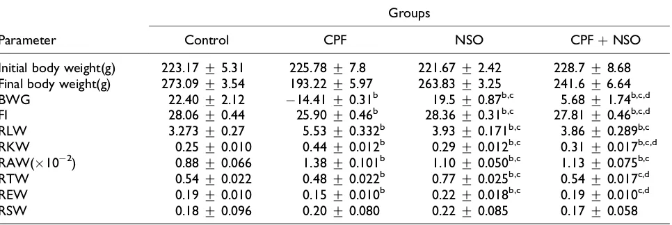
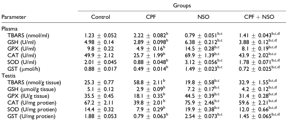
The histological investigations of testes of control (Figures 1(a) and 2(a)) and rats treated with CPF (Figures 1(b) and 2(b)), NSO (Figures 1(c) and
2(c)), and CPF þ NSO (Figures 1(d) and 2(d)) are
presented in Figures 1 and 2. Testes of CPF-treated rats showed a decrease in the number of seminifer-ous tubules along by degenerative aspect of semi-nal epithelium, vacuolization, poor/scarce sperm in lumen of some tubules, enlargement of the con-nective tissue, and depletion of a variety of cell types. Coadministration of NSO and CPF showed that NSO mitigates and modulates the structural alterations toward normal status.
Table 4.Changes in the levels of TBARS and GSH, and the activities of the antioxidant enzymes (GPX, CAT, SOD, GST) in plasma and testis after treatment of male rats with CPF, NSO, and/or their combination for 4 weeks.a
Parameter
Groups
Control CPF NSO CPFþNSO
Plasma
TBARS (nmol/ml) 1.23+0.052 2.22+0.082b 0.79+0.051b,c 1.41+0.043b,c,d
GSH (U/ml) 4.98+0.14 2.89+0.098b 6.38+0.212b,c 3.88+0.12b,c,d
GPX (U/ml) 9.8+0.22 4.9+0.16b 14.5+0.28b,c 8.1+0.19b,c,d
CAT (U/ml) 49.9+2.12 25.7+1.99b 69.9+1.39b,c 43.9+2.02b,c,c
SOD (U/ml) 2.01+0.045 0.88+0.048b 3.12+0.056b,c 1.78+0.071b,c,d
GST (mmol/h) 0.88+0.017 0.49+0.014b 1.49+0.023b,c 0.72+0.025b,c,d
Testis
TBARS (nmol/g tissue) 25.3+0.77 58.8+2.11b 19.8+0.58b,c 32.9+1.55b,c,d
GSH (mmol/g tissue) 5.1+0.12 2.9+0.09b 7.2+0.17b,c 4.2+0.12b,c,d
GPX (IU/g tissue) 35.5+0.45 18.1+0.35b 44.5+0.39b,c 31.4+0.28b,c,d
CAT (U/mg protien) 67.2+2.11 39.8+2.01b 75.9+2.46b,c 59.6+2.21b,c,d
SOD (U/mg protien) 14.4+0.32 7.9+0.29b 19.9+0.38b,c 12.0+0.66b,c,d
GST (U/mg protien) 1.88+0.053 0.79+0.063b 2.54+0.073b,c 1.45+0.065b,c,d
TBARS: thiobarbituric acid reactive substances; GSH: glutathione; GPX: glutathione peroxidase; CAT: catalase; SOD: superoxide dis-mutase; GST: glutathione-Stransferase; CPF: chlorpyrifos; NSO:Nigella sativaoil.
aValues are given as mean+SD, significance atp< 0.05. bValues differ significantly from control group.
Discussion
Chlorpyrifos
Previous studies were focused on the adverse effects of CPF on the main functional and structural aspects of the most vital organs but not on the reproductive systems, sperm quality, and fertility. Cholinesterase inhibition and oxidative stress are considered as the chief mechanism of action by which these effects may be occurring. Recent studies have identified that CPF induces oxidative stress in different organs and spe-cies of living organisms by the generation of free radi-cals which in turn can lead to protein and lipid peroxidation, DNA damage, and apoptosis, and like-wise decrease the antioxidant enzymes (SOD, CAT, and GPX) activities (Kalender et al., 2012; Saulsbury et al., 2009; Verma et al., 2007). In this study, CPF confirms to induce an increase in the levels of TBARS and a decrease in the levels of GSH, and the activities of the antioxidant enzymes (GPx, CAT, SOD, and GST) in plasma and testis (Table 4) at dose level with clear general toxicity.
The present results showed that treatment with CPF caused significant decline in semen quality (Table 2). Meeker et al. (2006), Pin˜a-Guzma´n et al. (2006), El Mazoudy et al. (2011), and Shittu et al. (2012) have found that OP compounds alter male reproductive function, particularly semen quality. Epidemiological study showed that in Chi-nese pesticide factory workers, OP exposure was associated with a decrease in sperm concentration, motility, and testosterone level and an increase inLH and higher sex chromosome aneuploidy in semen (Perry et al., 2007).
OP pesticides such as parathion, methyl parathion, dimethoate, and CPF are endocrine disrupters. In fact, they are structurally similar to various sexual mones and may interact with hormone receptors, hor-mone action, and metabolism by activation or inhibition of the enzyme activities that involved in steroid hormones synthesis and/or with induction of gene expression. This might alter the endogenous hor-mones levels and result in a deflection from normal male developmental programming and reproductive
tract growth and function (De Angelis 2009; Luccio-Camelo and Prins, 2011; Tait et al. 2009).
As a possible mechanism for the antigonadal action of OPs, they may change the concentration of neuro-transmitters, affecting the pituitary gland causing changes in gonadotropin concentrations and thus sub-sequent spermatogonic impairment (Sarkar et al., 2000; Tait et al. 2009).
In the current study, the detrimental effects of CPF on reproductive performance can be attributed to: (1) its effects on hormonal balance of hypothala-mic–pituitary–gonadal and/or –thyroid axis (FSH, LH, testosterone, T4) that are involved in the control of steroidogenesis, spermatogenesis, and spermiation as shown in Table 3, (2) its capacity to enhance the production of ROS that causes lipid peroxidation in spermatozoa membrane, sperm DNA damage and to inhibit the activities of antioxidant enzymes (Table 4), (3) to decrease the expression of testicular dogenic acute regulatory (StAR) protein and in
steroi-dogenic (3-HSD and 17-HSD) enzymes, which
help in the transfer of cholesterol in mitochondria and
testosterone biosynthesis (Manna et al., 2001). Indeed, the present results showed that the activity of 17-KSR that convert androstenedione to testoster-one was significantly decreased in rats treated with CPF compared with the control group (Table 3).
The significant decline in the absolute and relative reproductive organ weights (testes, epididymides, and seminal vesicles; Table 1), sperm and spermatid count, motility (Table 2), and testosterone level (Table 3), as well an elevation in dead and abnormal sperm rates (Table 2) are accompanied by histopatho-logical changes in testis that pronounced by altera-tions of spermatogenesis with a complete loss of all the stages of germ cell maturation, Sertoli cell toxi-city, mild to severe degenerative aspects of seminifer-ous tubules, and widening of interstitial spaces (Figures 1 and 2).
As shown in Table 3, hormone analysis showed that CPF caused significant reduction in testosterone and FT4 and induction of LH and FSH levels as com-pared to control. These may be responsible for impaired male reproductive system suggesting the
presence of CPF in rats. Rats subjected to prolonged thyroid hormone deficiency showed marked morpho-logical and functional testicular alterations. Thyroid hormone was shown to play a critical role in maintain-ing steroidogenesis, spermatogenesis, and metabolic processes in testis (Wagner et al., 2008) and increase the expression of StAR protein in the Leydig cells (Manna et al., 2001). Consistent with our results (Tables 2 and 3), it was reported that CPF induced several histopathological changes in the testes (Kalen-der et al., 2012), a depression of sperm parameters and plasma T4 level (De Angelis et al., 2009; Rawlings et al., 1998), gene expression of gonadotropin-releasing hormone in hypothalamic cell line GTI-7 (Gore, 2001), and may interfere with testosterone metabolism, potentially leading to hormonal unba-lance and poor SC (Meeker et al., 2006). In addition, antiandrogenic activity for CPF has been describedin vitro by using NIH3T3 cell line stably expressing human androgen receptor (Viswanath et al., 2010). Mariana et al. (2009) found that dimethoate (OP), gly-phosate, and zineb (carbamate), either alone or in combination induced increase in the oxidative dam-age in the plasma, liver, and testes, leading to a decrease in testosterone and an increase in the levels of FSH and LH in the treated rats, and this is a coin-cidence with the present results (Table 3). Therefore, oxidative stress may be partly responsible for the vital and reproductive organs injury and dysfunction.
N. sativa
oil
Our data (Table 1) showed that treatment with NSO alone caused significant increase in the relative weights of the reproductive organs. The coadministra-tion of NSO with CPF reversed markedly the adverse effects of CPF within control levels, and this may be due to the antioxidant activities of bioactive compo-nents ofN. sativa(Table 4). Recent studies were per-formed on the antioxidant activity of essential oil of
N. sativaand its effects on antioxidant enzyme status; it was found that TQ, carvacrol, 4-terpineol, anethol, dithymoquinone, and thymol have proper radical scavenging properties. It appears also that TQ (the major active constituent of seed oil extract) in turn exhibits cerebral, renal, liver, and cardiac protective effect against many xenobiotics through its antioxi-dant action and ability to boost antioxiantioxi-dant enzymes activities in animals (Mohamadin et al., 2010).
The results obtained in the present work indicated that treatment with NSO normalized all altered semen
parameters to the normal status levels in rats treated with CPF; again, this protective effect might be due to its antioxidant capacity (Table 4). Also NSO alone improved semen charactristics (Table 2). As a support to this data,N. sativais known for its action in ame-liorating reproductive performance in male rats (Al-Sa’aidi et al., 2009). Moreover, both the crude fixed oil ofN. sativaand TQ has been found to inhibit membrane lipid peroxidation (Kanter, 2011) and to enhance the antioxidant defense systems (Mohamadin et al., 2010).
Treatment with NSO significantly increased the
activities of 17-KSR and decreased 17-HSD
com-pared with the control. Indeed, NSO increased the process of steroidogenesis and hence testosterone pro-duction (Table 3), improving sperm proliferation (Table 2). Also the presence of NSO with CPF caused significant increase in the decline of 17-KSR com-pared with the group of CPF. The decline in the activ-ity of 17-KSR in animals treated with CPF indicates its adverse effects on the steroidogenic process for production of testosterone which in turn affects the process of fertility via decreasing testosterone hor-mone production (Table 3) and sperm production, and proliferation and hence motility, count, and abnormal-ity (Table 2).
Recent subchronic studies were performed on the possible ameliorating role of the ethanolic and aqu-eous extracts of N. sativa seeds or their fixed oil on male fertility; the results revealed a significant increase in the weight of reproductive organs, sperm motility, spermatids, and SC as well as decreased lipid peroxidation level and improved activities of antioxidant enzymes GPx, SOD, and CAT. Moreover, overall reproductive parameters showed increased testosterone and FSH levels and decreased excitation time of first mount, first ejaculation, and second trail (Al-Sa’aidi et al., 2009). El-Tohamy et al. (2010) found that N. sativa has the best promising role in improving the semen parameters and immunity as well as reducing free radicals generation more than
Raphanus sativusandEruca sativa.
of testosterone, FSH, and LH in rats. This may result from the activation of hypothalamic–pituitary–testi-cular axis, which stimulates steroidogenesis and sper-matogenesis processes (El Khasmi et al., 2011).
The histopathological examinations of testes showed that NSO coadministration alleviated evidently adverse effects induced by CPF. In accordance with our results, the histological investigation of testis and epididymis in rats treated by aqueous or alcoholic extract ofN. sativa
seeds showed an increase in the spermatogenesis activ-ity, seminiferous tubules thickness, and diameters as well as in the diameter of Leydig cells, sperm density in the lumen of seminiferous tubules, and epididymis ducts, which in turn presented higher epithelial cells (Al-Sa’aidi et al., 2009). Our data supported those obtained by Wahba (2011) who reported that pretreat-ment of male rats withN. sativa, linseed, and celery oils for 4 weeks produced a protective effect against testicu-lar injury induced by sodium valproate. This effect was manifested by increased weight of the testis, improved semen quality and quantity, elevated serum testosterone level, decreased lipid peroxidation in the testis as well as alleviation of degenerative changes in testes of rats given sodium valproate. The mechanism for this protec-tive effect of NSO against the toxic effects of CPF is due its free radical scavenging activity and increased antiox-idant enzymes in rats.
Conclusion
This study indicated that coadministration of NSO and CPF reverses changes partly or completely in the relative reproductive, vital organs weights, semen characteristics, hormone levels, oxidative damage, antioxidant enzymes, and the histopathological inju-ries of testes within the normal status and thereby improved semen quality. Also, NSO can improve semen picture and moderate CPF-induced reproduc-tive toxicity by its antioxidant properties.
Highlights
OP compounds such as CPF induced
reproduc-tive toxicity.
Oxidative stress is the main mechanism of
adverse effects.
The oxidative stress is known to be a key factor in several diseases.
Herbalism is an alternative or folk medicine for medicinal purposes.
NGO moderates CPF-induced reproductive
toxicity.
Conflict of interest
The authors declared no conflicts of interest.
Funding
This research received no specific grant from any funding agency in the public, commercial, or not-for-profit sectors.
References
Ait Mbarek L, Ait Mouse H, Elabbadi N, et al. (2007) Anti-tumor properties of black seed (Nigella sativa L.) extract. Brazilian Journal of Medical and Biological Research40: 839–847.
Al-Sa’aidi JAA, Al-Khuzai ALD and Al-Zobaydi NFH (2009) Effect of alcoholic extract of Nigella sativa on fertility in male rats. Iraqi Journal of Veterinary Sciences23(2): 123–128.
Amann RP, Johnson L, Thompson DL, et al. (1976) Daily spermatozoal production, epididymal spermatozoal reserves and transit time of spermatozoa through the epididymis of the rhesus monkey.Biology of Reproduc-tion15: 586–592.
Ambali SF, Akanb DO, Oladipo OO, et al. (2011) Subchro-nic chlorpyrifos-induced cliSubchro-nical, hematological and biochemical changes in Swiss albino mice: protective effect of vitamin E.International Journal of Biological and Medical Research2(2): 497–503.
Asperlin A (1994)Pesticide industry sales and usage-1992 and 1993 market estimates. Report no. 733-K-94-001. Washington DC: US Environmental Agency.
Beitens IZ, O’loughlin K, Ostrea T, et al. (1976) Gonado-tropin determination in timed 3-hour urine collections during menstrual cycle and LHRH testing.The Journal of Clinical Endocrinology and Metabolism43: 46–55. Blazak WF, Treinen KA and Juniewicz PE (1993) Male
reproductive toxicology, part A, In: Chapin RE, Hein-del JJ (eds)Methods in Toxicology. San Diego, Calif: Academic Press, pp. 86–94.
Burits M, Bucar F (2000) Antioxidant activity of Nigella sativaessential oil.Phytotherapy Research14: 323–328. Chiu DTY, Stults FH and Tappel AL (1976) Purification and properties of rat lung soluble glutathione peroxi-dase.Biochimica et Biophysica Acta445: 558–566. De Angelis S, Tassinari R, Maranghi F, et al. (2009)
Devel-opmental exposure to chlorpyrifos induces alterations in thyroid and thyroid hormone levels without other toxi-city signs in Cd1 mice.Toxicological Sciences108(2): 311–319.
El Khasmi M, Issaoub-Allah A, Farh M, et al. (2011) Effect of Nigella sativa fixed oil on the hormonal profile of androgens and circulating in male rats. Phytothe´rapie 9(6): 338–342.
El Mazoudy RH, Attia AA and El-Shenawy NS (2011) Pro-tective role of propolis against reproductive toxicity of chlorpyrifos in male rats. Pesticide Biochemistry and Physiology101: 175–181.
El-Tohamy MM, El-Nattat WS and El-Kady RI (2010) The beneficial effects of Nigella sativa, Raphanus sativus andEruca sativaseeds cakes to improve male rabbit fer-tility immunity and production. Journal of American Science6(10): 1247–1255.
Figa-Talamanca I, Traina ME and Urbani E (2001) Occu-pational exposures to metals, solvents and pesticides: recent evidence on male reproductive effects and biolo-gical markers. Occupational Medicine London 51: 174–188.
Giwercman A, Giwercman YL (2011) Environmental fac-tors and testicular function. Best practice and research. Clinical Endocrinology and Metabolism25: 391–402. Gore AC (2001) Environmantal toxicants effects on
neu-roendocrine function.Endocrinology14: 235–246. Gultekin F, Delibas N, Yasar S, et al. (2001) In vivo
changes in antioxidant systems and protective role of melatonin and a combination of vitamin C and vitamin E on oxidative damage in erythrocytes induced by chlorpyrifos-ethyl in rats. Archive of Toxicology 75: 88–96.
Habig WH, Pabst MJ and Jakoby WB (1974) Glutathione-S transferases. The first enzymatic step in mercapturic acid formation. The Journal of Biological Chemistry 249: 7130–7139.
Ji G, Gu A, Wang Y, et al. (2012) Genetic variants in anti-oxidant genes are associated with sperm DNA damage and risk of male infertility in a Chinese population.Free Radical Biology and Medicine52(4): 775–780. Jollow DJ, Mitchell JR, Zampaglione N, et al. (1974)
Bromobenzene-induced liver necrosis, protective role of glutathione and evidence for 3,4-bromobenzene oxide as the hepatotoxic metabolite. Pharmacology 11: 151–169.
Kalender Y, Kaya S, Durak D, et al. (2012) Protective effects of catechin and quercetin on antioxidant status, lipid peroxidation and testis-histoarchitecture induced by chlorpyrifos in male rats.Environmental Toxicology and Pharmacology33(2): 141–148.
Kanter M (2011) Thymoquinone reestablishes spermato-genesis after testicular injury caused by chronic toluene exposure in rats. Toxicology and Industrial Health 27(2): 155–166.
Katryna B, Anita PP (1980) Purification of rat testicular microsomal 17-ketosteroid reductase, evidence that 17-ketosteroid reductase and 17hydroxysteroid dehy-drogenase are distinct enzymes.The Journal of Biologi-cal Chemistry255: 5552–5559.
Khader M, Bresgen N and Eckl PM (2009)In vitro toxico-logical properties of thymoquinone.Food and Chemical Toxicology47: 129–133.
Linder RE, Klinefetter GR and Strader LF (1995) Dibro-moacetic acid affects reproductive competence and sperm quality in the male rats. Fundamental and Applied Toxicology28: 9–17.
Liobet JM, Colomina MT and Sirvent JJ (1995) Reproduc-tive toxicology of aluminum in male mice.Fundamental and Applied Toxicology25: 45–51.
Lowry OH, Rosebrough NJ, Farr AL, et al. (1951) Protein measurement with the Folin phenol reagent.The Journal of Biological Chemistry193: 269–275.
Luccio-Camelo DC, Prins GS (2011) Disruption of andro-gen receptor signaling in males by environmental che-micals. The Journal of Steroid Biochemistry and Molecular Biology127: 74–82.
Luck H (1974) Catalase. In: Bergmayer MV (ed)Method of Enzymatic Analysis. New York: Verlag Chemic Academic Press, p. 885.
Manna PR, Kero J, Tena-Sempere M, et al. (2001) Assess-ment of mechanisms of thyroid hormone action in mouse leydig cells: regulation of the steroidogenic acute regula-tory protein, steroidogenesis, and luteinizing hormone receptor function.Endocrinology142(1): 319–331. Mantovani A (2006) Risk assessment of endocrine
disrup-ters: the role of toxicological studies.The Annals of the New York Academy of Sciences1076: 239–252. Mantovani A, Maranghi F (2005) Risk assessment of
che-micals potentially affecting male fertility. Contracep-tion72: 308–313.
Mariana A, De Alaniz MJT and Marra CA (2009) The impact of simultaneous intoxication with agrochemicals on the antioxidant defense system in rat.Pesticide Bio-chemistry and Physiology94: 93–99.
Meeker JD, Ryan L, Barr DB, et al. (2006) Exposure to nonpersistent insecticides and male reproductive hor-mones.Epidemiology17(1): 61–68.
Misra HP, Fridovich I (1972) The role of superoxide anion in the autoxidation of epinephrine and a simple assay for superoxide dismutase.The Journal of Biological Chem-istry247(10): 3170–3175.
Perry MJ, Scott AV, Dana BB, et al. (2007) Environmental pyrethroid and organophosphates insecticide exposures and sperm concentration. Reproductive Toxicology23: 113–118.
Pin˜a-Guzma´n B, Solı´s-Heredia MJ, Rojas-Garcı´a AE, et al. (2006) Genetic damage caused by methyl–parathion in mouse spermatozoa is related to oxidative stress. Toxi-cology and Applied PharmaToxi-cology216: 216–224. Rawlings NC, Cook SJ and Waldbillig D (1998) Effects of
the pesticides carbofuran, chlorpyrifos, dimethoate, lin-dane, triallate, trifluralin, 2,4-D, and pentachlorophenol on the metabolic endocrine and reproductive endocrine system in ewes.Journal of Toxicology and Environmen-tal Health A54(1): 21–36.
Santner S, Santner R, Kulin H, et al. (1981) A model vali-dation of radioimmuno-assay kit reagents: measurement of follitropin and lutrupin in blood and urine. Clinical Chemistry27: 1892–1895.
Sarkar R, Mohanakumar KP and Chowdhury M (2000) Effects of an organophosphate pesticide, quinalphos, on the hypothalamo–pituitary–gonadal axis in adult male rats.Journal of Reproduction and Fertility18: 29–38. Saulsbury MD, Heyliger SO, Wang K, et al. (2009)
Chlor-pyrifos induces oxidative stress in oligodendrocyte pro-genitor cells.Toxicology259: 1–9.
Sharma NK, Ahirwar D, Jhade D, et al. (2009) A review: medicinal and phamacological potential of Nigella sativa.Ethnobotanical Review13: 946–955.
Shittu M, Ayo JO, Ambali SF, et al. (2012) Chronic chlorpyrifos-induced oxidative changes in the testes and pituitary gland of Wistar rats: ameliorative effects of vitamin C. Pesticide Biochemistry and Physiology 102: 79–85.
Tait S, Ricceri L, Venerosi A, et al. (2009) Long-term effects on hypothalamic neuropeptides after develop-mental exposure to chlorpyrifos in mice.Environmental Health Perspectives117(1): 112–116.
Tappel AL, Zalkin H (1959) Inhibition of lipid peroxida-tion in mitochondria by vitamin E.Archives of Biochem-istry and Biophysics80: 333–336.
Verma RS, Mehta A and Srivastava N (2007)In vivo chlor-pyrifos induced oxidative stress: attenuation by antioxi-dant vitamins. Pesticide Biochemistry and Physiology 88: 191–196.
Viswanath G, Chatterjee S, Dabral S, et al. (2010) Anti-androgenic endocrine disrupting activities of chlorpyri-fos and piperophos.The Journal of Steroid Biochemistry and Molecular Biology120: 22–29.
Wagner MS, Wajner SM and Maia AL (2008) The role of thyroid hormone in testicular development and function. Journal of Endocrinology199: 351–365.
Wahba HMA (2011) Protective effect of Nigella Sativa, linseed and celery oils against testicular toxicity induced by sodium valproate in male rats.Journal of American Science7(5): 687–693.
World Health Organization (WHO) (2008) Traditional medicine.Fact sheet 134. Geneva: WHO.
Yaman I, Balikci E (2010) Protective effects of Nigella sativaagainst gentamicin induced nephrotoxicity in rats. Experimental and Toxicologic Pathology62: 183–190. Yousef MI (2004) Protective role of ascorbic acid to enhance reproductive performance of male rabbits treated with stannous chloride.Toxicology207: 81–89. Yousef MI (2010) Vitamin E modulates reproductive toxi-city of pyrethroid lambda-cyhalothrin in male rabbits. Food and Chemical Toxicology48: 1152–1159. Yousef MI, Salama AF (2010) Propolis protection from
reproductive toxicity caused by aluminium chloride in male rats. Food and Chemical Toxicology 47: 1168–1175.
Yousef MI, Abdallah GA and Kamel KI (2003a) Effect of ascorbic acid and vitamin E supplementation on semen quality and biochemical parameters of male rabbits. Animal Reproduction Science6: 99–111.
Yousef MI, El-Demerdash FM and Al-Salhen KS (2003b) Protective role of isoflavones against the toxic effect of cypermethrin on semen quality and testosterone lev-els of rabbits. Journal of Environmental Science and HealthB38: 463–478.
Yousef MI, El-Morsy AMA and Hassan ME (2005) Aluminium-induced deterioration in reproductive per-formance and seminal plasma biochemistry of male rab-bits: protective role of ascorbic acid. Toxicology 215: 97–107.
Yousef MI, Ismail AM and Baghdadi HH (2004) Effect of isoflavones on reproductive performance, testosterone levels, lipid peroxidation and seminal plasma biochem-istry of male rabbits.Journal of Environmental Science and HealthB39: 819–833.
Yousef MI, El-Demerdash FM, Kamil KI, et al. (2006) Ameliorating effect of folic acid on chromium(VI)-induced changes in reproductive performance and semi-nal plasma biochemistry in male rabbits.Reproductive Toxicology21: 322–326.
Yousef MI, Kamil KI, El-Guendi MI, et al. (2007) Anin vitrostudy on reproductive toxicity of aluminium chlor-ide on rabbit sperm: the protective role of some antiox-idants.Toxicology239: 213–223.