Orthodontic treatment with skeletal anchorage system
(Case report)
Arya Brahmanta *, Jusuf Sjamsudin **
* Department of Orthodontics Faculty of Dentistry University of Hang Tuah Surabaya ** Department of Orthodontics Faculty of Dentistry University of Airlangga
Abstract
Background: The correction of Class I malocclusion with bimaxillary dental protrusion and unilateral free end right upper ridge in adult patient is one of difficult
biomechanical case in orthodontics. Because this case needs proper anchorage for upper
incisor retraction with missing teeth in the right posterior segment Purpose: This article presented a case of an adult patient with severe bimaxillary protrusion treated with Skeletal
Anchorage System (SAS). Case: A female patient, 36 year old complaining for the difficulty of lip closure due to severe bimaxillary protrusion with incompetence lip. Case Management: Firstly correction of the maxillary and mandibular incisor proclination were done by extraction of the mandibular first premolar, the maxillary second premolar on left
side and finally placement of miniplates implant in the zygomatic process on right side as an
absolut anchorage. Conclusion: The results of this treatment indicated that Skeletal Anchorage System (SAS) can be considered as an effective therapy choice for corection
bimaxillary protrusion with unilateral free end ridge.
Key words : Skeletal Anchorage System (SAS), bimaxillary protrusion, unilateral free end ridge
Abstrak
35 tahun datang dengan keluhan utama kesulitan untuk menutup mulut oleh karena gigi rahang atas dan rahang bawahnya maju dan bibirnya tidak kompeten. Manajemen kasus: Koreksi pada gigi insisivus rahang atas dan insisivus rahang bawah yang protrusi dilakukan dengan melakukan pencabutan terlebih dahulu pada gigi premolar pertama dirahang bawah sisi kanan dan sisi kiri serta pencabutan pada gigi premolar kedua dirahang atas sisi kiri dan pemasangan miniplate implant di regio prosesus zigomatikus di sisi kanan sebagai penjangkar absolut. Kesimpulan : Hasil dari perawatan ini menunjukkan bahwa sistem penjangkar absolut pada perawatan ortodonti merupakan pilihan terapi perawatan yang efektif pada kasus penderita dewasa dengan protrusi bimaksiler dan kehilangan gigi posterior pada regio kanan atas.
Correspondence : Arya Brahmanta, Bagian Ortodonti Fakultas Kedokteran Gigi Universitas Hang Tuah. Jl. Arif Rahman Hakim 150 Surabaya. Telp. (031) 5912191.
Email: [email protected] orthodontics treatment generally depend on the anchorage protocol, planned for the particular case. To prepare good anchorage, the clinician must be realistic to predict the possibility of anchorage loss. Anchorage loss is a consequences from unstable construction and lack of patient cooperation as well. Type of anchorage based on the type of tooth movements 1,2.
Anchorage has been one of the greatest problems in the field of orthodontic. Orthodontists always faced with difficulties in trying to achieve maximal anchorage due to the orthodontic movements in response to orthodontic forces. Therefore, in maximal anchorage case, patients need to using headgear as additional anchorage. Reinforced anchorage with extraoral appliances has severe limitations because it requires excellent patient compliance 2,3.
made it possible to use implants for orthodontic anchorage. Since the implant is known like an ankylosed tooth, it can be used as a reliable anchorage unit for orthodontic tooth movements. It is reported experimental biomechanic studies on animal models and clinical investigations shown that dental implants placed in the alveolar bone is resistant to orthodontic force 3.
In correction of severe maxillary protrusion it is important to achieve maximal anchorage especially in cases with excesive molar anchorage loss on free end ridge. Skeletal Anchorage System (SAS) was developed for correcting Class II malocclusions with maxillary protrusion. Using this system, the anterior retraction without unfavorable side effects became possible 4.
The purpose of this article is to deliver a case of an adult patient with severe bimaxillary protrusion, treated with Skeletal Anchorage System (SAS).
CASE REPORT
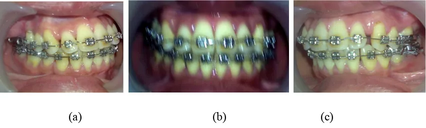
A patient 36 year old woman, presented a bimaxillary dental protrusion with class I malocclusion and unilateral free end ridge on upper arch came to the orthodontic specialist clinic Airlangga university dental hospital. She complaining about the difficulty of lip closure due to severe bimaxillary dental protrusion. Her facial profile was convex with a protrusive upper lip and no facial asymmetry. Over jet and over bite 2 mm (Figure 1 b). Occlusal contact recognized only at the premolar and molar on the left side (Figure 1 c). No occlusal contact at premolar and right molar. The upper left first molar, second molar and lower first molar were missing (Figure 1 a,e).
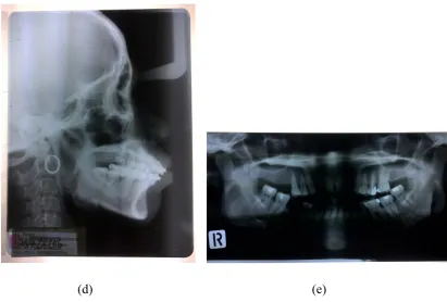
(d) (e)
Figure 1. Intra oral photographs (a) right side, (b) front side, (c) left side, (d) Cephalometric and (e) Panoramic photographs before treatment
Cephalometric analysis showed a skeletal class I jaw base relationship SNA 88º; SNB 84º; ANB 4 º. The facial profile was convex FH-NP 88º; NAP10 º ; Y-Axis 62,5º; The upper and lower incisor were labially inclined I -NA line 11,5 mm; I- NA angle 35 º ; I- NB line 16 mm ; I- NB angle 45 º ; Interinsisal angle 96º. The mandibular plane angle was steep 30 º and the gonial angle was large 110 º ( Figure 1 d ).
CASE MANAGEMENT
A patient diagnosed with Angle Class I malocclusion with bimaxillary dental protrusion and unilateral free end right upper ridge, skeletal class I jaw base relationship.
Before starting orthodontic treatment, the patient received periodontal treatment. Periodontal treatment involved oral hygiene instruction and scaling. The upper left second molar was extracted because of poor condition caries. Bilateral mandibular lower first premolar were extracted to gain space for retraction.
Firstly levelling with 0.012 inch round NiTi archwire was initiated. After leveling with a 0.016 inch NiTi arch wire, the miniplates implanted onto the zygomatic process of the maxilla through buccal mucosa (Figure 2 a,b). The miniplates contoured to fit the bone surface. The head portion of miniplates intraorally exposed and positioned outside the dentition (Figure 2 c).
After a month for healing, integration and adaptation retraction of the anterior teeth was started with elastic chain. An elastic chains was applied from the upper right premolar region to the miniplate as absolute anchorage for retraction with sliding mechanic (Figure 3 a, b, c).
(a) (b) (c)
Figure 2. (a) Miniplates implant placement, (b) miniplates implant, (c) suturing
(a) (b) (c)
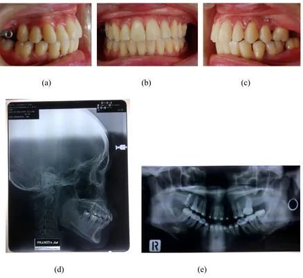
The results of this treatment showed space in the upper dentition were closed. The upper incisor inclined palatally and the lower incisor lingually inclined. Acceptable occlusion achieved and the overjet and overbite come to normal. The caninus relation were Class I on the both sides (Figure 4 a, b, c).
(a) (b) (c)
(d) (e)
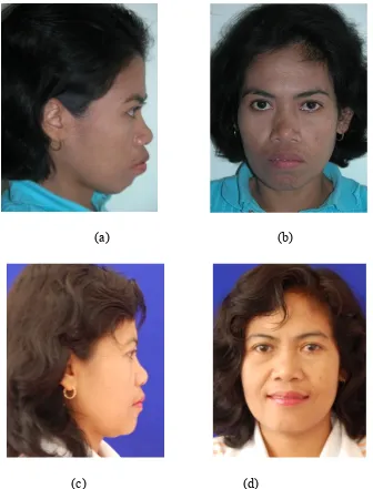
(a) (b)
(c) (d)
Figure 5. Facial photographs (a) right side, (b) front side before treatment. (c) right side, (d) front side after treatment
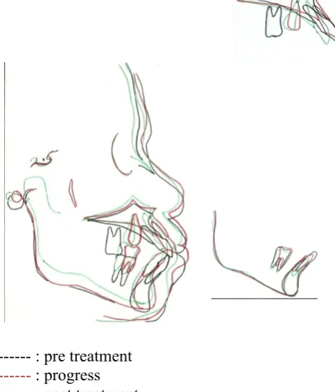
--- : pre treatment
--- : progress --- : post treatment
Figure 6. Superimposing cephalometric tracing
Cephalometric superimposing analysis showed a normal SNA 86º; SNB 84º; ANB 2 º . The facial profile was becoming straight FH-NP 85º; NAP10 º ; Y-Ax 62,5º; The upper and lower incisor have been corected I -NA line 7 mm;21 º ; I- NB line 12 mm; 40 º ; Inter Insisal 114º . Comparing the superimposing pretreatment and posttreatment cephalometric tracings showed maxillary and mandibular incisor crown had moved posteriorly (Figure 6).
(a) (b) (c)
During retention with Hawley retainer acceptable occlusion and facial profile also maintained, indicating a stability of the occlusion (Figure 7).
DISCUSION
Facial esthetics as a major concern of many orthodontic patients. The negative impacts on the facial profile with upper lip protrusion often lead patients to seek orthodontic treament. Increased upper lip procumbency is commonly associated with protrusive maxillary dentition in Angle Class II division 1 malocclusions and Class I malocclusions with bimaxillary protrusion 2.
In such circumtances, the major orthodontic treatment goal is to reduce the proclination of the maxillary incisors. Consequently, the treatment plan often includes extraction of the bilateral maxillary premolars, followed by retraction of the anterior teeth with maximum anchorage. Maximum anchorage was added to prevent forward movement of the maxillary posterior teeth during anterior teeth retraction and can be provided with different approaches 5,6.
Extraoral headgear appliances are commonly used to reinforce posterior anchorage during anterior tooth retraction or are directly applied to retract anterior teeth. Wearing headgear full time is demanding for most patients, extraoral appliances are often rejected by adults for social reasons. Patients cooperation is an important factor for the effectiveness of extraoral appliances 6,7.
Application of bony anchorage for tooth movements are more effficiently, because not depending on patient cooperation in wearing extraoral appliance. Several method of bone anchorage have been reported, are : dental implants, titanium screws and miniplates. The use of the miniplate implant for absolute anchorage has proved to have many advantages. Absolute anchorage leads to more reliable treatment plan and enables a reduction in the treatment time. This anchorage system obviates the dependency on patient compliance 5,6 8.
In this case, orthodontic treatment was performed in adult patient diagnosed as Angle Class I malocclusion with bimaxillary dental protrusion and unilateral free end upper right posterior segment. The only option to correct the proclination of anterior teeth was to move the anterior teeth to the distal is use of absolute anchorage. Therefore, skeletal anchorage system (SAS) offered the best benefit therapy choice. Miniplates were placed in the zygomatic process in the maxilla. The titanium L – shaped (hook of miniplates) facilitated adjustment of the direction of force to retract the upper incisors 9,10.
Patients with bimaxillary dental protrusion have specific characteristics, including incisor proclination and facial profile. To correct dentoalveolar protrusion, extraction of the premolar is indicated. The treatment mechanic for space closure of the extraction sites by anterior tooth retraction was closed by sliding mechanics. The use of miniplates as skeletal anchorage system for patient with insufficient teeth for anchorage is almost 100% succesfull,if the right type of implant selected and the clinical situation properly evaluated. 10,11,12
.
The type of retraction movement of the maxillary incisor can assesed by comparing the superimpose pre treatment and post treatment on cephalometric tracings. The tipping control assigned by maxillary incisor crown moved posteriorly with the center of rotation at the root of the tooth. With uncontrolled tipping, the maxillary central incisor crown moved posteriorly either the root moved anteriorly. The mechanotherapy control were important for satisfactory correction of dentoalveolar protrusion, leading to a positive soft - tissue response, with reductions of lip protrusion 11,12.
The patient’s main complaints, in which difficulty of lip closure due to severe bimaxillary dental protrusion with incompetence lip was improved by the treatment. Since the proclination was corected, the upper lip became more relaxed and the lips showed less tension.
REFERENCE
1. Keles A, Erverdi N. Bodily molar distalization with absolute anchorage. Angle Orthod 2003;73:471- 82.
2. Nanda R. Biomechanics and esthetic strategies in clinical orthodontics. St.Louis, Saunders Elsevier 2005; 295-309.
3. Sugawara J. JCO interviews, Dr. Junji Sugawara on the skeletal anchorage system. J Clin Orthod 2000;33:689 - 96.
4. Tanaka E, Sasaki-Nishi A, Hasegawa T, Nishio C, Nobuhiko K, Tanne K. Skeletal anchorage for orthodontic correction of severe maxillary protrusion after previous orthodontic treatment. Angle Orthod 2008;78: 181- 88
5. Sugawara J, Nagasaka H, Umemori M, Mitani H, Kawamura H. Skeletal Anchorage System (SAS). Dental Outlook 2002;99:397-406
6. Lee J S. Application of Orthodontic Mini – Implants. Hanover Park, Quintessence 2007; 7,179,202-06
7. Fukunaga T, Kuroda S, Kurosaka H, Yamamoto T. Skeletal anchorage for
orthodontic correction of maxillary protrusion with adult periodontitis. Angle Orthod 2006;76: 148 - 55
8. Wahl N. Orthodontics in 3 millennia. Chapter 15 : Skeletal anchorage.American Journal of Orthod and Dentofacial Orthopedics 2008;134; 707- 10
9. Bong KC, Dong SC, Jang I, Brinkmann J, Peter Ngan. Maxillary protraction using miniplates as skeletal anchorage. Hong Kong Dent J 2010;7:87-93
10.Yao CJ, Hua lai EH, Chang C, Chen I. Comparison of treatment outcomes between skeletal anchorage and extraoral anchorage in adults with maxillary dentoalveolar protrusion. Am J Orthod Dentofac Orthop 2008;134:615 – 24. 11.Labanauskaite B, Jankauskas G, Vasiliauskas A, Haffar N. Implants for
orthodontic anchorage. Meta – analysis. Stomatologija Baltic and maxillofacial Journal 2005;7:128- 32
12.Lee K, Leung C, Wong K, Rabie. Versatility of skeletal anchorage in orthodontics. World Journal of orthodontics. 2007;9:221-32.