VOL. 7, NO. 2, pp. 102 - 107, April 2017 Submitted June 2016; Revised October 2016; Accepted January 2017
Role of Antibody Anti-AGE on the Expression of Nephrin and Rage in Primary Glomerulus Cell
Culture Exposed to AGE
Rudy Salam 1
*, Diana Lyrawati , Nursamsu 2
1
Department of Pharmacy, Faculty of Medicine, University of Brawijaya, Malang, Indonesia
2 Division of Nephrology and Hypertension, Department of Internal Medicine, Dr. Saiful Anwar Public Hospital, Malang, Indonesia
ABSTRACT
Nephrin is associated with the initial stage of the loss of the permeability barrier in diabetic nephropathy. Interaction AGERAGE in creases angiotensin II on Renin AngiotensinAldosterone System (RAAS) and activation of protein kinase c (PKC) which induce al -terations in nephrin mRNA expression. Al-terations of nephrin expression induce transformation of slit membrane structure and the permeability changes at the glomerular filtration barrier. Anti-AGE vaccination once may cause the changes of nephrin and RAGE expression and can prevent progression of diabetic nephropathy. This study used primary glomerulus cell culture obtained from renal of Wistar rat aged 3 months, weighing 200300 grams and assigned into negative control group that exposed to BSA 100 µg/mL, pos -itive control group that exposed to AGE-BSA 100 µg/mL, treatment group 1 that exposed to polyclonal anti-AGE antibody 5 µg/mL and AGE-BSA 100 µg/mL and treatment group 2 that exposed to monoclonal anti-CML antibody 5 µg/mL and AGE-BSA 100 µg/mL. Paired ttest with a 0.05 level of confidence results showed that there were significantly different v in level of RAGE expres -sion between experimental groups with control groups. Administration of polyclonal anti-AGE antibody decreased RAGE expres-sion compared to negative control (p = 0.188) and positive control (p = 0.000). RAGE expression did not differ significantly in administra tion of monoclonal antiCML antibody compared to negative control but significant with positive control. Administration of mono -clonal anti-CML antibody inhibited increasing of nephrin expression compared to negative and positive control (p = 0.73; 0.125). In conclusion, this study suggested that administration of polyclonal antiAGE or monoclonal antiCML antibody could inhibit increas -ing of RAGE and nephrin expression in glomerulus primary culture that exposed to AGE which is expected to prevent the progres-sion of diabetic nephropathy.
Keywords: Anti-AGE antibody, AGE, RAGE, nephrin, primary glomerulus cell culture
Diabetic nephropathy is one of diabetic mellitus complication leading to thickening of glomerular basal membrane, glomerular hypertrophy and mesangial ex-pansion [1]. The pathogenesis of diabetic nephropathy involve various mechanism and include hyperglycaemic condition, polyol pathway activation, renin-angiotensin system, reactive oxygen species (ROS), activation of protein kinase C (PKC) pathway, increase of advanced glycation end-product (AGE) and glomerular hyperfil-tration [1, 2]. Interaction of extracellular AGE with Re-ceptor for Advanced Glycation End Products (RAGE) increases angiotensin II on Renin Angiotensin-Aldos-terone System (RAAS) and activation of protein kinase
c (PKC) which induce alterations in nephrin mRNA expression [3, 4].
Nephrin is required for renal development process for podocyte maturation and formation of SD [5]. Downregulation of nephrin expression occurred on glomerular disease condition. Interestingly, upregula-tion of nephrin expression has been reported at early stage of glomerular injury and decreased at late stages of nephropathy (follow-up period up to 6 months us-ing STZ model) [1]. Activation of PKC causes substan-tial increase of nephrin mRNA and protein expression [6].
Blocking AGE by amino guanidine, pyridoxamine, alagebrium and monoclonal antibody anti-TGF-β con-INTRODUCTION
*Corresponding author: Rudy Salam
Department of Biomedicine, Faculty of Medicine, Brawijaya University,
Jalan Veteran, Malang, Indonesia 65145 E-mail: rudy_salam@ub.ac.id
How to cite:
tinuously could protect diabetic patients from glomeru-losclerosis and renal failure [7, 8]. Such treatment is costly if applied lifetime to manage diabetic vascular complications. Anti-AGE vaccination once may inhibit diabetic complication progression. AGE consist of gly-cation protein antigenic properties which could be used to develop antibody [9]. Administration of human RAGE antibody increases survival and cytoskeleton dy-namicity of podocyte [10]. Anti-AGE antibody induces formation of immune complex with AGE. The correla-tion of decreasing AGE level with increasing of im-mune complex in vascular circulation indicate the role of anti-AGE antibody in decreasing of AGE level by in-hibits signalling activation of factors that causes DN [11]. However, the role of antibody anti-AGE in nephrin and RAGE expression is still unclear whether upregulation or downregulation. The aims of this study were to examine the effects of anti-AGE antibody on RAGE and nephrin expression on primary glomerulus cells culture after incubated with AGE.
Primary glomeruli cell culture
Primary glomeruli cell culture obtained from renal of Wistar rat aged 3 months, weighing 200-300 grams from Laboratory Bioscience University of Brawijaya. Antibody anti-AGE used Anti-Carboxymethyl Lysine/ Anti-CML (Circulex, cy-m1028) and polyclonal anti-body anti-AGE (Abcam, ab23732). Antianti-body for RAGE used monoclonal antibody anti-RAGE (Circulex, cy-m1038) and nephrin used monoclonal antibody anti-nephrin (Bioss, bs-0513r).
Kidney male Wistar rat at ages 3 months (Labora-tory Bioscience University of Brawijaya) were dissected and cut into small pieces (1–2 mm cubes) with a surgi-cal blade in PBS solution. The tissues were digested in collagenase solution containing 1 mg/mL collagenase A (Roche Diagnostics GmbH, Mannheim, Germany) and 0.2 mg/mL deoxyribonuclease I (Roche Diagnostics GmbH) in Hanks’ Balanced Salt Solution at 37°C for 60 min. The collagenase-digested tissues were gently pressed through a 100 mm cell strainer (BD Bio-sciences, Stockholm, Sweden) using a flattened pestle. Digested tissues was sentrifugated with 800 rpm for 4 min. Supernatant was removed out and collected pellet was resuspended using deionized water.
Resuspended pellet were cultured in culture dishes or glass coverslips (Asahi techno glass, Tokyo, Japan) using RPMI 1 × medium that’s containing 5% fetal bovine serum (Cansera International, Canada) supple-mented with 1% Insulin–Transferrin–Selenium-A,
liq-uid media supplement (Invitrogen), 100 U/ml peni-cillin, and 100 mg/mL streptomycin. Cultures were in-cubated in a 37°C humidified incubator with 5% CO2.
Treatment monoclonal antibody Anti-CML and poly-clonal antibody Anti-AGE
Primary glomerulus cells incubated for 48 hours and they were divided into 4 groups. Group 1 were treated with polyclonal anti-AGE antibody 5 µg/mL and AGE-BSA 100 µg/mL. Group 2 were treated with monoclonal anti-CML antibody 5 µg/mL and AGE-BSA 100 µg/mL. For negative control, cells at group 3 were treated with BSA 100 µg/mL and AGE-BSA 100 µg/mL for positive control group. Group 1 and group 2 were incubated for 30 minutes after the administra-tion of antibody, followed by administraadministra-tion of AGE-BSA then incubated for 24 hours. Negative control was incubated for 24 hours after the administration of BSA and positive control incubated for 24 hours after ad-ministration of AGE-BSA. Analysis of nephrin and RAGE expression was performed after 24 hours treat-ment.
Immunofluorescence microscopy
After treatment, primary glomeruli cells culture was fixed in 2% paraformaldehyde in PBS for 10 minutes, permeabilized with 0.3% Triton X-100 in PBS for 2 minutes, and stained with antibodies. Rabbit anti-nephrin mouse and mouse anti-RAGE antibody was applied as primary antibodies for double labelling. Af-ter washing with PBS, the specimens were stained with Goat anti-rabbit IgG-FITC (Santa Cruz; sc-2012) and Rabbit anti-mouse IgG-R (Santa Cruz; sc-2092), re-washed with PBS, and subsequently reacted. Im-munofluorescences of the specimens were observed with a laser scanning confocal microscope (MRC-1024; Bio-Rad Laboratories). Visualization of expression of nephrin and RAGE was performed on three fields of view of each slide. Fluorescent density of nephrin and RAGE were measured using image J version 1.49.
Statistical analysis
All data were analysed by SPSS 20.0 software and expressed as mean ± standard deviation (SD). The sig-nificance of difference was determined by paired t-test. A value of p > 0.05 was considered statistically signifi-cant.
Average values of RAGE expression in negative and positive control groups were 262,923.50 ± 29,997.98
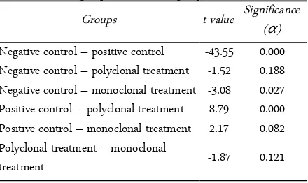
Table 1. Results of paired t-test analysis of RAGE expression among control groups and treatment groups
Groups t value Significance (α)
Negative control – positive control -43.55 0.000 Negative control – polyclonal treatment -1.52 0.188 Negative control – monoclonal treatment -3.08 0.027 Positive control – polyclonal treatment 8.79 0.000 Positive control – monoclonal treatment 2.17 0.082 Polyclonal treatment – monoclonal
treatment -1.87 0.121
Table 2. Results of paired t-test analysis of nephrin expression among control groups and treatment groups
Groups t value Significance (α)
Negative control – positive control 13.59 0.000 Negative control – polyclonal treatment 2.14 0.086 Negative control – monoclonal treatment 2.27 0.073 Positive control – polyclonal treatment 11.21 0.000 Positive control – monoclonal treatment 1.84 0.125 Polyclonal treatment – monoclonal
treat-ment 2.68 0.044
and 942,532.33 ± 19,081.42; 363,53 ± 158,126.25. RAGE expression on experimental groups after treated by AGE continued with antibody polyclonal and mon-oclonal were 363,528.67 ± 158,126.25 and 654,396.83 ± 325,322.83.
Table 1 showed the results of paired t-test of treat-ment groups with control groups with a 0.05 level of confidence and the results showed that there were sig-nificant differences in level of RAGE expression. Ad-ministration of polyclonal antibody decreased RAGE expression among negative control (p = 0.188) but not in positive control (p = 0.000). In contrast to mono-clonal anti-AGE antibody, RAGE expression had no different significantly compared to negative control but significant than positive control. This result indicated that antibody anti-AGE blocked expression of RAGE.
Nephrin expressions in negative control group were 284,514.67 ± 52,644.92 and 615,802.00 ± 10,390.73 for positive control group. Average values of nephrin ex-pressions in polyclonal AGE and monoclonal anti-CML antibody treatment groups were 205544.00 ± 86,150.45 and 451,740.17 ± 214,140. Completely paired t-test of nephrin expression results showed in Table 2. Administration of monoclonal anti-AGE antibody
in-hibited decreasing of nephrin expression compared to negative and positive control (p = 0.73; 0.125). Nephrin expressions in polyclonal anti-AGE antibody treatment groups were significantly different compared to nega-tive control groups (p < 0.05) in contrast with posinega-tive control. This result showed that nephrin expressions inhibited by administration of polyclonal anti-AGE or monoclonal anti-CML antibody.
The role of antibody anti-AGE in nephrin and RAGE expression is still unclear whether increasing or decreasing. To examine the effects of AGE anti-body treatment on RAGE and nephrin expression on primary glomerulus cells culture, we exposed cultured glomerulus primary cells with anti-AGE antibody and AGE. In normal condition, podocytes and glomerular endothelial cells, among other renal cell types express RAGE [12]. Interaction of AGE-RAGE induce the ac-tivation of inflammatory signalling [10]. Signalling pathways which activated by AGE-RAGE are ERK (ex-tracellular signal-regulated kinase)1/2, p38 MAPK (mi-togen-activated-protein-kinase)-JNK (c-Jun N-terminal kinases), JAK (Janus-kinase)-STAT (signal transducer and activator of transcription), and Rac-Cdc42 [12]. Activation of inflammatory signalling pathways in-creasing of reactive oxygen species (ROS) and leads positive feed-forward loop of NF-KB activation which is induces RAGE expression [12]. In this study, we hy-pothesized that anti-AGE antibody inhibits RAGE ex-pression. Indeed, it had been demonstrated in adminis-tration of polyclonal antibody decreased RAGE expres-sion among negative control (p = 0.188) but not in positive control (p = 0.000). In contrast to monoclonal anti-AGE antibody, RAGE expression did not differ significantly compared to negative control but signific-ant than positive control. This result indicated that both of polyclonal and monoclonal anti-AGE antibody could inhibit RAGE expressions. The possibility of mechanisms that are involved in inhibition of RAGE expression is polyclonal and monoclonal anti-AGE an-tibody inhibit interaction of AGE-RAGE that cause in-hibition of NF-KB activation and other signalling path-ways and leads to inhibition of RAGE expression [12].
downregula-tion of mRNA nephrin expression with development of proteinuria in DN [13]. Interestingly, upregulation of nephrin expression has been reported at early stage of glomerular injury and decreased at late stages of neph-ropathy (follow-up period up to 6 months using STZ model) [1]. In this study, nephrin expression increase in positive control compared with negative control.
eration in glomeruli and structural damages in podocytes [1, 3]. Studies from Wang et al.[6] have pro-posed that nephrin specific mRNA level was upregu-lated in PMA (phorbol-12-myristate-13-acetate) groups compared with normal and PKC has determined for the upregulation of nephrin mRNA. This finding sup-port our results of increased nephrin expression in nor-mal primary glomerulus cell which exposed to AGE (positive control).
Administration of monoclonal anti-AGE antibody inhibited increasing of nephrin expression compared to negative and positive control (p = 0.73; 0.125). De-creasing of nephrin expression does not differ signifi-cantly by administration of polyclonal AGE anti-body compared to positive control. Alteration of neph-rin expression associated with the activation of PKC. Activation of PKCs are mediated by higher concentra-tions of ROS then generated following AGE-RAGE in-teraction [15]. PKCs are divided into three major classes in order of their enzymatic qualities: the con-ventional PKC/ cPKC (α, βI, βII and γ isoforms)
which are activated dependently of calcium and diacyl-glycerol (DAG), novel PKC/ nPKC (δ, ε, η, θ iso-forms) which are activated independently of calcium and dependently of DAG and atypical PKC/ aPKC (ζ,
ι isoforms) which are activated independently of cal-cium and DAG [16]. Upregulation of PKCα which ara activated by DAG and/or calcium lead to enhanced en-docytosis of nephrin and instability of the slit dia-phragm. Atypical PKC is required for foot process formation, cell polarity and nephrin exocytosis [15]. Hoyer et al. showed that the expression and location of cPKC isozymes α and βII were unchanged but atyp-ical PKC isozyme ζ activity increased up in early dia-betes [16]. The results showed that inhibition of AGE using antibody anti-AGE can inhibit interaction of AGE-RAGE and prevent the activation of PKC.
This study suggested that administration of poly-clonal anti-AGE or monopoly-clonal anti-CML antibody could inhibit RAGE and nephrin expression in glomerulus primary culture that exposed to AGE.
The authors thank to Nurona Azizah and Musthika Wida Mashitah who have allowed me to joined on Health Professional Education Quality (HPEQ) project and also technical assistance of staffs at Central Laboratory of Life Sciences, especially Helly and Choirunil Chotimah for their excellent laboratory skill
guide and advice as well.
1. Aaltonen P, Luimula P, Aström E et al. (2001) Changes in the expression of nephrin gene and protein in experimen-tal diabetic nephropathy. Laboratory Investigation 81 (9): 1185-1190.
2. Maezawa Y, Takemoto M, Yokote K (2015) Cell biology of diabetic nephropathy: Roles of endothelial cells, tubu-lointerstitial cells and podocytes. Journal of Diabetes In-vestigation 6 (1): 3-15. doi: 10.1111/jdi.12255.
3. Jia J, Ding G, Zhu J et al. (2008) Angiotensin II infusion induces nephrin expression changes and podocyte apopto-sis. American Journal of Nephrology 28 (3): 500-507. doi: 10.1159/000113538.
4. Menne J, Meier M, Park JK et al. (2006) Nephrin loss in experimental diabetic nephropathy is prevented by dele-tion of protein kinase C alpha signaling in-vivo. Kidney International 70 (8): 1456-1462. doi: 10.1038/sj.ki.5001830.
5. Li X, Chuang PY, D'Agati VD et al. (2015) Nephrin pre -serves podocyte viability and glomerular structure and function in adult kidneys. Journal of the American Society of Nephrology 26 (10): 2361-2377. doi: 10.1681/ASN.2014 040405.
6. Wang SX, Menè P, Holthofer H (2001) Nephrin mRNA regulation by protein kinase C. Journal of Nephrology 14 (2): 98-103.
7. Goldin A1, Beckman JA, Schmidt AM, Creager MA (2006) Advanced glycation end products: Sparking the de-velopment of diabetic vascular injury. Circulation 114 (6): 597-605. doi: 10.1161/CIRCULATIONAHA.106.621854 8. Goh SY, Cooper ME (2008) Clinical review: The role of
advanced glycation end products in progression and com-plications of diabetes. The Journal of Clinical Endocrinol-ogy and Metabolism 93 (4): 1143-1152. doi: 10.1210/jc.2007-1817.
9. Reddy S, Bichler J, Wells-Knecht KJ et al. (1995) N ep-silon-(carboxymethyl)lysine is a dominant advanced glyca-tion end product (AGE) antigen in tissue proteins. Bio-chemistry 34 (34): 10872-10878.
10. Müller-Krebs S, Kihm LP, Madhusudhan T et al. (2012) Human RAGE antibody protects against AGE-mediated podocyte dysfunction. Nephrology Dialysis Transplanta-tion 27 (8): 3129-3136. doi: 10.1093/ndt/gfs005.
11. Turk Z, Ljubic S, Turk N, Benko B (2001) Detection of autoantibodies against advanced glycation endproducts and AGE-immune complexes in serum of patients with di-abetes mellitus. Clinica Chimica Acta 303 (1-2): 105-115. 12. Win MT, Yamamoto Y, Munesue S et al. (2012)
Regula-tion of RAGE for attenuating progression of diabetic vas-ACKNOWLEDGMENT
REFERENCES
cular complications. Experimental Diabetes Research 2012: 894605. doi: 10.1155/2012/894605.
13. Toyoda M, Suzuki D, Umezono T et al. (2004) Expression of human nephrin mRNA in diabetic nephropathy. Nephrology Dialysis Transplantation 19 (2): 380-385. 14. Vidotti D, Casarini DE, Cristovam PC et al. (2004) High
glucose concentration stimulates intracellular renin activ-ity and angiotensin II generation in rat mesangial cells. American Journal of Physiology-Renal Physiology 286 (6): F1039-F1045. Doi: 10.1152/ajprenal.00371.2003
15. Teng B, Duong M, Tossidou I et al. (2014) Role of pro-tein kinase C in podocytes and development of glomerular damage in diabetic nephropathy. Frontiers in Endocrinol-ogy 5: 179. doi: 10.3389/fendo.2014.00179.