COMPARATIVE LEAF ANATOMY OF TETRASTIGMA SPECIES (VITACEAE) FROM GUNUNG MULU NATIONAL PARK, SARAWAK
NUR AISHAH BINTI AMIR (42999)
Bachelor of Science in Honor
Comparative leaf anatomy of Tetrastigma species (Vitaceae) from Gunung Mulu National Park, Sarawak
Nur Aishah binti Amir
This dissertation is submitted in partial fulfilment of requirements for the Degree Bachelor of Science with Honours in Plant Resource Science and Management
Department of Plant Science and Environmental Ecology Faculty of Resource Science and Technology
I
Approval sheet
Name of candidate: Nur Aishah binti Amir
Title of thesis: Comparative leaf anatomy of Tetrastigma species (Vitaceae) from Gunung Mulu National Park, Sarawak
_____________________________________ (Dr. Aida Shafreena binti Ahmad Puad) Supervisor
Plant Resource Science and Management Faculty of Resource Science and Management Universiti Malaysia Sarawak
Date:
_____________________________________ (Dr. Freddy Yeo Kuok San)
Programme coordinator
Plant Resource Science and Management Faculty of Resource Science and Management Universiti Malaysia Sarawak
II
DECLARATION
No portion of the work referred to in this report has been submitted in support of an application for another degree of qualification of this or any other university or institution of higher learning.
_______________________________
Nur Aishah binti Amir
Plant Resources Science and Management Programmme Faculty of Resource Science and Technology
III
ACKNOWLEDGEMENTS
Alhamdulillah, thanks to Allah SWT because I have managed to complete this thesis although had through many ups and downs in this project until the end.
First and foremost I would like to express my gratitude to my research supervisor, Dr. Aida Shafreena binti Ahmad Puad for her kind guidance and dedication to me in completing this project. Thanks also to postgraduate students, Wan Nur Fatiha bt Wan Zakaria and Siti Noor Aishah for helping me to get the project samples and shared knowledges.
To my mother, this thesis I have dedicated specially for you. Thank you for your endless moral and financial support. Without you, I might not overcome this hardship and made me who I am right now. Many thanks also to my siblings for their help and encouragement in my study. Lastly, many thanks to my fellow systematic friends Zarifah bt. Zainal Abidin, Janice Manyie, and Noor Hazwan Fahmy that helped me in labwork. Special gratitudes also to Azra Hamzi b. Hasani, Vievianna anak Charlie, Nurul Fahana bt. Abidin, Clora anak Jilan and other coursemates for your supportive help thorughout this research.
IV 3.1: Specimen preparation leaf blade and petiole... 7
3.2: Sectioning and tissue clearing... 7
3.3: Double staining………. 7
3.4: Dehydration, mounting and drying……… 9
V 4.0: Result and discussion
4.1: Leaf anatomical descriptions of Tetrastigma species……… 10
4.1.1: Tetrastigma pedunculare (Wall) Planch………... 11
4.1.2: Tetrastigma rafflesiae (Miq.) Planch……… 13
4.1.3: Tetrastigma dichotomum (Blume) Planch……… 15
4.1.4: Tetrastigma diepenhorstii (Miq.) Latiff………... 17
4.1.5: Tetrastigma megacarpum Latiff……… 19
4.2: Leaf midrib………. 21
4.3: Leaf lamina………. 21
4.4: Leaf margin……… 24
4.5: Taxonomic implication……….. 27
Conclusion and Recommendation……….. 29
VI
LIST OF ABBREVIATION
% = Percentage
µm = micrometre
o
C = Temperature (degree Celcius)
cm3= cubic centimetre CO2 = Carbon dioxide
ha = hectare
km = kilometre
mm = Millimetre
NP = National park
sq = square
TS = transverse section
VII
LIST OF TABLES
Page
Table 1
Table 2
Table 3
Specimen collected from Gunung Mulu NP
Anatomical characteristic of midrib between Tetrastigma species
Anatomical features of lamina and leaf margin between Tetrastigma species
8
22
VIII
Presence of vascular bundles in the midrib of T. pedunculare (medullary and abaxial part)
Presence of druses in the adaxial side of the midrib of T. pedunculare
TS of midrib of T. pedunculare TS of lamina of T. pedunculare TS of leaf margin of T. pedunculare TS of midrib of T. rafflesiae
TS of lamina of T. rafflesiae TS of midrib of T. rafflesiae TS of midrib of T. dichotomum TS of midrib of T. dichotomum TS of midrib of T. dichotomum TS of midrib of T. diepenhorstii TS of midrib of T. diepenhorstii TS of midrib of T. diepenhorstii TS of midrib of T. megacarpum TS of midrib of T. megacarpum TS of midrib of T. megacarpum
Midrib for Tetrastigma species studied under light microscopy
IX
Comparative leaf anatomy of Tetrastigma species (Vitaceae) from Gunung Mulu National Park, Sarawak
Nur Aishah binti Amir
Plant Science and Management Resource Programme Faculty of Resource Science and Technology
Universiti Malaysia Sarawak
ABSTRACT
Tetrastigma is a genus from the Vitaceae family that consist about 122 species worldwide. This species can be found in subtropical regions of South East Asia and Australia. Study on morphology of Tetrastigma has been done extensively. However, anatomical study on this genus is still limited. Therefore, the objective of this study was to investigate the anatomical variations in leaves of Tetrastigma species from Gunung Mulu National Park in order to see if anatomical characteristics from leaves can be used to identify species. In this study, the specimens collected from Gunung Mulu National Park, Miri were used to observed anatomical characteristics of the midrib, lamina, and leaf margin. Sliding microtome was used in leaf sectioning and double-stained in Safranin and Alcian green. Then, the dehydration was done in an alcohol series. The section mounted in Euparal before left the section to dry in the oven at 60ºC. The sections were observed under the light microscope and images captured using a digital camera fitted on a microscope and processed using Motic Image Plus 2.0 software and saved as JPEG file. The anatomical characteristics that have been observed in this study are vascular bundles, outline of midrib as well as the presence of the druses and trichomes in theTetrastigma species.
Keywords: Gunung Mulu National Park, leaf anatomy, species identification, Tetrastigma, Vitaceae
ABSTRAK
Tetrastigma adalah genus dari keluarga Vitaceae yang merangkupi 122 spesies dari seluruh dunia. Spesies ini boleh dijumpai di kawasan subtropical Asia Tenggara dan Australia. Kajian morfologi Tetrastigma telah dilakukan secara meluas. Walaubagaimanapun, kajian variasi anatomi untuk genus ini agak terhad. Oleh itu, objektif kajian ini adalah untuk mengkaji variasi anatomi daun Tetrastigma spesies dari Taman Negara Gunung Mulu bagi melihat sama ada ciri-ciri anatomi ini dapat menyumbang kepada pengenalpastian spesies. Dalam kajian ini, spesimen telah diambil dari Taman Negara Gunung Mulu, Miri bagi memerhati ciri-ciri anatomi di bahagian tulang daun, lamina dan tulang tepu. Mesin mikrotom gelungsur telah digunakan untuk memotong bahagian daun manakal pewarnaan berganda menggunakan Safranin dan Alcian Hijau. Dehidrasi telah dilakukan mengikut siri alcohol. Bahagian daun dilekap menggunakan Euparal sebelum dikeringkan dalam oven pada 60ºC. Daun diperhatikan menggunakan mikroskop majmuk dan diproses melalui aplikasi Motic Image 2.0 Plus dan disimpan sebagai fail JPEG. Ciri-ciri anatomi yang telah diperhatikan dalam kajian ini adalah berkas vascular, garis kasar tulang daun serta kehadiran druses dan trikom dalam species Tetrastigma.
1
1.0 INTRODUCTION
1.1 Background
Tetrastigma (Miq.) Planch is the largest genus in the family Vitaceae (grape family)
and consists of about 122 species (Latiff, 1984). The genus distributed in subtropical region of South East Asia and Australia (Nais, 2001). Tetrastigma is recognised due to its 4-lobed stigma features in the pistillate flower and is classified under the section Vitis (Yeo et al., 2012).
In this study, leaf samples of Tetrastigma were taken from Gunung Mulu National Park. Gunung Mulu National Park, the largest national park in Sarawak is situated in Miri, Sarawak. It is known as a UNESCO World Heritage Site due to its conservation value. Gunung Mulu National Park covers an area of 544 sq. km gives a breathtaking scenery of Mother Nature to be experienced and enjoyed. This park is a home to various species flora and fauna and spectacular attractions including limestone pinnacles and magnificent caves such as Deer Cave (Hazebroek & Abang Kashim, 2002).
2 1.2 Problem statement and objective
Leaf anatomical study on Tetrastigma species in Sarawak is poorly studied. Previous study by Najmaddin et al. (2013) only limited to a few species from Vitaceae of which only one species studied was from Tetrastigma. The identification of Tetrastigma species based on sterile materials alone may sometimes results in wrong identification. Therefore, there is a need for the use of anatomical evidence in addition to leaf morphological characteristics in the absence of reproductive material for species identification. Hence, the objective of this study was to investigate the anatomical differences in leaves of Tetrastigma species in order to see whether anatomical characteristics in addition to morphological characteristics can be used to identify the species.
3
2.0 LITERATURE REVIEW
2.1 Vitaceae Juss.
Vitaceae is known as a grape family which consist about 900 species from 15 genera which have the liana growth habit that produces profuse stem-derived adventitious roots (Chen, 2009; Nikolov et al., 2014). Some of known genera in this family are Vitis, Cayratia, Tetrastigma and Cissus. This family distributed mostly throughout tropical and temperate areas in Asia and Australia.
The members of this family are woody climber with vines. The leaves are simple, digitately to 1-3 pinnately compound, alternate or opposed arrangement, with multicellular,
caducous spherical structures known as “pearl glands” on leaf surface. The flowers are
small, 4-7 merous, and axillary in panicles. Fruits are berry and contain about 1-4 seeds. Seeds are endoskeleton with dorsal chalazal characteristics that can be extended to different genera (Zhiduan et al., 2007). Each of the genus in this family has its own distinct characteristics that could distinguished from one genus to another. For example, genus Tetrastigma can be identified by its 4-lobed stigma in pistillate flower, while Vitis is identified by its united petals at the apex and shed as a cap-like calyptra. Other genera such as Parthenocissus, Yua and Ampelopsis are identified as having 5-merous petals (Zhiduan et al., 2007)
Vitaceae has its own importance economically and medicinally. Vitis is the most economically important genus that produced mainly great wines in Europe region such as Vitis vinifera and V. labrusca through fermentation process. In medicinal uses, Parthenocissus leaf and roots can be used to treat ailments for human (Chen, 2009). Another study on leaves extract of Tetrastigma leucastophylum (Densst.) done by Krishna et al. (2014) stated that the investigation of leaf morphology showed its importance in
4 2.2 Tetrastigma Miq. Planch
Tetrastigma is known to be the largest genus in Vitaceae which comprised about
122 species (Latiff, 1984). This genus is mostly distributed in sub-tropic Asian and expanding to Australia as well (Chen et al., 2011).
Due to its 4-lobed stigma characters in the pistillate flower, Tetrastigma gained its genus name which derived from Latin word, tetra meaning four (Latiff, 1991). For the leaf morphological features of Tetrastigma, they are commonly described as simple type, rarely mixed with 2 or 3-foliate leaves (Ren & Wen, 2007). Tetrastigma species are climbers with large vines growing by its tendrils. It has striate and terete stems with very prominent lenticels. Its inflorescence is pedunculate with cymose structure in which peduncle length differs in different sex. The plants are dioecious, and the fruits are pulpy berries type (Nais, 2001). Some species of Tetrastigma can be identified by the type of leaf. For example T. scortechinii has simple leaves but other species such as T. cruciatum, T. dubium and T.
curtisii are predominantly trifoliate leaves (Latiff, 1984).
In the phylogenetic analysis, this genus is well-supported clade with close affinity to Cayratia and Cysphostemma of which both Tetrastigma and Cysphostemma are monophyletic while Cayratia is paraphyletic (Trias-Blasi et al., 2012).
Tetrastigma is known as the host plant to Rafflesia species, the biggest flower in the
world. According to Mat-Salleh (1991), it is remained unknown on how Rafflesia seeds could germinate and grow inside the vine of Tetrastigma. The seeds of Rafflesia may disperse to host through dispersal agent such as squirrel, ants, pigs, and elephants (Adam et al., 2013). The seeds will germinate and forms new plant on its host. The characteristic of
Tetrastigma to reproduce vegetatively by lateral runners enables Rafflesia to spread by
5 2.3 Leaf anatomical studies
Leaf is the primary organ of plant that holds an important role in the basic physiological function in order for plant to live. Leaves are function to manufacture food materials through photosynthesis process, transport of assimilated material and also transpiration process (Cutler et al., 2008).
All cell composition in the leaf structure is initiated to develop and grow. Generally, leaf anatomy consists of epidermis cell with stomata, mesophyll cell and vascular tissue with their own specialized functions (Cutler et al., 2008). For example, stomata forms and provide with specialized opening of guard cell for transpiration process, while bulliform cells to function in rolling and unrolling of leaves due to the loss or uptake of water (Dickison, 2000).
Leaf anatomical study also helps in species identification in order to compare with other species in taxonomic classification. Although the leaf is made up of the same tissue systems, the arrangements of leaf tissue in each species are different due to the physical and environmental factor that contributes to the evolution of leaf structure in anatomy and morphology such as water availability, light intensity, ecological niche and herbivores (Cutler et al., 2008)
6
simple and unicellular glandular trichomes, while C. hastata has “V” shape on abaxial surface and N. spicifera has “U” shape occasionally (Najmaddin et al., 2013).
2.4 Gunung Mulu National Park
Gunung Mulu National Park is located in Miri with the size of about 544 sq. km. Its unique features with high biodiversity gained its recognition as UNESCO World Heritage Place (Hazebroek & Abang Kashim, 2002).
Gunung Mulu, 2,376 m above sea level is the highest mountain in the park with massive sandstone features. It also acts as the home to one of the longest network of caves in the world. The Sarawak Chamber is the largest underground chamber in the world.. Deer Cave is the biggest cave passage in the world and Clear Water Cave is the longest cave in South East Asia and can be found in this chamber. (Hazebroek & Abang Kashim, 2002).
Gunung Mulu National Park covers an evergreen tropical forest that exhibit more than 3500 species of floras and also being habitat for wildlife animals. This forest gives the most luxuriant of all plant communities and probably has the greatest formation due to the changes and continuation of forest types in specific zone and altitude. Alluvial forest, dipterocarp forest, heath forest and forest over limestone are the types of forest found in this park (Proctor et al., 1983). In dipterocarp forest, it occurs on the lower slopes up to 800 m where most of the big trees belonging to the family Dipterocarpaceae such as
7
3.0 MATERIALS AND METHODS
3.1 Leaf sample
In this study, mature leaf samples obtained from Gunung Mulu National Park were fixed in a mixture of alcohol and glacial acetic acid in a ratio of 3:1 (Table 1). Three replicates were sectioned for each species studied. The leaves were sectioned transversely through the middle region of midrib, lamina, and leaf margin.
3.2 Sectioning and tissue clearing
Leaf samples were washed by running it with tap water for 20 minutes to clean from acetic acid and sectioned on the sliding microtome. The specimen held firmly in a universal clamp with polystyrene supporter and the position of specimen was set up in upright position. The microtome was set up to section about 20-30 μm thick or following the suitability of the specimen and were cut into 15-20 sections of leaf tissues enough to prepare for 3 slides for each section. The sections were transferred into a petri dish using a wet brush. Tissue clearing was done in the fume cupboard. All tissues were bleached in Clorox solution for 1-3 minutes before staining process.
3.3 Double staining
8 Table 1: Specimen collection from Gunung Mulu National Park, Miri
Species Collection number Elevation Collector's names T. dichotoum (Blume) Planch.
T. pedunculare (Wall) Planch.
T. pedunculare (Wall) Planch.
T. pedunculare (Wall) Planch.
T. rafflesiae (Miq.) Planch.
Summit trail Camp 4 (2297 m) Along traiil to HQ (88 m)
9 3.4 Dehydration, mounting and drying
Dehydration were done in series of ascending alcohol concentration of 50%, 75% (with additional of HCl solution), 95% and 100% alcohol solution. This process was done in a fume cupboard.
After dehydration process, mounting process was done by transferred sections from petri dish on to slide by using a small and soft paintbrush that was wetted with 100% alcohol. Each slide was consisting of 3-4 sections. A drop of Euparal was put on the section and cover slip was put on the slide slowly. Bubbles were avoided in the sections by pressing the cover slip slowly.
For drying process, the slide (with temporary labels) was dried in the oven with temperature of 40o C to 60o C for about two weeks. Finally, the slide was labelled with permanent label on the right side of the slide.
3.5 Data analysis
Lamina, leaf margin and midrib transversely sectioned were observed under the light microscope. Photomicrographs of sections were taken using compound microscope with a digital camera attached to it. Images were processed using Motic Image Plus 2.0
10
4.0 RESULTS AND DISCUSSION
4.1 Leaf anatomical descriptions of Tetrastigma species
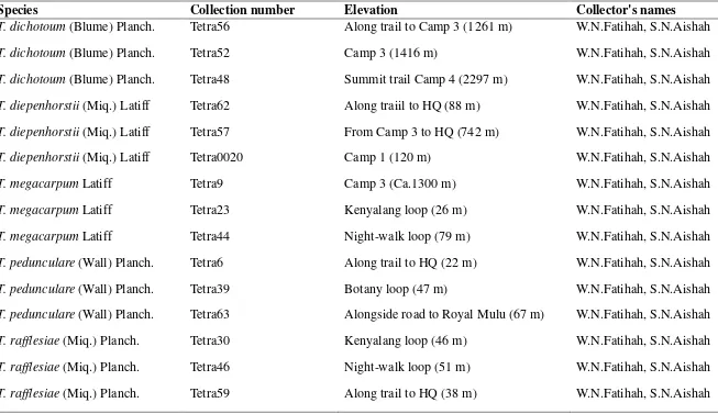
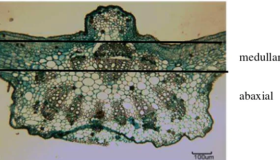
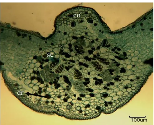
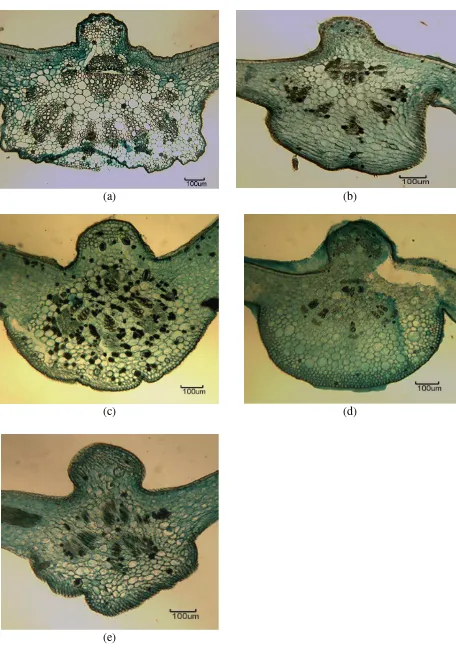
Leaf anatomical variations of the midrib, lamina and leaf margin of five Tetrastigma species are shown in Figure 1-15. Figure 1-3 show the transverse sections of T. pedunculare (Wall) Planch. while Figure 4-6 show the transverse sections of T. rafflesia (Miq.) Planch. Figure 7-9 show the transverse section of T. dichotomum (Blume) Planch and Figure 10-12 show the transverse section of T. diepenhorstii (Miq.) Planch while Figure 13-15 shows the transverse section of T. megacarpum Latiff. Midrib of Tetrastigma species in this study can be divided into two parts: medullary and abaxial side (Figure 1). Presence of druses also can be observed in Tetrastigma species studied (Figure 2).
Figure 1: Presence of vascular bundles in the midrib of T. pedunculare (medullary and abaxial part)
Figure 2: Presence of druses in the adaxial side of the midrib of T. pedunculare medullary
abaxial
11 4.1.1 Tetrastigma pedunculare (Wall) Planch
Midrib: The outline for adaxial is convex while the outline for abaxial is wide “U” shape with prominent and truncated bottom. Vascular bundle is discontinuous closed system with
“O” shape that consist of outer vascular bundles and medullary bundles. Outer vascular
bundle consists of discontinuous of several vascular bundle arranged in ¾ circle shape. Medullary vascular bundle consists of three vascular bundle at the adaxial side. Secretory canal, druses and collenchyma are present in the midrib (Figure 3).
12
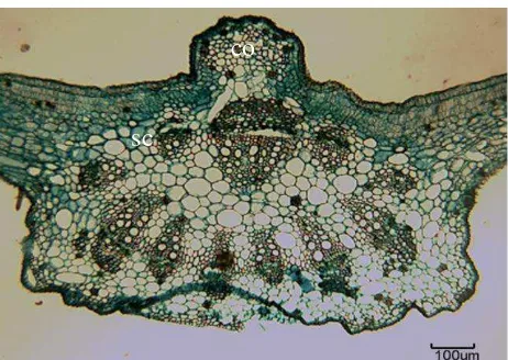
Figure 3: TS of midrib of T. pedunculare. Scale = 100 µm
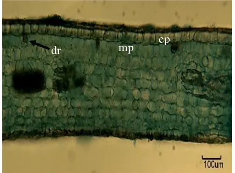
Figure 4: TS of lamina of T. pedunculare. Scale = 100 µm
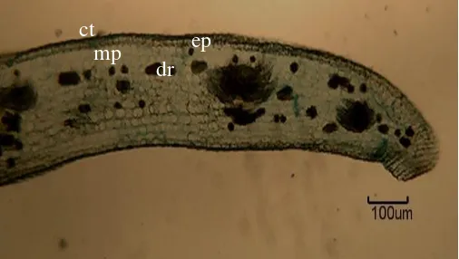
Figure 5: TS of leaf margin of T. pedunculare. Scale = 100 µm
Note: co: collenchyma; sc: secretory canal; mp: mesophyll palisade; ct: cuticle; ep: epidermis; dr: druses; tr: trichome
co
sc
mp ep
tr dr
13 4.1.2 Tetrastigma rafflesiae (Miq.) Planch
Midrib: The outline for adaxial is convex while the abaxial outline shows wide “U” shape with prominent and rounded convexity. Vascular bundle is closed system with “O” shape that consists of outer vascular bundles and medullary bundles. Outer vascular bundle consists of discontinouous of several vascular bundle arranged in ¾ circle shape. Medullary vascular bundle consists of single vascular bundle at the adaxial side. Druses and collenchyma are present (Figure 6).
14
Figure 6: TS of midrib of T. rafflesiae. Scale = 100 µm
Figure 7: TS of lamina of T. rafflesiae. Scale = 100 µm
Figure 8: TS of leaf margin of T. rafflesiae. Scale = 100 µm
Note: co: collenchyma; mp: mesophyll palisade; ct: cuticle; ep: epidermis; dr: druses co
ep
mp dr
ct
dr
15 4.1.3 Tetrastigma dichotomum (Blume) Planch
Midrib: The outline for adaxial is convex while the abaxial is “U” shape with prominent and rounded convexity. Vascular pattern closed system consist of outer and medullary vascular bundle. Outer vascular bundle consist of discontinuous of several vascular bundle arranged in ¾ circle shape. Medullary vascular bundle consists of single vascular bundle at the adaxial side. Druses are present in abundance (Figure 9).
16
Figure 9: TS of midrib of T. dichotomum. Scale = 100 µm
Figure 10: TS of lamina of T. dichotomum. Scale = 100 µm
Figure 11: TS of leaf margin of T. dichotomum. Scale = 100 µm
Note: co: collenchyma; sc: secretory canal; mp: mesophyll palisade; ct: cuticle; ep: epidermis; dr: druses
ep dr
co
sc
dr
mp
ct
17 4.1.4 Tetrastigma diepenhorstii (Miq.) Latiff
Midrib: The outline for adaxial is convex while the abaxial outline shows “U” shape with prominent and rounded convexity. Vascular bundle closed system consist of outer vascular bundle and medullary bundles. Outer vascular bundle consists of discontinuous of several vascular bundle arranged in ¾ circle shape. Medullary vascular bundle consists of single vascular bundle at the adaxial side. Druses, collenchyma and secretory cells are present (Figure 12).
18
Figure 12: TS of midrib of T. diepenhorstii. Scale = 100 µm
Figure 13: TS of lamina of T. diepenhorstii. Scale = 100 µm
Figure 14: TS of leaf margin of T. diepenhorstii. Scale = 100 µm
Note: co: collenchyma; sc: secretory canal; mp: mesophyll palisade; ct: cuticle; ep: epidermis; dr: druses
co
ct
dr
dr
19 4.1.5 Tetrastigma megacarpum Latiff
Midrib: The outline for adaxial is convex while the abaxial outline shows “U” shape with prominent and irregular convexity. Vascular bundle closed system consits of outer vascular bundles and medullary bundles. Outer vascular bundle consits of discontinuous of few vascular bundle arranged in ½ circle shape. Medullary vascular bundle consists of single vascular bundle at the adaxial side. Druses are present (Figure 15).
20
Figure 15: TS of midrib of T. megacarpum. Scale = 100 µm
Figure 16: TS of lamina of T. megacarpum. Scale = 100 µm
Figure 17: TS of leaf margin of T. megacarpum. Scale = 100 µm
Note: co: collenchyma; mp: mesophyll palisade; ct: cuticle; ep: epidermis; dr: druses co
dr
dr
dr ct
ct
ep
ep mp
21 4.2 Leaf midrib
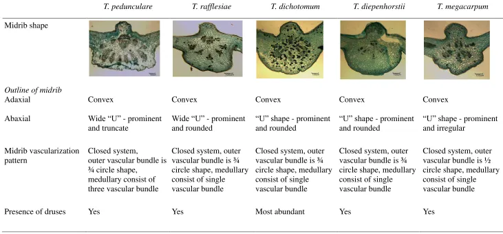
The transverse section of the midrib of Tetrastigma species studied present several characteristics and shapes as summarized in Table 2 and Figure 18. The outline of the midrib of Tetrastigma varies among each species studied. Generally, Tetrastigma species have convex shape on the adaxial side. However some Tetrastigma species have similar outline that is having prominent and rounded convexity on the abaxial such as shown in Tetrastigma rafflesiae (Fig.18b), T. dichotomum (Fig.18c), and T. diepenhorstii (Fig.18d). Tetrastigma pedunculare has an arc shape on the abaxial (Fig.18a), and T. megacarpum is prominent with an irregular projection on abaxial side (Fig.18e). Druses are found in abundance only in T. dichotomum. In general, druses are randomly scattered in the midrib in all Tetrastigma species studied.
4.3 Leaf lamina
All Tetrastigma species studied show a uniseriate epidermis covered by thin cuticle on the adaxial surface (Fig.4, 7, 10, 13 and 16). Trichomes only presents on the abaxial surface of T. pedunculare (Fig. 4).
The layers of mesophyll palisades varies in the Tetrastigma species studied. Tetrastigma pedunculare (Fig.4), T. rafflesiae (Fig.7) and T. dichotomum (Fig.10) consist two layers of mesophyll palisade, while both T. diepenhorstii (Fig.13) and T. megacarpum (Fig.16) consist one layer of mesophyll palisade.
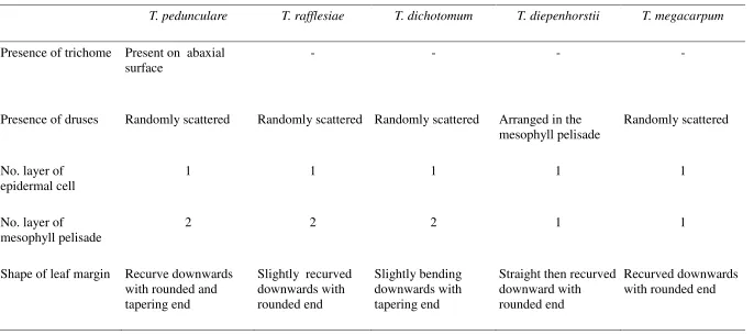
22 Table 2: Anatomical characteristics of midrib between Tetrastigma species
T. pedunculare T. rafflesiae T. dichotomum T. diepenhorstii T. megacarpum
23
(a) (b)
(c) (d)
(e)
24 4.4 Leaf margin
25
Table 3: Anatomical features of lamina and leaf margin between Tetrastigma species
T. pedunculare T. rafflesiae T. dichotomum T. diepenhorstii T. megacarpum
Presence of trichome Present on abaxial surface
- - - -
Presence of druses Randomly scattered Randomly scattered Randomly scattered Arranged in the mesophyll pelisade
26
(a) (b)
(c) (d)
(e)
27 4.2 Taxonomic Implication
Morphological characteristics are very important for species identification. However, the morphological characteristics are limited and fewer of them support analysis. Therefore, anatomical characteristics can help in adding more characters for species identification and analysis. Several studies by Najmaddin et al. (2011; 2013), Talip & Cutler (2009) and Wosch et al. (2015) have proven anatomical characteristics as useful in taxonomic purposes.
In the present study, not many diagnostic characteristics are observed. Transverse section of the midrib does show some diagnostic characteristics especially on the outline of the midrib and the vascular bundles as shown in Figure 17. From the previous study by Najmadin et al. (2013), four species under Vitaceae (Tetrastigma cruciatum, Pterisanthes miquelii, Cissus hastata and Nothocissus spicifera) showed good diagnostic characters by
possessed druses and rhapides. However in this study, rhapides are not observed. This concludes that druses are common feature found in Vitaceae family as to compare both previous and present study.
This study has shown the presence of trichomes can differentiate among species in this species. As an example, the presence of trichomes only present in the leaf lamina of T. pedunculare that can differentiate this species from other four Tetrastigma species that
28
29
5.0 CONCLUSION AND RECOMMENDATION
In conclusion, anatomical characteristics of Tetrastigma species in this study such as outline of midrib, vascular bundle pattern and presence of trichome is proven to help in species identification to a certain extent. The presence of druses is also a common characteristic for Tetrastigma species.
30
REFERENCES
Adam, J.H., Mohamed, R., Juhari, M.A.A, Nik Ariff, N.N.F, & Wan, K.L. (2013). Rafflesia sharifa-hapsahiae (Rafflesiaceae), a new species from Peninsular Malaysia. Turkish Journal of Botany, 37, 1038-1044.
Chen, I. (2009). History of Vitaceae inferred from morphology based on phylogeny and the fossil record of seeds. Published dissertation for the degree Doctor of Philosophy. University of Florida Unites States of America.
Chen, P., Chen I., & Wen J. (2011). The first phylogenetic analysis of Tetrastigma (Miq.) Planch, the host of Rafflesiaceae. Taxon, 60(2), 499-512.
Cutler, D.F., Botha, T., & Stevenson, D.W. (2008). Plant anatomy: An applied approach. MA, USA: Blackwell Publishing.
Dickison, W. (2000). Integrative Plant Anatomy. CA, USA: Hardcourt Academic Press. Hazebroek, H. P. & Abang Kashim b. Abang Morshidi. (2002). A Guide to Gunung Mulu
National Park. Sabah, Malaysia: Natural History Publications (Borneo) Sdn. Bhd.
Krishna, T.P.A., Ajeesh Krishna, T.P., Chithra, N.D., Deepa, P.E., Darsana, U., Sreelekha, K.P., Juliet, S., Naish, S.N., Ravindran, R., Kumar, K.G.A., & Ghosh, S. (2014). Acaricidal activity of petroleum ether extract of leaves of Tetrastigma leucostaphylum (densst.) Alston against Rhipicephalus (Boophilus) annulatus. The Scientific World Journal, 1-6.
Latiff, A. (1984). Studies in Malesian Vitaceae VII. The genus Tetrastigma in the Malay Peninsula. Garden Bulletin Singapore, 36(2), 213-216.
Latiff, A. (1991). Studies in Malesian Vitaceae X. Two new species of Tetrastigma from Borneo. Blumea, 35, 559-564.
Mat Salleh, K. (1991). Rafflesia magnificent flower of Sabah. Kota Kinabalu, Malaysia: Borneo Publishing Company.
Nais, J. (2001). Rafflesia of the world. Sabah, Malaysia: Sabah Parks & National History Publications (Borneo) Sdn. Bhd.
Najmaddin, C., Hussin, K. & Maideen, H. (2011). Comparative study on anatomy and palynology of the three variety of Vitis vinefera varity (family Vitaceae). African Journal of Biotechnology, 10(74), 16866-16874.
Najmaddin, C., Hussin, K. & Maideen, H. (2013). Comparative leaf anatomy of selected species in Vitaceae and Leeaceae. American Journal of Applied Science, 10(4), 414-417.
31
Proctor, J., Anderson, J.M., Chai, P. & Vallack, H.W. (1983). Ecological studies in four contrasting lowland rain forests in Gunung Mulu National Park, Sarawak: Forest environment, structure and floristics. The Journal of Ecology, 7(1), 237-260. Ren, H. & Wen. J. (2007). Tetrastigma. Flora of China, 12, 195-208.
Talip, N. & Cutler, D.F. (2009). Leaf anatomical and micromorphological characters of Malaysian Parashorea (Dipterocarpaceae). Journal of Tropical Science, 21(2), 156-157.
Trias-Blasi, A., Parnell, J.A.N. & Hodkinson, T.R. (2012). Multi-gene region phylogenetic analysis of the grape family (Vitaceae). BioOne Biology, Ecology and Environmental Science, 37(4), 941-950.
Wosch, L., Imig, D.C., Cervi, A.C., Moura, B.B., Budel, J.M., & Santos, C.A.M. (2015). Comparative study of Passiflora taxa leaves: I. A morpho-anatomic profile. Revista Brasileira de Farmacognosia, 25, 328-343.
Yeo, C.K., Ang, W.F., & Lok, A.F.S.L. (2012). Tetrastigma Planch. (Vitaceae) of Singapore. Nature in Singapore, 5, 263-270.