Changes of bovine blood lipid peroxides and some antioxidants in the
course of growth
Dubravka Križanović1*, Velimir Sušić1, Pero Božić2, Igor Štoković1, and Anamaria Ekert-Kabalin1
1Department of animal Breeding and husbandry, Faculty of Veterinary Medicine, University of Zagreb, Zagreb, Croatia
2Centre for animal reproduction, Zagreb, Croatia
KRIŽANOVIĆ, D., V. SUŠIĆ, P. BOŽIĆ, I. ŠTOKOVIĆ, A. EKERT-KABALIN: Changes of bovine blood lipid peroxides and some antioxidants in the course of growth. Vet. arhiv 78, 269-278, 2008.
ABSTRACT
Blood lipid peroxides (TBARS) and naturally present blood antioxidants glutathione (GSH), haemoglobin (Hb) and plasma albumin (ALB) were determined in nine Simmental young bulls. Blood samples were taken
five times. The animals were intensively fattened. Mean TBARS content showed a biphasic pattern in the course of growth with the highest value at 11.7 months of age. GSH content increased until the age of 8 months when the highest concentration was determined. Albumin and Hb concentrations were highest at 15.2 months of age. The main contributor to the significant increase and decrease of TBARS values could be the development of skeletal muscle tissue. Correlations among the parameters investigated were not significant in the course of growth, with the exception of GSH vice Hb at 5.4 months of age and TBARS vice Hb and TBARS vice ALB at 15.2 months of age. It might be concluded that the indicators of oxidant stress in young bulls were within the physiological range and that, possibly, blood natural antioxidants complemented each other.
Key words: beef cattle, blood, lipid peroxides, glutathione, albumin, haemoglobin
Introduction
During the animals’ growth proper nutrient and oxygen supply is a prerequisite for normal biochemical and physiological processes. Under physiological conditions the use of oxygen (O2) by cells of aerobic organisms generates potentially deleterious reactive oxygen species (ROS) which include superoxide anion (∙O
2¯, a free radical), hydroxyl radical (∙OH¯) and hydrogen peroxide (H
2O2). Mitochondria are a permanent
*Contact address:
and significant source of ROS. About 95% of all O2 used by the body is processed in mitochondria through oxydative phosphorilation (KIDD, 1997), and up to 5% of total mitochondrial O2 consumed is incompletely reduced and leads to ROS production. The primary ROS generated is superoxide (∙O
2¯) which is then converted to H2O2 by spontaneous dismutation or by superoxide dismutase (MILLS and HIGGINS, 1997; BATANDIER et al.,
2002; BROOKES, 2005).
Lipid peroxidation is one of the most important phenomena caused by the ROS. In order to counteract this living organisms have developed antioxidant defence systems which protect the functional and structural integrity of the cells. Glutathione (GSH) is a central constituent of this system and is considered to be a multifunctional antioxidant which links the different scavenger systems. Additionally, GSH can react directly with a number of other oxidizing compounds by nonenzymatic mechanisms (THOMAS et al.,
1995; CHIARADIA et al., 1998; PASTORE et al., 2003). Blood contains numerous antioxidants
and generally speaking proteins with the SH group may function as antioxidants. Albumin (ALB) is considered the major circulating antioxidant, which protects cells scavenging ROS by trapping them (CASTILLO et al., 2005; FUKUZAWA et al., 2005). With respect to this, haemoglobin (Hb) may also remove ∙O
2 − and H2O2, since it can function catalytically as an oxidase and peroxidase (WINTERBOURN and STERN 1987; EDWARDS and FULLER,
1996).
In spite of numerous studies of lipid peroxidation and naturally present antioxidants involving various species (GAMBHIR et al., 1997; MILLS et al., 1997; CHIARADIA et al.,
1998; AVELLINI et al., 1999; YAMATO et al., 1999; TAVAZZI et al., 2000; McARDLE et al., 2001;
SILVEIRA et al., 2003; MOSONI et al., 2004), there is a lack of data of lipid peroxidation in
meat animals during fattening. The extent of lipid peroxidation in living tissue of meat animals may cause muscle deterioration resulting in changes in texture and nutritive value during processing and storage. The aim of this study was to investigate the extent of lipid peroxidation during the growth of young bulls from the age of several months until early maturity. Naturally present blood antioxidants -GSH, albumin and Hb - were also examined. The relationship between the investigated parameters was also a matter of consideration.
Materials and methods
The study was performed on nine intensively fattened Simmental young bulls aged 3.2 to 15.2 months. They all were progeny of one sire. The bulls had been brought to the station at the age of about two months and were allowed to adapt to the feedlot conditions for about one month. Thereafter the fattening started and the average starting body mass (BM) was 173.8 ± 12.0 kg. The bulls received 4-6 kg/bull/day feed mixture (min. 14% crude proteins (CP), up to 12% roughage) up to 250 kg. Thereafter up until the end they
received 6-10 kg/bull/day feed mixture (min.12% CP, up to 15% roughage). During fattening the animals were offered hay or silage and water ad libitum. They were under health control and were in very good condition. At the age of 15.2 months they were slaughtered at an average BM of 619.6 ± 43.4 kg.
Blood samples were taken five times in the course of fattening by puncture of the jugular vein. First collection started at 3.2 months of age. Blood was collected in heparinized glass tubes always at the same time in the morning. The samples were immediately transferred to the laboratory in a hand refrigerator. The parameters were analyzed the same day or within 24 hours. For albumin determination, plasma was separated by blood centrifugation for 15 min (3000g).
Lipid peroxides were measured in the whole blood according to PLACER et al. (1966)
and TROTTA el al. (1982). It is generally accepted that an index of lipid peroxidation
rate is malondialdehyde (MDA). Peroxidation generates MDA and several MDA like aldehydes and ketones (DUMASWALA et al., 1999), all of which react with thiobarbituric acid (TBA).
Because MDA is a major reactive product generated, TBA reactivity is equated with MDA and the results are presented as thiobarbituric acid reactive substances (TBARS). The concentration of reduced glutathione (GSH) was determined in the whole blood by measurement of the 5,5′-dithiobis-(2-nitrobenzoate) derivative (BEUTLER et al., 1963). The contents of both parameters are presented in relative values - nmol and mg per gram of haemoglobin, respectively. The haemoglobin concentration was measured by the cyanohaemoglobin method and plasma albumin concentration was determined with brom-cresol green (local commercial kits were used -Herbos, Sisak). The first albumin determination is missing because of an accident during centrifugation and plasma separation. SAS statistical software was used for all data analyses (SAS, 1999-2003). For testing the significance of mean differences the Friedman test ANOVA and nonparametric sign test were applied. The strength of the relationship between the two variables was measured with the Spearman correlation coefficient. The results are presented as mean
± SD.
Results
The TBARS content in the course of growth shows a biphasic pattern (Table 1) being relatively high in young animals (3.2 mos), then followed by a significant decrease (5.4 mos), and thereafter up to the age of about twelve months a significant increase. By the end of fattening again a significant decrease occurred. The highest value was determined at the age of about one year. Glutathione content increased until the age of eight months (Table 1) when the highest value was determined. Thereafter up to the bulls’ age of 15.2 months, a significant decrease was observed. The values are in accordance with the range
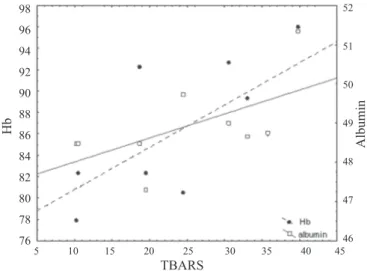
of values in literature (PASTORE et al., 2003). The correlation coefficients between TBARS and GSH (Table 2) were not significant and showed a weak relationship. The concentration of albumin, after a significant decrease from 5.4 until 8 months of age, significantly increased in the subsequent period with the highest value at 15.2 months of age (Table 1). At that age a positive and significant correlation with TBARS was determined (Table 2; Fig. 1). Haemoglobin concentration was lowest in young bulls (3.2 months) and highest at the age of 15.2 months (Table 1). After a significant increase up to 5.4 months, a significant decrease followed up to about 12 months of age. A significant correlation of Hb with GSH and TBARS was established at 5.4 and 15.2 months of age (Table 2; Fig. 2 and Fig. 1).
Table 1. Mean values ( ± SD) of bovine blood lipid peroxides and some antioxidants in the course
of growth (n = 9) Parameter Age (months) 3.2 5.4 8.0 11.7 15.2 TBARS (nmol/g Hb) 46.59 ± 19.50a 19.82 ± 10.11b 54.53 ± 11.82a 58.14 ± 15.79a 24.71 ± 10.79b GSH (mg/g Hb) 3.76 ± 1.32ac 4.74 ± 0.75ab 4.86 ± 1.15a 3.77 ± 0.73bc 3.74 ± 0.56dc Albumin (g/L) - 42.90 ± 1.32a 41.54 ± 1.69b 46.28 ± 1.35c 48.91 ± 1.11d Hb (g/L) 68.60 ± 5.94a 76.64 ± 4.70b 73.64 ± 5.70a 72.56 ± 3.27ab 86.61 ± 6.25c
abcd Means within row with different superscripts significantly differ (P<0.05)
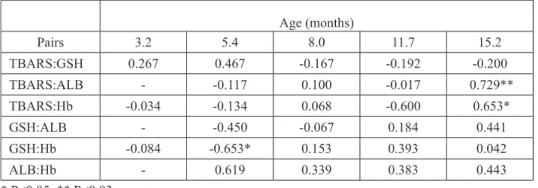
Table 2. Correlations (r) among investigated parameters in the course of bovine growth
Age (months) Pairs 3.2 5.4 8.0 11.7 15.2 TBARS:GSH 0.267 0.467 -0.167 -0.192 -0.200 TBARS:ALB - -0.117 0.100 -0.017 0.729** TBARS:Hb -0.034 -0.134 0.068 -0.600 0.653* GSH:ALB - -0.450 -0.067 0.184 0.441 GSH:Hb -0.084 -0.653* 0.153 0.393 0.042 ALB:Hb - 0.619 0.339 0.383 0.443 * P<0.05; ** P<0.03
Fig. 2. Regression of haemoglobin (y = 94.00 - 3.66 x; r = -0.653; P<0.05 ) on glutathione at 5.4 months of age
Fig. 1. Regression of albumin (y = 47.40 + 0.06x; r = 0.729; P<0.03 ) and haemoglobin (y = 76.84 + 0.40x; r = 0.653; P<0.05 ) on TBARS at 15.2 months of age
TBARS GSH 98 96 94 92 90 88 86 84 82 80 78 76 Hb Albumin Hb 86 84 82 80 78 76 74 72 70 3.6 3.8 4.0 4.2 4.4 4.6 4.8 5.0 5.2 5.4 5.6 5.8 6.0 5 10 15 20 25 30 35 40 45 52 51 50 49 48 47 46
Discussion
Generation of ROS takes place continually. Healthy cells homeostatically oppose ROS through the use of naturally present antioxidants. In spite of that, once formed, ROS initiate lipid peroxidation whereby an unpaired electron reacts with an unsaturated double bond in lipid biomembranes (CHAN and DECKER, 1994; MILLS and HIGGINS, 1997). Therefore the cells with high content of unsaturated lipids in the membrane are more exposed to peroxidation (KIDD, 1997). The biphasic pattern of blood TBARS content may imply that either there exist two regions in the body which generate an increased amount of ROS depending on the stage of development, or some tissues develop biphasicly. The actual blood TBARS content originates from different body tissues but we suppose mostly from skeletal muscles. There are many reasons for this assumption. Muscles are a large O2 consumer, they have a high proportion of unsaturated fatty acids in their membranes
(CHAN and DECKER, 1994), they have low levels of antioxidants compared with other
tissues (AVELLINI et al., 1999) and muscle tissue comprises a large portion of the animal’s body. A characteristic of muscle tissue development could perhaps explain the biphasic pattern of blood TBARS content, The greatest changes in muscle weight distribution occur in the post-natal phase before five months of age. This phase continues in some muscles to a quadrupling of weight (BERG and BUTTERFIELD, 1976). Besides, growth generally implies an increase in energy demand which is produced via the aerobic metabolism and an increase in dietary energy intake enhances mitochondrial ROS production (KIDD,
1997; FANG et al., 2002). Significant decrease of TBARS content from three to five months
of age might indicate the end of the first phase of muscle development. Another period of cattle growth, which sets off a whole series of changes in bulls aimed at producing the mature muscle weight distribution, is puberty (BERG and BUTTERFIELD, 1976). Our results show that in that period (6-12 months of age) a significant increase of TBARS occurred again. Thereafter a significant decrease happened and TBARS value at 15 months of age is similar to that at five months. Possibly these values are the baseline values. Further, recent studies indicate that skeletal muscle cells continually release multiple ROS during non-damaging contraction (REID and DURHAM, 2002; SILVEIRA et al., 2003; ARBOGAST
and REID, 2004; McARDLE et al., 2004). In addition to muscle cells, muscle tissue in vivo
contains endothelial cells, lymphocytes and fibroblasts that may also contribute to the generation of ROS (McARDLE et al., 2001; PATWELL et al., 2004). It has been shown that skeletal muscle releases H2O2 during contractile activity which together with ∙O
2¯diffuse outside the cell and enter the bloodstream. Leaking of H2O2 thanks to the high permeability of the muscle cell membrane, may constitute an important defence mechanism against the formation of more dangerous ∙OH¯ (SILVEIRA et al., 2003; PATWELL et al., 2004). In
blood they are likely to be removed by many constituents but erythrocytes (E) seem to be the most important. Erythrocyte membrane is permeable to ∙O
2¯ and H2O2 and could efficiently remove extracellular H2O2 (WINTERBOURN and STERN, 1987; BROWN et al.,
1989). Erythrocyte GSH is an important component of antioxidant defence and under physiological conditions all H2O2 encountered by E is detoxified by GSH peroxidase, an enzyme that uses GSH as substrate (THOMAS et al., 1995; GAMBHIR et al., 1997; YAMATO
et al., 1999; KURATA et al., 2000; SEO et al., 2004).
The blood GSH we determined is actually E GSH, since it is most abundant inside cells (mmolar levels) and relatively lacking outside cells (µmolar levels) (KIDD, 1997;
PASTORE et al., 2003). Regulation of GSH levels in vivo should be considered in terms
of the entire organism in which some organs are net synthesizers whereas others are net exporters. Increase of GSH content until the age of eight months could be the result of increased synthesis in E, but also its delivery from the liver as a part of a complicated inter-organ transport network (MILLS et al., 1997; SIES, 1999). In both cases it is a consequence of the increased needs which should not be only scavenging of ROS. There is a variety of specific reactions where GSH participates in essential aspects of cellular homeostasis where the protein SH groups are reversibly bound to GSH, regulating protein function (significant correlation with Hb at 5.4 mos of age) (PASTORE et al., 2003; POMPELLA et
al., 2003). Lack of the significant correlation between TBARS and GSH may indicate
that ROS are homeostatically maintained in the physiological range. There is evidence that the indices of oxidative stress significantly correlate under severe pathological conditions (GAMBHIR et al.,1997; DOTAN et al., 2004). The correlations in Table 2 should be understood in this context. Due to the contribution of GSH to a number of cellular processes it is possible that in different growth phases different functions predominate. Significant decrease of GSH content from the bulls’ age of eight months up to the end of the trial might indicate that GSH acts as an antitoxic agent when GSH-S-conjugates are being generated and this is accompanied by GSH lowering (PASTORE et al., 2003). In that period some other blood antioxidant might partly take over the role of scavenger. This could be ALB or Hb. Their highest concentrations were at the bulls’ age of 15.2 months. At that age a significant correlation of ALB and Hb with TBARS was established. High concentration of these thiol proteins, together with a significant decrease of GSH content after the bulls’ age of eight months may imply that antioxidant systems complement each
other, when the oxidative challenge has decreased (lower TBARS value at 15.2 mos).
Conclusion
This work shows that during the growth of young bulls the blood TBARS values significantly increase and decrease. We suppose that the main cause of these changes could be the development of skeletal muscle tissue, which, with its increasing mass and abundant double bonds in cell membranes, might greatly influence the blood TBARS values. In spite of the significant change of the TBARSs, they are within the physiological range. That means that the natural blood antioxidants homeostatically maintain the peroxidation under control. One may expect that the muscle texture and nutritive value
should be protected as a result. According to our results it seems that blood antioxidants - GSH on the one hand, and ALB and Hb on the other - complement each other during the bulls’ growth.
References
ARBOGAST, S., M. B. REID (2004): Oxidant activity in skeletal muscle fibers is influenced by
temperature, CO2 level and muscle-derived nitric oxide. Am. J. Physiol. 287, R698-R705.
AVELLINI, L., E. CHIARADIA, A. GAITI (1999): Effect of exercise training, selenium and
vitamin E on some free radical scavengers in horses (equus caballus). Comp. Biochem.
Physiol. B, 123, 147-154.
BATANDIER, C., E. FONTAINE, Ch. KÉRIEL, M. LEVEVRE (2002): Determination of mitochondrial reactive oxygen species: methodological aspects. J. Cell. Mol. Med. 6, 175-187.
BERG, R. T., R. M. BUTTERFIELD (1976): New Concepts of Cattle Growth. Sydney University Press, First edition, Sydney, pp. 66-80.
BEUTLER, E., O. DURON, B. MIKUS KELLY (1963): Improved method for the determination of blood glutathione. J. Lab. Clin. Med. 61, 882-888.
BROOKES, P. S. (2005): Mitochondrial H+ leak and ROS generation: An odd couple. Free Radical
Biol. Med. 38, 12-23.
BROWN, J. M., M. A. GROSSO, L. S. TERADA, C. J. BEEHLER, K. M. TOTH, G. J. WHITMAN, A. H. HARKEN, J. E. REPINE (1989): Erythrocytes decrease myocardial hydrogen peroxide levels and reperfusion injury. Am. J. Physiol. 256, H584-H588.
CASTILLO, C., J. HERNANDEZ, A. BRAVO, M. LOPEZ-ALONSO, V. PEREIRA, J. L. BENEDITO (2005): Oxidative status during late pregnancy and early lactation in dairy cows. Vet. J. 169, 286-292.
CHAN, K. M., E. A. DECKER (1994): Endogenous skeletal muscle antioxidants. Crit. Rev. Food Sci. Nutr. 34, 403-426.
CHIARADIA, E., L. AVELLINI, F. RUECA, A. SPATERNA, F. PORCIELLO, M. T. ANTONIONI, A. GAITI (1998): Physical exercise, oxidative stress and muscle damage in race horses. Comp. Biochem. Physiol. B. 119, 833-836.
DOTAN, Y., D. LICHTENBERG, I. PINCHUK (2004): Lipid peroxidation cannot be used as a universal criterion of oxidative stress. Prog. Lipid Res. 43, 200-227.
DUMASWALA, U. J., L. ZHUO, D. W. JACOBSEN, S. K. JAIN, K. SUKALSKI (1999): Protein
and lipid oxidation of banked human erythrocytes: role of glutathione. Free Radical Biol. Med.
27, 1041-1049.
EDWARDS, C. J., J. FULLER (1996): Oxidative stress in erythrocytes. Comp. Haematol. Int. 6, 24-31.
FANG, Y-Zh., S. YANG, G. WU (2002): Free radicals, antioxidants, and nutrition. Nutrition. 18,
FUKUZAWA, K., Y. SAITOH, K. AKAI, K. KOGURE, S. UENO, A. TOKUMURA, M. OTAGIRI, A. SHIBATA (2005): Antioxidant effect of bovine serum albumin on membrane lipid peroxidation induced by iron chelate and superoxide. Biochim. Biophys. Acta 1668, 145-155.
GAMBHIR, J. K., P. LALI, A. K. JAIN (1997): Correlation between blood antioxidant levels and lipid peroxidation in rheumatoid arthritis. Clin. Biochem. 30, 351-355.
KIDD, P. M. (1997): Glutathione: Systemic protectant against oxidative and free radical damage. Alter. Med. Rev. 2, 155-176.
KURATA, M., M. SUZUKI, N. S. AGAR (2000): Glutathione regeneration in mammalian erythrocytes. Comp. Haematol. Int. 10, 59-67.
McARDLE, A., D. PATTWELL, A. VASILAKI, R. D. GRIFFITHS, M. J. JACKSON (2001): Contractile activity-induced oxidative stress: cellular origin and adaptive response. Am. J. Physiol. 280, C621-C627.
McARDLE, A., J. VAN DER MEULEN, G. L. CLOSE, D. PATTWELL, H. VAN REMMEN, T-T. HUANG, A. G. RICHARDSON, Ch. J. EPSTEIN, J. A. FAULKNER, M. J. JACKSON (2004): The role of mitochondrial superoxide dismutase in contraction-induced generation of reactive oxygen species in muscle extracellular space. Am. J. Physiol. 286, C1152-C1159. MILLS, P. C., A. J. HIGGINS (1997): Oxidant injury, nitric oxide and pulmonary vascular functions:
implications for the exercising horse. Vet. J. 153, 125-148.
MILLS, P. C., N. C. SMITH, R. C. HARRIS, P. HARRIS (1997): Effect of allopurinol on the formation of reactive oxygen species during intense exercise in the horse. Res. Vet. Sci. 62, 11-16.
MOSONI, L., D. BREUILLE, C. BUFFIÈRE, Ch. OBLED, Ph. P. MIRAND (2004): Age-related
changes in glutathione availability and skeletal muscle carbonyl content in heal rats. Exp. Gerontol. 39, 203-210.
PASTORE, A., G. FEDERICI, E. BERTINI, F. PIEMONTE (2003): Analysis of glutathione: implication in redox and detoxification. Clin. Chim. Acta 333, 19-39.
PATWELL, D. M., A. McARDLE, J. E. MORGAN, T. A. PATRIDGE, M. J. JACKSON (2004): Release of reactive oxygen and nitrogen species from contracting skeletal muscle cells. Free Radical Biol. Med. 37, 1064-1072.
PLACER, Z. A., L. L. CUSHMAN, B. C. JOHNSON (1966): Estimation of product of lipid peroxidation (malonyl dialdehyde) in biochemical systems. Anal. Biochem. 16, 359-364. POMPELLA, A., A. VISVIKIS, A. PAOLICCHI, V. DeTATA, A. F. CASINI (2003): The changing
faces of glutathione, a cellular protagonist. Biochem. Pharmacol. 66, 1499-1503.
REID, M. B., W. J. DURHAM (2002): Generation of reactive oxygen and nitrogen species in contracting skeletal muscle. Ann. N. Y. Acad. Sci. 959, 108-116.
SAS, 1999-2003. Release 8. 1 (SAS Institute Inc, Cary, NC, USA)
SEO, Y. J., J. W. LEE, E. H. LEE, H. K. LEE, H. W. KIM, Y-H. KIM (2004): Role of glutathione in
SIES, H. (1999): Glutathione and its role in cellular function. Free Radical Biol. Med. 27, 916-921.
SILVEIRA, L. R., L. PEREIRA-DA-SILVA, C. JUEL, Y. HELLSTEN (2003): Formation of hydrogen peroxide and nitric oxide in rat skeletal muscle cells during contractions. Free Radical Biol. Med. 35, 455-464.
TAVAZZI, B., D. DiPIERRO, A. M. AMORINI, G. FAZZINA, M. TUTTOBENE, B. GIARDINA, G. LAZZARINO (2000): Energy metabolism and lipid peroxidation of human erythrocytes as a function of increased oxidative stress. Eur. J. Biochem. 267, 684-689.
THOMAS, J. A., B. POLAND, R. HONZATKO (1995): Protein sulfhydryls and their role in the antioxidant function of protein S-thiolation. Arch. Biochem. Biophys. 319, 1-9.
TROTTA, R. J., S. G. SULLIVAN, A. STERN (1982): Lipid peroxidation and haemoglobin
degradation in red blood cells exposed to t-butyl hydroperoxide. Biochem. J. 204, 405-415.
WINTERBOURN, Ch. C., A. STERN (1987): Human red cells scavenge extracellular hydrogen peroxide and inhibit formation of hypochlorous acid and hydroxil radical. J. Clin. Invest. 80, 1486-1491.
YAMATO, O., M. HAYASHI, E. KASAI, M. TAJIMA, M. YAMASAKI, Y. MAEDE (1999):
Reduced glutathione accelerates the oxidative damage produced by sodium n
-propyl-thiosulfate, one of the causative agent of onion-induced hemolytic anemia in dogs. Biochim.
Biophys. Acta 1427, 175-182.
KRIŽANOVIĆ, D., V. SUŠIĆ, P. BOŽIĆ, I. ŠTOKOVIĆ, A. EKERT-KABALIN: Promjene lipidnih peroksida i nekih antioksidansa u krvi goveda tijekom tova. Vet. arhiv 78, 269-278, 2008.
SAŽETAK
U punoj krvi devet simentalskih bičića određivani su lipidni peroksidi (TBARS), prirodni antioksidansi glutation (GSH) i hemoglobin (Hb), te albumin u plazmi. Životinje su bile u intenzivnom tovu. Prosječna koncentracija TBARS pokazivala je tijekom tova bifazno kretanje s najvišom vrijednosti u dobi životinja od 11,7 mjeseci. Koncentracija GSH je rasla do dobi od 8 mjeseci kada je utvrđena najviša vrijednost. Koncentracije albumina i Hb bile su najviše u dobi od 15,2 mjeseca. Pretpostavlja se da je razvoj i rast skeletnih mišića znatno pridonio signifikantnom porastu i smanjenju TBARS vrijednosti. Korelacije među istraživanim pokazateljima nisu bile signifikantne s izuzetcima GSH i Hb u dobi od 5,4 mjeseca, te TBARS i Hb uz TBARS i albumin u dobi životinja od 15,2 mjeseca. Zaključuje se da su se određivane vrijednosti pokazatelja oksidativnog stresa u bičića kretale unutar fizioloških granica, te da se prirodni antioksidansi u krvi nadopunjuju u djelovanju tijekom tova.
Ključne riječi: tovno govedo, krv, lipidni peroksidi, glutation, albumin, hemoglobin Received: 8 July 2007 Accepted: 2 July 2008