Hepatoprotective Effect of
Trigona spp.
Bee Propolis against Carbon
Tetrachloride-Induced Liver Injury in Rats
Rachel Amelia,1 Achadiyani,2 Begawan Bestari3
1Faculty of Medicine Universitas Padjadjaran, 2Department of Anatomy and Cell Biology Faculty
of Medicine, Universitas Padjadjaran, 3Department of Internal Medicine Faculty of Medicine
Universitas Padjadjaran/Dr. Hasan Sadikin General Hospital Bandung
Abstract
Background: Oxidative stress reaction can cause liver injury. This process can be prevented by antioxidant activities which can break the destructive chain caused by free radical substances in the liver. Propolis produced by Trigona spp. bee is known to have a high level of antioxidant. The aim of this study was to examine the effect of Trigona spp. bee propolis on liver histological toxicity caused by carbon tetrachloride-induced oxidative stress.
Methods:This experimental study was conducted in September 2013 at the Animal Laboratory of Departement of Pharmacology and Therapy, Faculty of Medicine Universitas Padjadjaran. Twenty-four healthy male Wistar rats as objects were adapted for one week and randomly divided into 3 groups. Group I was the control negative, group II was given carbon tetrachloride on day 14, group III was given Trigona spp. bee propolis on day 1-14. On day 14, group III was injected CCl4 intraperitoneally. The quantitative data were statistically analyzed using the one way ANOVA and Tukey test with p value < 0.05.
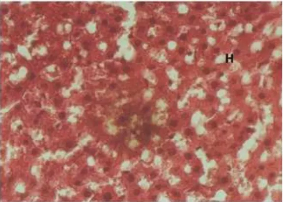
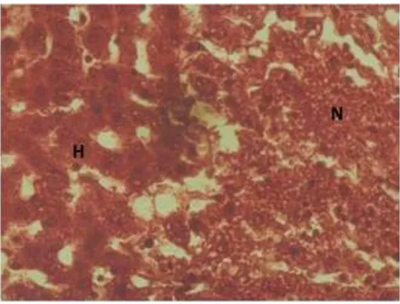
Results: Group I showed the liver contained normal cells, without significant injury of the membrane, round and complete nucleus. The average number of liver cell was 464 ± 9.59281 cells/field; group II underwent necrosis and the average of the cells was 146 ± 7.56885 cells/field; group III showed some normal liver cells, and some necrotic area with the normal liver cells average was 263 ± 14.10860 cells/field. The p-value=0.00. Conclusions: Trigona spp. bee propolis has a hepatoprotective effect against CCl4-induced liver injury histologically. [AMJ.2016;3(3):482–6]
Keywords: Carbontetrachloride, hepatoprotective, propolis, Trigona spp.
Correspondence: Rachel Amelia, Faculty of Medicine, Universitas Padjadjaran, Jalan Raya Bandung-Sumedang Km.21, Jatinangor, Sumedang, Indonesia, Phone: +6281222475898 Email: [email protected]
Introduction
Hepatitis is still a serious health problem in Indonesia. According to Riset Kesehatan Dasar (Riskesdas) Nasional 2007, Indonesia was placed as the second largest country among other SEARO countries with hepatitis patients.1 In October 2007–2009, the Ministry
of Health of the Republic of Indonesia reported about 17.999 positive hepatitis C cases. Infection with hepatitis C is associated with increased levels of Reactive Oxygen Species (ROS)/Reactive Nitrogen Species (RNS) and decreased antioxidant levels.2 ROS/RNS is a
cause of oxidative stress.
Oxidative stress is a condition where there
is a high level of free radicals; one of the
potential substance is carbon tetrachloride which has strong connection with pathologic
process like cell destruction.3 Currently, it is
commonly used for inducing hepatitis model in laboratory experimental animals. When carbon tetrachloride enters the body, this substance will be metabolized in the liver and will produce free radical such as trichloromethyl radical (CCl3) and trichloromethyl peroxy radical (CCl3O2) that induce lipid peroxidation and death of the cell.4 One substance that can
prevent lipid peroxidation is antioxidant. Furthermore, propolis is a natural product, which is a mixture of resin and beeswax collected from plants particularly from
flowers and leaf buds by honey bees (Apis spp.
propolis produced by the Apis mellifera.5,6 Until
now, there has been only few studies about its hepatoprotective effect.7 The objective of this
study was to examine whether the Trigona spp. bee propolis has a hepatoprotective effect against oxidative stress mechanism, hence after further preclinical and clinical studies has been performed, it expected that this propolis could be used effectively by hepatitis high risk patients and also by patients with chronic hepatitis.
Methods
This experimental research was conducted at the Animal Laboratory of the Pharmocology Department, Faculty of Medicine Universitas Padjadjaran, in September 2013. Twenty-four healthy male Wistar rats as objects were used.4 All laboratory experimental
animals in this study was approved by the Health Research Ethics Committee Faculty of Medicine Universitas Padjadjaran. The
Table 1 Average Number of Normal Hepatocyte from Each Group
Group Number of Normal Hepatocyte (mean±SD)
p *
Group I 463.50±9.59281 0.00
Group II 146.07±7.56885 0.00
Group III 263.24±14.10860 0.00
Note: p* = one way ANOVA
animals were randomly divided into 3 groups and given standard food and water. This study
was performed in fifteen days, excluded from
the one week of adaptation. Group I was the control negative, which was given standard food and water. Group II was the control positive which was given 1ml/kgBW CCl4 50% intraperitoneally at day 14 to induce necrosis of hepatocytes.8 Group III was given 1ml/
kgBW Trigona spp. propolis on day 1–14, and on day 14, three hours after given propolis, the rats were injected with 1 ml/kgBW CCl4 50% intraperitoneally.9 Twenty-four hours
after the last treatment, all rats from all
groups were sacrificed. First the animals were
anaesthetized using ketamine intramuscularly, and then laparotomy was performed to expose the liver organ. Afterward, the liver was perfused using NaCl 0.9% through the left ventricle, and blood was cleared from the body through inferior vena cava. The liver was also washed with 10% formalin, then excised
and transferred to a 10% formalin fixative
Figure 2 Liver Histologic View from Group II Note: H=normal hepatocyte, N=necrotic area
Figure 3 Liver Histologic View from Group III Note: H=normal hepatocyte, N=necrotic area
solution. The liver tissues were processed for
paraffin embedding and sections (5 µm thick)
were taken in a microtome. After staining with hematoxylin and eosin, slides were examined under the microscope (400x) for histopathological changes and counted for the average of the normal cells in each group The existence of a necrotic area on hepatocytes, was characterized by rupture of the cell membrane,
vacuolization of the cytoplasm, karyolisis, pyknosis, and karyorrhexis. Moreover, the quantitative data were statistically analyzed using the One Way Analysis of Varian (ANOVA) followed by the Tukey test using the Statistical Product and Service Solutions (SPSS) for Windows version 13.0. A p-value of less than
solution. The liver tissues were processed for
paraffin embedding and sections (5 µm thick)
were taken in a microtome. After staining with hematoxylin and eosin, slides were examined under the microscope (400x) for histopathological changes and counted for the average of the normal cells in each group The existence of a necrotic area on hepatocytes, was characterized by rupture of the cell membrane, vacuolization of the cytoplasm, karyolisis, pyknosis, and karyorrhexis. Moreover, the quantitative data were statistically analyzed using the One Way Analysis of Varian (ANOVA) followed by the Tukey test using the Statistical Product and Service Solutions (SPSS) for Windows version 13.0. A p-value of less than
0.05 was considered as statistically significant.
Results
The data analyzed by the one way ANOVA
statistical test showed p = 0.00; it means, the
difference of the average number of normal
hepatocytes between groups was significant.
The Tukey test also showed the difference of the average number of normal hepatocytes
between groups were significant one by
another. The result from group I showed the liver contained normal cells, without
significant injury of the membrane, round and
complete nucleus. The average number of liver cell in this group was 464 ± 9.59281 cells /
field (Figure 1); group II underwent necrosis
and the average of the cells was 146 ± 7.56885
cells / field (Figure 2); group III showed some
normal liver cells, and some necrotic area with the normal liver cells average was 263 ±
14.10860 cells / field (Figure 3).
Discussion
The effect of CCl4 in 24 hour acute exposure in the control positive group was marked by the existence of necrotic area on hepatocytes, characterized by the rupture of cell membrane, vacuolization of the cytoplasm, karyolisis,
pyknosis, and karyorrhexis. These findings are
further supported by earlier reports by Girish et al.8 that CCl
4 produced various histological
changes in the hepatocytes after 24 hours of administration.8,9 The analytic study of
the average number of normal hepatocytes showed the difference between the control negative and control positive group were
significantly different (p=0.00). It could be
concluded that CCl4 that was given to group
II made significant injury to the liver cell. This
process began from CCl4 turned into CCl3 and
reacted with the oxygen forming more reactive substance CCl3O2 and then, it reacted with membrane cell phospholipid and initiated an extensive intrahepatic peroxidation reaction chain. In the end, there was necrosis of the hepatocytes.4
Group III had more normal hepatocytes compared to group II. The data were
significant after being analyzed statistically.
The protective effect of this propolis was about 37%. According to Chanchao et al.10, the
propolis itself is non-toxic to the normal cells. The condition of oxidative stress is established
due to insufficient defense capacities against
reactive oxygen species (ROS). The ROS also affect the antioxidant defense mechanism, and reduce intracellular concentration of antioxidant produced by the body. Although our body has its own antioxidant and regulation to this substance capacity physiologically, other source of antioxidant is important as well for scavenging free radicals that are formed in the body. Additionally, propolis consists of more than 300 different compounds including
flavonoids, phenolics, wax, vitamins, and
minerals.5 It has been proved that bees do not
change its chemical composition. In propolis,
the major antioxidant is flavonoids. It could
give protection to the liver cells by decreasing formation of CCl3 and CCl3O2.11 It has already
been studied that Trigona spp. bee propolis has
a high level of antioxidant, especially flavonoid
4% which is higher than in another propolis that is produced by Apis mellifera bee is 1.5%.5,6 Besides, the propolis contains another
substance that is known for its antioxidant and
antiinflammatory activities such as caffeic acid
phenethyl ester (CAPE) where it showed a free radical scavenging effect.12
It can be concluded that Trigona spp. bee propolis has a hepatoprotective effect against CCl4-induced liver injury in male Wistar rats histopathologically.
References
1. Kementerian Kesehatan Republik Indonesia. Masyarakat Dunia Peringati Hari Hepatitis. [cited 28 July 2010]. Available from: www.depkes.go.id/index.
php?vw=2&id=1156.
2. Muriel P. Role of free radicals in liver
diseases. Hepatol Int. 2009;3(4):526–36.
Philadelphia: Elsevier Saunders; 2010. p.
20–2.
4. U.S Environmental Protection Agency Washington D. Toxicological review of carbon tetrachloride. Washington DC: United States Environmental Protection
Agency; 2010.
5. Surendra NS, Bhushanam M, Ravikumar H. Antimicrobial Activity of Propolis of Trigona sp. and Apis mellifera of Karnataka, India. Prime Journal of Microbiology
Research. 2012;2(2):80–5.
6. Mahani, Karim R, Nurjanah N. Keajaiban propolis trigona. Jakarta: Pustaka Bunda
Grup Puspa Swara; 2011.
7. Umthong S, Puthong† S, Chanchao C. Trigona laeviceps Propolis from Thailand: antimicrobial, antiproliferative and cytotoxic activities. Am J Chin Med.
2009;37(05):855–65.
8. Girish C, Koner BC, Jayanthi S, Rao KR, Rajesh B, Pradhan SC. Hepatoprotective activity of six polyherbal formulation in
CCl4-induced liver toxicity in mice. Indian
J Exp Biol. 2009;47(4):257–63.
9. Xu J-Y, Su Y-Y, Cheng H-S, li S-X, Liu R, Li W-X, et al. Protective effects of fullerenol on carbon tetrachloride-induced acute hepatotoxicity and nephrotoxicity in rats.
Carbon. 2010;48(5):1388–96.
10. Chanchao C, Umthong S, Phuwapraisirisan P, Puthong S. Trigona laeviceps propolis: chemical compositions and antiproliferative activity on cancer cell
lines. Planta Med. 2011;77:49.
11. Shukla S, Bhadauria M, Jadon A. Effect of propolis extract on acute carbon tetrachloride induce hepatotoxicity. Indian
J Exp Biol. 2004;42(10):993–7.
12. Wu W-M, Lu L, Long Y, Wang T, Liu L, Chen Q, et al. Free radical scavenging and antioxidative activities of caffeic acid phenetyl ester (CAPE) and its related compounds in solution and membranes: a structure-activity insight. Food Chem.