Yohanes Buang
* Corresponding author. Tel/Fax : +62-380-831613/881557 Email address : [email protected]
DIETARY ADENINE ALLEVIATES FATTY LIVER INDUCED BY OROTIC ACID
Yohanes Buang1,2 1
Laboratory of Applied Biochemistry, Department of Applied Biological Science, Saga University, Saga Shi, Honjo Machi-1, Saga, Japan
2
Department of Chemistry, Faculty of Science and Engineering, Nusa Cendana University, Jln. Adisucipto Penfui-Kupang
Received March 18, 2010; Accepted June 8, 2010
ABSTRACT
The effects of dietary adenine in fatty liver induced by orotic acid (OA) were studied. Rats were paired-fed 1% OA-supplemented diets with/or without 0.25% adenine or a diet without OA for 10 days. Serum lipid profiles were measured using enzyme assay kits. Lipids of liver tissues were extracted and liver lipid contents were determined. A peach of liver was prepared to determine the activities of fatty acid synthase (FAS) and fatty acid β-oxidation. The results showed that liver TG content of OA-fed rats increased markedly in comparison to basal group. However, the addition of adenine to the diet reversed promotion of liver TG content to basal level. It was also found that FAS activities decreased. Furthermore, these diets reversed the inhibition of fatty acid β-oxidation to basal level and induced the serum lipid levels secretion. Therefore, the alleviation of fatty liver in OA-treated rats given dietary adenine is associated with the inhibition of FAS activities accompanied with the promotion of mitochondrial fatty acid β-oxidation and the promotion of serum lipid secretion from the hepatic tissue into the bloodstream.
Keywords: adenine, orotic acid, serum lipid, triglyceride, fatty liver, hepatic steatosis
INTRODUCTION
Cells of non-adipose tissues have a limited capacity for storage of lipids. Liver, a main organ of lipid metabolism [1-2], has a limited capacity for storage of those lipids. The liver lipids include triacylglycerol (TG), cholesterol, and phospholipids (PL), in which TG is a non structural lipid. If TG content inside liver cells makes up more than 5-10% of liver’s weight, the subject hold simple fatty liver called hepatic steatosis [3-4]. The certain chemical compounds, nutritional, and endocrine disorders can cause hepatic steatosis. Drugs or poisons include orotic acid (OA) could induce hepatic steatogenic. The steatogenic effects of dietary OA in model animals has been detected by numerous authors [5-7], in which Raisonnier et al. [5] found the development of hepatic steatosis induced by OA was due to the inhibition of very-low density lipoprotein (VLDL) secretion from hepatic tissues, whereas Miyazawa et al. [6] discovered that OA inhibited capacities of mitochondrial β-oxidation. The related study conducted by Cha et al. [7] showed that OA promoted the activities of phosphatidate phosphohydrolase, a key enzyme in TG biosynthesis, and the liver TG level markedly increased. Overall, those findings indicate that dietary OA inhibits VLDL secretion, reduces capacities of mitochondrial β-oxidation accompanied with promotion of lipogenic enzyme
activities. As a result, dietary OA develops fatty liver at a certain given concentration [5-7].
The OA is a parent compound of pyrimidine base and an intermediate metabolism of pyrimidine nucleotides. Adenine however is known as purine nucleotides. Both pyrimidine and purine nucleotides are essential components of nucleic acids. The adenine nucleotides also play as an integral part of the structure of many coenzymes such as nicotinamide adenine dinucleotide (NAD) and flavine adenine dinucleotide (FAD), and the adenosine diphosphate (ADP). The NAD, FAD, and ADP molecules play major roles in cellular respiration. Lehninger et al. [8] express the red-ox reactions between OA and adenine-derived compounds in cellular metabolism as shown in Fig. 1. The addition of adenine might be possible to promote concentration of NAD+ or NADP+ oxidator and therefore push the reaction from the right to the left sides. It was therefore dietary adenine might be able to reverse the effects of dietary OA in development of fatty liver in rats.
Fatty liver, for a long time, was considered to be a benign condition. However, recent data have indicated a wide spectrum of clinical and pathological manifestations
OA + NADH + H (S)-dihydroorotate + NAD and
OA + NADPH + H (S)-dihydroorotate + NADP
+ +
+ +
R R
Fig 1. The redox reactions between OA and
Yohanes Buang
that subjects with the developments of non-alcoholic fatty liver, which together are termed non-alcoholic fatty liver disease (NAFLD). The manifestations of NAFLD are similar to those seen in patients with alcoholic liver disease and range from mild hepatic steatosis, steatohepatitis, fibrosis to cirrhosis [9-10] and, rarely, to hepatocellular carcinoma [11]. These fatty liver-related diseases strongly cause disorders in the liver functions, both human and animals [12-15]. Fatty liver also induces the pathogenesis of heart failure, obesity, and diabetes (10). Therefore, the discovery of nutrients that ameliorate fatty liver disease is of interest. We and the other authors previously reported that OA-induced fatty liver was partly alleviated by treatments with phosphatidylcholine and ω-3 polyunsaturated fatty acid-containing fat [7,16-17]. However, there were no studies to explore the effect of dietary adenine in fatty liver induced by OA. It was therefore the present study was conducted to elucidate the effects of dietary adenine in OA-induced fatty liver in rats.
EXPERIMENTAL SECTION
Materials
Casein, safflower oil, vitamin mixture, mineral mixture, choline bitartrate, DL-methionine, cellulose, α -cornstarch, orotic acid, adenine, and sucrose, methanol, chloroform, double distillated water, petroleum ether, KOH, silica gel G, isopropanol, NaIO4, acetyl acetone,
silica gel H, HClO4, ammonium molibdate,
2,4-diaminophenol, NaHSO3, kits (Wako Pure Chemical
Industries), Na2CO3, CuSO4, NaOH, Na2C4H4O6.2H2O,
CH3COOH, phenol reagent, KH2PO4, EDTA, acetyl-CoA,
malonyl-CoA, NADPH, tris-HCl, triton-x, DTNB, palmitoyl-CoA, and L-carnitine. All other chemicals and reagents were of the best commercial grade available.
Instrumentation
Analytical balance (ABT 320-4M), food mixer (Meiying, Model Number: XNJBJ-5L/7L), centrifuge (Model: EUtdl-12/25), ultracentrifuge (Beckman Coulter: Optima™ L-100XP ultracentrifuge), rotor (model Z601144 centrifuge (Sigma) and TFT 50.38 ultracentrifuge (Sorvall) rotors), lipid separator equipment (evaporator, vacuum pump, refrigerator), UV-VIS spectrophotometer (Shimadzu BioSpec-mini), and branson electric (Branson 250 sonifier, USA).
Procedure
Animal and experimental design
All aspects of the experiment were conducted according to guidelines provided by the ethical
committee of experimental animal care at Saga University (Saga, Japan). Male Sprague-Dawley rats aged 5 weeks were housed individually in an air-conditional room (24 °C) with a 12-h light/dark cycle. After a 1-week adaptation period, rats were assigned to three groups (five rats each). Basal diet (as basal group) was prepared according to recommendations of the American Institute of Nutrition (AIN) and contained (in weight %) 20 of casein, 10 of safflower oil, 1 of vitamin mixture 93), 3.5 of mineral mixture (AIN-93), 0.20 of choline bitartrate, 0.3 of DL-Methionine, 5 of cellulose, 15 of α-cornstarch, and sucrose to make 100. The orotic acid diet (as OA group) was prepared by supplementation of 1.0% orotic acid to the basal diet at the expense of sucrose. The diet of OA+Ad group was prepared by supplementation of 0.25% adenine to the orotic acid diet at the expense of sucrose. The animals received the diets for 10 days. At the end of the feeding period, rats were killed by decapitation after a 9-h starvation. Livers were excised immediately, and serum was separated from the blood.
Analyses of Serum and Liver Lipids
Liver lipids were extracted according to the method of Folch et al. [18] and concentrations of TG and PL were measured by the methods of Fletcher [19] and Bartlett [20], respectively. The total cholesterol content of liver tissues and the serum TG, PL, and cholesterol were measured using enzyme assay kits from Wako Pure Chemicals according to the manufacture’s instructions.
Preparation of Liver Subcellular Fractions
The mitochondrial and cytosol of liver sub cellular fractions were prepared as previously reported by Nagao et al. [21]. Protein concentration was determined by the method of Lowry et al. [22].
Assays of Hepatic Enzyme Activity
The lipogenic enzyme determined was fatty acid synthase (FAS; EC2.3.1.85). The enzyme activities of FAS were determined as previously described by Nagao et al. [21]. The lipolytic enzyme determined was carnitine palmitoyl transferase-1 (CPT; EC2.3.1.23), a rate-limiting enzyme of fatty acid β-oxidation. The enzyme activities of CPT were also measured as previously reported by Nagao et al. [21].
Statistical Analyses
Table 1. Growth parameters of the treatment groups*
Group Basal diet OA OA + Ad
Initial body weight (g) 166 ± 4 167 ± 4 167 ± 4
Final body weight (g) 210 ± 3 208 ± 6 210 ± 5
Food intake (total, in gram) 154 ± 0 154 ± 2 153 ± 3 Liver weight (g/100 g body weight) 4.4 ± 0.2a 5.8 ± 0.2b 4.4 ± 0.1a
OA, orotic acid; Ad, adenine, *Rats were paired-fed OA supplemented diet with/or without adenine or a diet without OA (basal diet) for 10 days. Rats were killed by decapitation after a 9-h starvation. Values are expressed as mean± SEM of five rats. See Experimental Section for composition of diets.
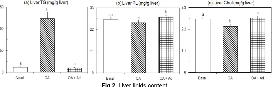
Fig 2. Liver lipids content
Rats were paired-fed OA supplemented diet with/or without adenine or a diet without OA (basal diet) for 10 days. Rats were killed by decapitation after a 9-h starvation. Values are expressed as mean± SEM of five rats. See EXPERIMENTAL SECTION for composition of diets. abDifferent letters indicate significant differences at P < 0.05.
RESULT AND DISCUSSION
Dietary adenine reversed the body weight and liver weight growths of OA-treated rats
The daily food intake was paired-fed for each animal in order to get the same quantity of macronutrient, vitamin, and mineral ingested. The macronutrient as sources of caloric food of each group was prepared in excess amount. It was therefore, the addition of food supplements, OA or adenine compound, to the basal diet provided the same quantity of the energy needed by each animal’s homeostasis metabolisms in each group during the time course. Thus, the effect of dietary adenine in OA-induced fatty liver by the given concentration could be elucidated from this experimental design.
The daily adjusted food intake is shown in Table 1. As shown in the table, those food intakes were nearly similar among the groups; there were however the final body weights decreased by the feeding OA food supplement compared to basal group, although the magnitude failed to reach significant level (P<0.05). Dietary adenine however reversed the inhibition of body weight gain in those OA-treated rats. Furthermore, the growth of liver showed that dietary OA significantly promoted the liver weight (P<0.05) but these promotions were reversed to basal level by additional adenine supplement. Dietary adenine therefore reversed the
body weight and liver weight growths of OA-treated rats to basal level.
Dietary OA induced fatty liver but these OA-treated
animals receiving additional adenine supplement
reversed the liver TG promotion
Fig. 2 shows the differences in liver lipid contents. The liver TG content of OA group markedly increased in comparison to the other groups (P<0.05), whereas the liver TG levels of OA-treated rats given dietary adenine was nearly similar with the basal group. As previously reported that if TG content inside the liver cells make up more than 5-10% of liver’s weight, the subject hold fatty liver [3-4]. This is further confirmed by the reports of Jou et al. [24] that hepatocyte accumulation of TG is a hallmark of fatty liver. The results of present study showed that liver TG content of OA group involved 12.35% of the liver weight, but basal and OA+Ad groups involved 1.14%, and 1.03%, respectively. The OA-treated rats, therefore, held fatty liver. Those liver TG contents are consistent with the tendencies of liver weight changes (Table 1). The enhancement of liver TG content of OA group compared to basal group was consistent with our previous studies [16-17] and the other [5-7]. The development of this fatty liver induced by OA might be associated with an over expression of sterol-regulatory element binding protein-1c (SREBP-1c) as reported by numerous authors [25-29] that over expression of
Yohanes Buang
SREBP-1c produces a pronounced elevation of hepatic TG concentrations leading to the development of NAFLD. Indeed, this result is constantly consistent with the studies as reported [5-7,11,16]. Both basal and adenine-containing groups, however, were free from fatty liver. Dietary adenine therefore reversed promotion of liver TG content in fatty liver induced by OA.
Data shown in Fig. 2 indicate that liver PL and Chol contents decreased slightly in OA-treated rats (P<0.05). These data were in agreement with the reports of Cha et al. [7] that liver PL and liver Chol contents decreased in OA-treated rats. However, those OA-treated rats receiving additional adenine supplement promoted significantly these lipids content (P<0.05). Both latter data were in agreement with the report of Windmueller [30] that both cholesterol and PL contents increased in liver tissue of rats that were given OA and received additional adenine supplement. Dietary adenine therefore reversed inhibition of liver PL and cholesterol contents in OA-induced fatty liver.
Dietary adenine reversed inhibition of serum lipids secretion in OA-induced fatty liver
The excessive accumulation of liver lipids can result from two mechanisms: 1) the impairment of secretion of VLDL from liver tissues into the bloodstream, and 2) increased expression of lipogenic enzymes combined with the impaired entry of fatty acids into the mitochondrial β-oxidation pathway as reported [16]. VLDL is synthesized in the liver and function as lipids transporter particles of the liver [8]. The lipids majority contained by the VLDL is neutral lipids such as TG and therefore VLDL plays a major role in the transportation of TG droplet from the liver inside cells into the bloodstream [8,31]. The increased VLDL secretion depends directly on the matured VLDL levels at the final step of VLDL synthesis.
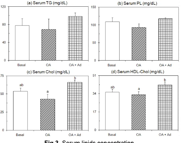
The results of the present study shown in Fig. 3 indicated that dietary food supplemented with OA caused decrease the serum TG, PL, total cholesterol, and the high-density lipoprotein (HDL)-cholesterol levels in comparison to the basal group (P<0.05). Their decreases were approximately by 12%, 15%, 20%, and 7%, respectively. Those results were in agreement with numerous studies previously [5-7, 16-17]. Furthermore, Pottenger et al. [32] reported that OA treatment interfere the secretion of VLDLs rather than with the synthesis of their protein. The reductions of serum lipid levels in OA-treated rats of the present study therefore were possible related to the intervention of VLDL secretion by the OA treatment. However, these OA-treated rats receiving additional adenine supplement promoted the serum lipid levels (P<0.05), in which those serum levels approximately increased by 26%, 8%, 23%, and 20%,
respectively, than that of the basal group. Overall, dietary OA-supplemented food inhibited serum lipid secretions; however those OA-treated rats receiving additional supplement adenine reversed the inhibition of those serum lipid secretions.
Dietary adenine reversed promotion of lipogenic enzyme and the inhibition of lipolytic enzyme in OA-induced fatty liver
Fatty acid is one of the substrates in lipids biosyntheses and fatty acid degradation. The biosyntheses of those fatty acids are catalyzed by FAS, a key enzyme of fatty acid synthesis, and their degradations are catalyzed by CPT-1, a key enzyme of fatty acid β-oxidation. Although, the increased fatty acid concentrations is possible to promote the liver lipid levels; however the lipid levels of the liver depend on the rates of VLDL secretion and mitochondrial fatty acid
β-oxidation. Numerous studies [5-7,16-17] constantly reported the reduction of mitochondrial fatty acid β -oxidation accompanied with excessive accumulation of liver TG content in OA-treated rats. The enhancement of the liver TG content in OA-induced fatty liver accompanied with decrease liver PL and cholesterol levels is in agreement with the reports of Cha et al. [7].
The result of present study shown in Fig. 4 indicated that FAS activity of OA group increased approximately by 33% compared to the basal group (P<0.05). These data suggested that dietary OA induced fatty acid biosynthesis in their liver cells and therefore enhanced the fatty acid concentration. Hence, the OA intake might cause promotion of FAS gene transcription because these intakes economically provide a short pathway in pyrimidine nucleotides biosynthesis and therefore promoted FAS enzyme content. As the result, FAS activity increased. Although, these mechanisms are not clear yet but our previous study found that dietary OA modulated the FAS gene transcription and its enzyme activity [16]. The promotion of FAS activity in OA-treated rats was constantly consistent with the previous studies [5-7,16-17]. However, the present study found that dietary adenine inhibited FAS activity in OA-induced fatty liver.
Lehninger et al. [8] reported that adenine is metabolized through the salvage pathway, in which it is directly reacted with phosphoribosyl pyrophosphate to generate adenosine monophosphate (AMP). Thereafter, AMP kinase catalyzes the phosphorilation of AMP to generate adenosine diphosphate (ADP) and regulates the metabolic hepatic disorders as reported [33]. Both AMP and ADP trigger the respiration reactions and therefore induced mitochondrial fatty acid
Fig 3. Serum lipids concentration
Rats were paired-fed OA supplemented diet with/or without adenine or a diet without OA (basal diet) for 10 days. Rats were killed by decapitation after a 9-h starvation. Values are expressed as mean± SEM of five rats. See EXPERIMENTAL SECTION for composition of diets. abDifferent letters indicate significant differences at P < 0.05.
Fig 4. The activities of lipogenic and lipolytic enzymes
Rats were paired-fed OA supplemented diet with/or without adenine or a diet without OA (basal diet) for 10 days. Rats were killed by decapitation after a 9-h starvation. Values are expressed as mean± SEM of five rats. See Experimental Section for composition of diets. abDifferent letters indicate significant differences at P < 0.05.
and converted to nucleotides intracellular and this is supported by the reports of Karasawa et al. [35] that dietary food fortified with adenine did not find free adenine compound in serum and liver tissues. The results of the present study showed that OA-treated rats receiving additional adenine supplement promoted CPT-1 activities (P<0.05). This data indicated that the activities of fatty acid β-oxidation were promoted by the treatment and possible increase the respiration rates to generate adenosine triphosphate. The activities of CPT-1 enzyme of OA-induced fatty liver however decreased significantly in comparison to basal group (P<0.05). The reduction of CPT-1 activity indicated that mitochondrial fatty acid β-oxidation decreased in OA-induced fatty liver. The reduction of this CPT-1 activity might be associated with promotions of long-chain fatty acid level in the OA-treated rats (data not shown). This was
because the phenomenon in agreement with the reports of Ventura et al. [36] that long-chain acyl-coenzyme A (CoA) esters are potent inhibitors of several enzymes in the oxidative phosphorylation system, in which CPT-1 is a key enzyme in production of acetyl-CoA before reach the oxidative phosphorylation pathway. Those results also were in agreement with the reports of Boer et al. [37] that long-chain 3-hydroxyacyl-CoA dehydrogenase deficiency induced long-chain fatty acid oxidation disorders and carnitine palmitoyl-CoA transferase II deficiency.
Furthermore, the alleviation of OA-induced fatty liver by dietary adenine might be associated with stimulations of nuclear transcription factors such as peroxisome proliferators-activated receptor α (PPARα). Although the latter mechanism was not elucidated yet but Hu et al. [29] recently reported that alleviation of
Yohanes Buang
orotic acid-induced fatty liver in rats is through PPARα signaling. Overall, the modulation of FAS accompanied with inhibition of CPT-1 enzymes (Fig. 4) and the reduction in serum lipid secretions (Fig. 3) yielded excessive accumulation of TG in the liver. As a result, dietary OA promoted liver TG content as shown in Fig. 2. Indeed, the attenuation of FAS and the stimulation of CPT-1 activities in OA-induced fatty liver given dietary adenine indicated that adenine interfere the fatty acid biosynthesis accompanied with promotions of mitochondrial fatty acid β-oxidation and therefore ameliorates accumulation of liver TG content.
CONCLUSION
Dietary OA promoted fatty acid biosynthesis whereas reduced capacities of mitochondrial fatty acid β -oxidation. However, the OA-treated rats given dietary adenine inhibited fatty acid biosynthesis and reversed mitochondrial fatty acid β-oxidation to basal level. Therefore, dietary adenine alleviates fatty liver induced by OA which is associated with the inhibition of lipogenic enzyme activity accompanied with a promotion of mitochondrial fatty acid β-oxidation and the stimulation of lipid secretion from hepatic tissue into the bloodstream.
ACKNOWLEDGEMENT
The author wishes to express high appreciation for the suggestions and the continued encouragement from Prof. Teruyoshi Yanagita, PhD, the Professor of Saga University, and excellent assistance of Dr. Koji Nagao in enzymatic determinations and the useful assistance from Dr. Yu-Ming Wang in handling instruments and animals. The appreciation is also given to all of the members of Laboratory of Applied Biochemistry-Saga University for the contributions. The author also gives thanks to the Japanese Monbukagakusho for providing the fund of the research.
REFERENCES
1. Li, Z., Clark, J., and Diehl, A.M., 2002, Clin. Liver Dis., 6, 4, 867–877.
2. Harrison, S.A., and Diehl, A.M., 2002, Semin. Gastrointest. Dis., 13, 1, 3–16.
3. Website. http://www.patient.co.uk/health/Fatty-Liver-Disease.htm. Accessed July 10, 2009.
4. Sherlock, S., and Dooley, J., 1997, Diseases of the liver and biliary system, Blackwell Science, Oxford. 5. Raisonnier, A., Bouma, M.E., Salvat, C., and Ifante,
R., 1981, Eur. J. Biochem., 118, 3, 565-569.
6. Miyazawa, S., Furuta, S., and Hashimoto, T., 1982,
Biochim. Biophys. Acta, 711, 3, 494–502.
7. Cha, J.Y., Cho, Y.S., Kim, I., Anno, T., Rahman, S.M., and Yanagita, T., 2001, Plant Foods Hum. Nutr., 56, 4, 349–358.
8. Lehninger, A.L., Nelson, D.L., and Cox, M.M., 1993, Principles of Biochemistry, 2nd ed., Worth Publishers, Inc. USA.
9. Dixon, J.B., Bhathal, P.S., and O’Brien, P.E., 2001,
Gastroenterology, 121, 1, 91–100.
10. Schwimmer, J.B., Deutsch, R., Rouch, J.B., Behling, C., Newbury, R., and Lavine, J.J., 2003, J. Pediatr., 143, 4,. 500–505.
11. Cotrim, H.P., Paraná, R., Braga, E., and Lyra, L., 2000, Am. J. Gastroenterol., 95, 10, 3018–3019. 12. Rolo, A.P., Teodoro, J.S., Peralta, C.,
Rosello-Catafau, J., and Palmeira, C.M., 2009, Transpl. Int.., 22, 11, 1081–1090.
13. Tilg, H., and Hotamisligil, G.S., 2006,
Gastroenterology, 131, 3, 934–945.
14. Pagano, C., Soardo, G., Pilon, C., Milocco, C., Basan, L., Milan, G., Donnini, D., Faggian, D., Mussap, M., Plebani, M., Avellini, C., Federspil, G., Sechi, L.A., and Vettor, R., 2006, J. Clin. Endocrinol. Metab., 91, 3,1081–1086.
15. Tilg, H., and Moschen, A.R., 2008, Mol. Med., 14, 3-4, 222–231.
16. Buang, Y., Wang, Y.M., Cha, J.Y., Nagao, K., and Yanagita, T., 2005, Nutrition, 21, 7-8, 867–873. 17. Buang, Y., Cha, J.Y, Nagao, K., Wang, Y.M.,
Inoue, N., and Yanagita, T., 2004, J. Nutr. Sci. Vitaminol., 50, 4 272–276.
18. Folch, J., Lees M., and Sloane-Starley, G.H., 1957,
22. Lowry, O.H., Rosebrough, N.J., Farr, A.L., and Randal, R.J., 1951, J. Biol. Chem., 193, 1, 265–
27. Matsuzaka, T., Shimano, H., Yahagi, N., Amemiya-Kudo, M., Okazaki, H., Tamura, Y., Iizuka, Y., Ohashi, K., Tomita, S., Sekiya, M., Hasty, A., Nakagawa, Y., Sone, H., Toyoshima, H., Ishibashi, S., Osuga, J., and Yamada, N., 2004, Diabetes, 53, 3, 560–569.
Yohanes Buang
29. Hu, X.Q., Wang, Y-M., Wang, J-F., Xue, Y., Li, Z-J., Nagao, K., Yanagita, T., and Xue, C-H. , 2010,
Lipids in Health and Disease, 9, 25, 1-9.
30. Windmueller, H.G., 1964, J. Biol. Chem., 239, 2, 530–537.
31. Stipanuk, M.H. 2006, Biochemical, Physiological, and Molecular Aspects of Human Nutrition, 2nd ed., Saunders-Elsevier, New York, 666–672.
32. Pottenger, L.A., and Gets, G.S., 1971, J. lipid Res., 12, 450–459.
33. Viollet, B., Foretz, M., Guigas, B., Horman, S., Dentin, R., Bertrand, L., Hue, L., and Andreelli, F., 2006, J. Physiol.,574, 1, 41–53.
34. Yu, V.Y.H, 1998, HK J. Paediatr., 3,122-126. 35. Karasawa Y., Takasaki, K., and Koh, K., 2002, J.
Poult. Sci., 39, 4, 285–291.
36. Ventura, F.V., Tavares, D.A.I., and Wanders R.J.A., 2007, Biochem. Biophys. Res. Commun., 352, 4, 873–878.
37. den Boer, M.E.J., Wanders, R.J.A., Morris, A.A.M., IJlst, L., Heymans, H.S.A., and Wijburg, F.A., 2002,