Characteristics of nanosize spinel Ni
0.9Fe
2Co
0.1O
4prepared by sol-gel
method using pectin as an emulsifying agent
Septhian Try Sulistiyoaand Rudy Situmeangb
aGraduate student in Department of Chemistry, University of Lampung bDepartment of Chemistry, University of Lampung
Abstract
Ni0.9Fe2Co0.1O4nanomaterial have been prepared using a sol-gel method. Preparation of material was carried out by dissolving nitrate salts of iron, cobalt and nickel, in pectin solution and then the sample was stirred throughly using magnetic stirrer while adjusting pH to 11. After freeze-drying process, the sample was subjected to calcination treatment and subsequently characterized using the techniques of X-ray diffraction (XRD), Scherrer Methods, FTIR and DT-TG analysis. The results of XRD characterization indicated that materials consist of two crystalline phases, such as CoFe2O4and NiFe2O4. These two crystalline phases are superimposed. DT-TGA result showed that spinel Ni0.9Fe2Co0.1O4formed above 400oC. Then, PSA determination proved that the grain size of spinel ferrites is a range of 30 95.2 nm as much as 21%. Crystallite size calculation using Scherrer equation, proved that the size is 31.95 nm. Its size increased as temperature calcination augmaunted.
Keywords Nanomaterial, spinel ferrites, BrØnsted Lowry and Lewis acid sites, freeze-drying
1. Introduction
Spinel structured materials have an interesting physical and chemical characteristics therefore these materials have been utilized abroadly in industrial applications such as catalysts (Van der Laan & Beenackers, 1999; Rajput & Kaur, 2013; Mangrulkar et al., 2012), memory devices (Lu et al., 2007], sensors (Abdel-Latif, 2012; Tudorache & Petrila, 2013], magnetic materials (Mukherjee & Mitra, 2014; Shokrollahi & Sharifi, 2013) and materials for stealth technology (Pullar, 2012; Zhao et al., 2009). This interesting both chemical and physical properties is determined mainly by the occupations of cations at both octahedral
and tetrahedral sites in the spinel ferrites, M1M2Fe2O4 where M1and M2are +2 or +3 cations,
and M1 or M2 could be Co+2, Ni+2, Cu+2, Zn+2, Mo+3 and others (El-Sayed, 2003; Juang &
Mathew, 2007). One of the other factors determining its performance on the application is the method of preparation.
Preparation methods that have been used currently to produce spinel ferrites are co-precipitation (Murthy et al., 2009; Derakhshi et al., 2012), sol-gel (Singhal et al., 2005; Trisunaryanti et al., 2008), soft mechanochemical (Lazrevi et al., 2012), hydrothermal (Nejati & Rezvanh, 2012; Zhou et al., 2005) and precipitation (Yang et al., 1999; Yongvanich et al., 2010; Atashi et al., 2013). The aim of those preparations is to devote and focuse on two aspects e.g development of synthetic method and development of various materials which satisfy the continuous growing of utilization.
low temperature. The phases developed for nano-material is identified qualitatively by XRD analysis. Microstructural size is determined by Particle Size Analyzer and Scherrer equation. Then, FTIR analysis is done successively to confirm the bond formation and acid sites of those nano materials .
2. Materials and methods
2.1 Materials
Materials used in this work are pectin powder, Ni(NO3)2. 6H2O (Merck, 99%), Fe(NO3)3
. 9H2O (Merck, 99%), Co(NO3)3. 6 H2O (Merck, 99%), pyridine (C5H5N, J.T Baker), NH3(Merck,
99%), and aquades.
2.2 Instrumentations
The instruments used for characterization were Fourier Transform Infrared (FTIR) spectrometer (Shimadzu Prestige-21) for identifying the presence of functional groups, and Differential Temperature and Gravimetry analysis (DT-GA) for elucidating the formation process . Then, a Philips X-ray diffractometer (XRD) model PW 1710 with Cu-K radiation was used for structural and crystalline phases identification.
2.3 Preparation of
Ni
0.9Fe
2Co
0.1O
4nanomaterial
Stoichiometric amount of Ni (II) nitrate hydrates, Co (II) nitrate hydrates and Fe (III)
nitrate hydrates were dissolve in distilled water, having compositions Ni0.9Fe2Co0.1O4 under
magnetic stirring for 1 hour, respectively, followed by mixing each solution to make final solution molar ratio between nitrates to pectin is 3:2. Adjust the pH=11 in the above solution
by addition of ammonia, and heat it at 80oC with continue stirring to form viscous gel. Dried
the gel using freeze dryer for 7 hours to form the precursors networks and calcined at 600
and 800oC for 3hours. Finally Co doped Ni - ferrite nanoparticles has prepared.
2.4 Characterization of Ni0.9Fe2
Co0.1O4
nanomaterial
Differential temperature and gravimetry analysis
Sample of approximately 5 6 mg precursor was placed in Pt-sample pan and another pan was allowed blank as a reference. The experiment was carried out under a flowing nitrogen atmosphere (50 ml/min, SII TG/DTA 7300). Then, scan analysis was worked in the range of 25
to 600oC with a heating rate of 5oC min-1.
X- ray diffractogram analysis
X-ray powder diffraction pattern of the sample was recorded from 2= 10 to 90oon a Philips
diffractometer Model PW 1710 using Cu Kradiation at a step 0.02oper second. The phase
Acid sites analysis of Ni0.9Fe2Co0.1O4
After heating at 120oC, sample was transferred into a crucible and placed in vaccumized
dessicator. Pyridine was transferred into another crucible and placed in the dessicator to allow the vapour of the pyridine to contact with the sample. After 24 hours, the sample was taken from dessicator and left on open air for 2 hours to expell the physically adsorbed pyridine from the sample. Finally, the sample was analyzed using the FTIR spectroscopy. The analysis was conducted by grinding the sample with KBr of spectroscopy grade,and
scanned over the wave number range of 4000 400 cm1(Parry, 1963; Ryczkowski, 2001).
3. Results and discussion
Differential temperature and gravimetry analysis
The precursor of Ni0.9Fe2 Co0.1O4 nanomaterial prepared were characterized using
thermal analysis techniques such as differential temperature and thermal gravimetric analysis (DT-TGA). Thermal gravimetric analysis was used to measure the losses of weight as a function of temperature. The TGA results for the 4.8 h aged precursor containing [Ni]/[Co]
= 9/1 molar ratio is shown in Figure 1.
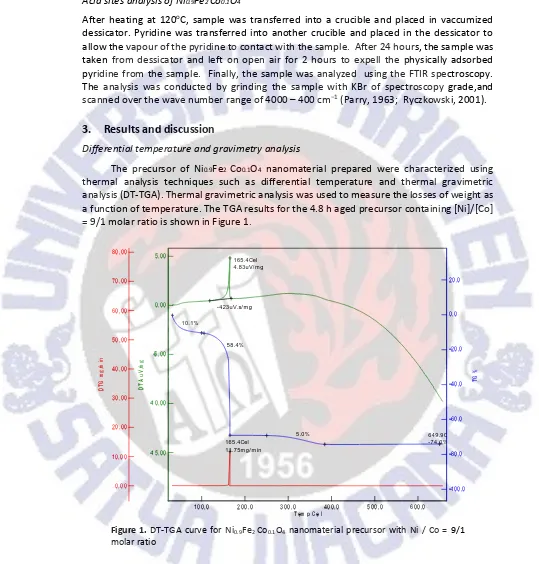
Figure 1.DT-TGA curve for Ni0.9Fe2Co0.1O4nanomaterial precursor with Ni / Co = 9/1 molar ratio
There is a gradual weight loss upon heating to about 200°C; above this temperature
there is a crucial weight loss until 400oC. During the first period of the weight loss, the
exothermic process is occured at 165.5oC and the energy of 4.83V/mg. The first weight loss
with temperatures range of 80 200°C and is due to the removal of absorbed water in pectin networks. The second period of weight losses with temperatures at around 200 400°C, are
considered to be due to the decomposition of pectin, and nitrate precursors, so that the decomposition at these high temperature regions produce water, carbon dioxides and
nitrogen oxides simultaneously. Above 400oC, there is no weight loss and the formation of
Ni0.9Fe2Co0.1O4nanomaterial is started.
X-ray diffractogram analysis
The prepared sample was characterized by Philips PW-1710 X-ray diffractometer using
Cu-Kradiation ( =1.5406 ) source. The X-ray diffraction patterns of the prepared sample
is shown in Figure 2.
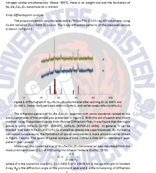
Figure 2.Diffractogram of Ni0.9Fe2Co0.1O4nanomaterial after calcining at: (a) 800oC and (b) 600oC. (Notes: black bar-peaks refers to CoFe2O4and red bar-peaks refers to NiFe2O4)
The difraction pattern of Ni0.9Fe2Co0.1O4 together with some standards related to the
predicted phases of the sample was presented in Figure 2. With the aid of search and match method, using Phase Identification from Powder Diffraction Files, it was found that the major
phase is spinel NiFe2O4 (JCPDF- 10-0325), CoFe2O4 (JCPDF-22-1086). In general, it can be
implied that both NiFe2O4and CoFe2O4crystalline phases are superimpossed. By increasing
calcination temperature, the formation of spinel compounds is more pronounced as shown in Figure 2 above. The peaks of spinel compoud were shifted a little bit to references peak pattern (bar - peaks).
Moreover, the crystallite size of Ni0.9Fe2Co0.1O4nanomaterial was calculated from the
most prominent peak (211) of XRD using the Scherer formula (Cullity, 1978):
=
λcos
,
whereDis the crystallite size (nm), =1.5406 or 0.15406 nm is the wavelength of incident
X-ray, is the diffraction angle at the prominent peak and is the broadening of diffraction
line measured at half maximum intensity, π
×
(in radian), and k is a constantwith range of 0.9 1.0 (in this calculation,k= 0.94).is the Bragg s angle in degree unit. The
As shown in Table 1, the crystallite sizes of Ni0.9Fe2Co0.1O4samples are in the range of
11 21 nm, demonstrating the efficacy of the proposed method to produce nano-size spinel
Ni0.9Fe2Co0.1O4. In relation with the calcination variation, it was found that the higher the
calcination temperature used the larger the crystallite size formed.
Table 1. Crystallite size calculation using Scherrer equation Ni0.9Fe2Co0.1O4
Calcined at 2, deg hkl
FWHM Size of Crystallite (D),
nm
degree ()/ radian
600oC 35.16 211 0.7510 0.013113 11.58
800oC 35.32 211 0.4265 0.007974 20.41
Acid sites analysis ofNi0.9Fe2Co0.1O4
Fourier transform infrared spectroscopy was applied to identify the functional groups present in the sample, primarily to identify the existence of Lewis and BrØnsted Lowry acid sites. The acid sites identification is of particular importance since the acidity is acknowledged as very important characteristic which determine the performance of a
material as a catalyst (Parry, 1963; Ryczkowski, 2001). The FTIR spectra of Ni0.9Fe2Co0.1O4
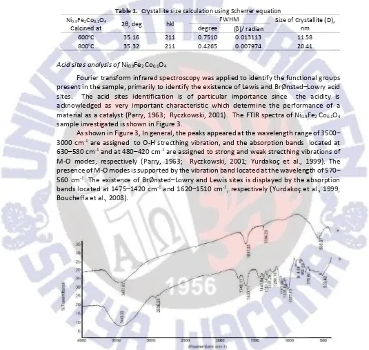
sample investigated is shown in Figure 3.
As shown in Figure 3, In general, the peaks appeared at the wavelength range of 3500
3000 cm-1are assigned to O-H strecthing vibration, and the absorption bands located at
630 580 cm-1and at 480 420 cm-1are assigned to strong and weak strecthing vibrations of
M-O modes, respectively (Parry, 1963; Ryczkowski, 2001; Yurdakoç et al., 1999). The presence of M-O modes is supported by the vibration band located at the wavelength of 570
560 cm-1. The existence of BrØnsted Lowry and Lewis sites is displayed by the absorption
bands located at 1475 1420 cm-1and 1620 1510 cm-1, respectively (Yurdakoç et al., 1999;
Boucheffa et al., 2008).
Figure 3.FTIR Spectrum of Ni0.9Fe2Co0.1O4after exposed to pyridine vapour: (a) calcined at 600oC and (b) calcined at 800oC.
In the samples investigated, calcined at 600 and 800oC, the presence of O-H functional
3451.80 and 3443.00 cm-1, respectively. The existence of Lewis acid sites in the sample
calcined at 800oC is displayed by the adsorption bands located at 1746.11 and 1636.71 cm-1
which indicate that the pyridine was bound to the surface of the sample by coordination
bond [Parry, 1963] and more pronounced than that of calcined at 600oC, while the existence
of BrØnsted Lowry acid sites is presented by the absorption bands located at 1445.64 and
1381,34 cm-1for the sample calcined at 800oC and 1384.29 cm-1for the sample calcined at
600 oC. By comparing the intensities of the absoprtion bands associated with Lewis and
BrØnsted Lowry acid sites, it can be concluded that the acid characteristic of the sample is dominated by Lewis acid. In the fingerprint region of the spectra, the absorption band representing strecthing vibration of Fe-O and bending vibration of Ni-O and Co-O was
detected at 588.97, 529.42 and 440 cm-1[Ryczkowski, 2001], suggesting the existences of
Fe-O-Ni and Fe-O-Co bond which confirms the formation of Ni0.9Fe2 Co0.1O4 structure as
expected. In addition, the peaks appeared which are displayed by the absorption bands
located in the range of 914.58 770 cm-1 refers to the possibility of metal-metal bond
formation.
4. Conclusion and remarks
This current study demonstrated the potential of pectin solution as a emulsifying agent for preparation of nano-size materials using sol-gel method. The XRD results revealed that
the crystallite size of the Ni0.9Fe2Co0.1O4sample prepared is in the range of 11 to 21 nm. The
samples were found to exhibit Lewis and Bonsted Lowry acid characteristics, with Lewis acid as the dominant site, as revealed by the FTIR analyses.
Acknowledgment
The authors wish to thank and appreciate the Directorate General Higher Education Republic of Indonesia for research funding provided through The National Grant for Competitive Research University of Lampung and Ministry of Indonesia Higher Education Program.
References
Abdel-Latif, I.A. (2012). Fabrication of nano-size nickel ferrites for gas sensors applications. J. Phys.,
1(2), 50 53.
Atashi, H., Sarkaria, M., Fazlollahib, F., Mirzaeid, A.A., & Heckerb, W.C. (2013). Using different preparation methods to enhance Fischer Tropsch Products over iron-based catalyst. Chem.
Biochem. Eng. Q.,27(3), 259 266.
Boucheffa, Y., Benaliouche, F., Ayrault, P., Mignard, S., & Magnoux, P. (2008). NH3-TPD and FTIR spectroscopy of pyridine adsorption studies for characterization of Ag- and Cu- exchanged X-zeolites.Microporous and Macroporous Material,113(1 3), 80 88.
Cullity, B.D. (1978).Elements of X-ray diffraction(2nd ed.). Addison-Wesley, London.
Derakhshi, P., Khorrami, S.A., & Lotfi, R. (2012). An Investigation on synthesis and morphology of nickel doped cobalt ferrite in the presence of surfactant in different calcination temperature by coprecipitation route.World Appl. Scie. J.,16(2), 156 159.
El-Sayed, A.M. (2003). Electrical conductivity of nickel zinc and Cr substituted nickel zinc ferrites. Mat.
Chem. Phys.,82, 583 587.
Lazrevi , Z. ., Jovaleki , ., Milutinovi , A., Rom evi , M.J., & Rom evi , N. . (2012). Preparation and characterization of nano ferrites.Acta Physica Polonica A,121(3), 682 686.
Mangrulkar, P.A., Polshettiwar, V., Labhsetwar, N.K., Varma, R.S., & Rayalu, S.S. 2012. Nano-ferrites for water splitting: unprecedented high photocatalytic hydrogen production under visible light.
Nanoscale,4(16), 5202 5209.
Mukherjee, S., & Mitra, M.K. (2014). Characterization of perovskite-spinel nanocomposites (BFO-ZFO) ferrites prepared by chemical route.J. Austr. Ceram. Soc.,50(2), 180 187.
Murthy, Y.L.N., Viswanath, I.V.K., Rao, T.K., & Singh, R. (2009). Synthesis and characterization of nickel copper ferrite.Int. J. ChemTech Research,1(4), 1308 1311.
Nejati, K., & Zabihi, R. (2012). Preparation and magnetic properties of nano size nickel ferrite particles using hydrothermal method.Chem. Centr. J.,6, 23.
Parry, E.P. (1963). An Infrared study of pyridine adsorbed on acidic solids characterization of surface acidity.J. Catal.,2(5), 371 379.
Pullar, R.C. (2012). Hexagonal ferrites: A review of the synthesis, properties and applications of hexaferrite ceramics.Prog. Mater Sci.,57(7), 1191 1334.
Rajput, J.K., & Kaur, G. (2013). CoFe2O4 nanoparticles: An efficien heterogeneous magnetically separable catalyst for click synthesis of arylidene barbituric acid derivatives at room temperature.Chin. J. Catal.,34, 1697 1704.
Ryczkowski, J. (2001). IR spectroscopy in catalysis.Catalysis Today,68, 263 381.
Singhal S., Singh, J., Barthwal, S.K., & Chandra, K. (2005). Preparation and characterization of nanosize nickel-substituted cobalt ferrites (Co1xNixFe2O4).J. Sol. Stat. Chem.,178(10), 3183 3189. Shokrollahi, H., & Sharifi, I. (2013). Structural, magnetic and mossbauer evaluation of Mn substituted
Co Zn ferrite nanoparticles synthesized by co-precipitation.J. Magn. Magn. Mat.,334, 36 40. Trisunaryanti, W., & Oktaviano, H.S. (2008). Sol gel derived Co and Ni based catalysts: Application for
steam reforming of ethanol.Indo. J. Chem., 8(1), 47 53.
Tudorache, F., & Petrila, I. (2013). Humidity sensor applicative material based on copper-zinc-tungsten spinel ferrite.Mater. Lett.,108, 129 133.
Valenzuela, R., Cruz-Franco, B., Gaudisson, T., Ammar, S., Bolarín-Miró, A.M., Mazaleyrat, F., Nowak, S., Vázquez-Victorio, G., Ortega-Zempoalteca, R., & de Jesús, F.S. (2014). Magnetic properties of nanostructured spinel ferrites.IEEE Trans. on Magn.,50(4), 280 286.
Van der Laan, G.P., and Beenackers, A.A.C.M. (1999). Kinetics and selectivity of the Fischer Tropsch synthesis: A literature review.Catal. Rev.: Sci. Eng.,41(3 4), 255 318.
Yang, J.M., Tsuo, W.J., & Yen, F.S. (1999). Preparation of ultrafine nickel ferrite powders using mixed Ni and Fe Tartrates.J. Solid State Chem.,145(1), 50 57.
Yongvanich, N., Visuttipitukkul, P., Leksuma, P., Vutcharaammat, V., & Sangwanpant, P. (2010). Sinterability and microstructure of bi-added SnO2 nanomaterials by precipitation method.
Journal of Metals, Materials and Minerals,20(3), 67 72.
Yurdakoç, M., Akçay, M., Tonbul, Y., & Yurdakoç, K. (1999). Acidity of silica-alumina catalysts by amine titration using Hammett indicators and FT-IR study of pyridine adsorption. Turk. J. Chem.,23, 319 327.
Zhao , D.-L., Lv, Q., & Shen, Z.-M. (2009). Fabrication and microwave absorbing properties of Ni Zn spinel ferrites.Journal of Alloys and Compounds,480(2), 634 638.