LABORATORY MANUALS
Fundamentals of Biomedical
Science I
Editors:
Dr. med. Tri Hanggono Achmad, dr. Gaga Irawan Nugraha, dr., M.Gizi., Sp.GK
Contributors (alphabetically): Abdullah Firmansah, dr., M.Kes., Sp.GK
Dr. Dhiah Dhianawaty Dr. med. Tri Hanggono Achmad, dr. Gaga Irawan Nugraha, dr., M.Gizi., Sp.GK
Freshmen Year Program
Faculty of Medicine Universitas Padjadjaran
Bandung
PREFACE
As a part of the ongoing evolution/revolution in medical education, the basic sciences and clinical sciences are to be integrated throughout the curriculum. Although there has been an orchestrated and organized integration of the clinical sciences, such has not been the case generally for the basic sciences. Student in most modern medical schools, especially in the basic science courses, are required to memorize a large number of “facts”. Often the sets of facts they are required to memorize are chosen by the instructor based on what he or she knows and is familiar with. The sets of facts may or may not be relevant to medical practice. Furthermore, the student, a passive recipient in this approach, may or may not learn the facts in such a way that they are useful in future practice, regardless of whether the student is able to demonstrate on examination that his or her memory is good.
Bridging the basic science years with the clinical years using problem-based learning will enhance the skills of creating a differential diagnosis and appropriately using diagnostic tests. Problem-based learning is the learning that results from the process of working toward the understanding or resolution of a problem. Learning through problem-solving is much more effective for creating in a student’s mind a body of knowledge useable in the future than is traditional memory-based learning. The physician skills most important for patients are problem-solving skills, not memory skills.
This book is design as the manual for the students, which will guide them in doing their laboratory practical work and assist them to understand better the theory they learn during the lecture. In its chapter it consists of general objective of the laboratory activity, patient’s case presentation, topics to be discussed, which are related to topics they learn during the class lecture, laboratory methods, short review of the clinical case and some assignments.
Without support, encouragement and participation of many people, this book would never been accomplished. Our ersonal p and very deep appreciation and acknowledgement goes to all of the contributors for accepting the challenge of preparing the contents, for sharing their ideas and recommendations to construct this manual, for accepting so readily suggestions to modify their contributions, and for their understanding and cooperating throughout the period of the preparation. To each we extend our sincerest thanks for a job well done.
In any project, somebody must accept the responsibility for the final product. The decisions concerning the selection of topics and format, reviewing the drafts, and responsibility for the final checking of the manual were entirely ours. We welcome comments, criticisms, and suggestions from the students, faculty, colleagues, and professionals who read this manual. It is our hope that this work will be of a value to those searching on the exciting experience of learning biochemistry where the basic of medical science begins and is expanding so rapidly.
GENERAL INSTRUCTION
Before starting any experiment the student should read the instructions carefully, paying attention to all details, and should be quite certain about what he/she is trying to do. Be prepared for the discussion, read a lot about the discussion topic. During the discussion, do not hesitate to ask or to express opinion. Try to think and speak logically. Try to listen to your friend’s opinion. The result of the experiment and discussion should be written and handed over to the tutor on the next meeting.
LABORATORY RULES
1. Do not replace any solution in a reagent bottle; take only the minimum amount required.
2. Replace the stopper immediately a solution has been taken from a bottle and take care not to mix stopper.
3. Do not remove bottles of special reagents from side shelves to your own benches. 4. Never mouth-pipette a corrosive fluid, always use pipette filler.
5. Cleanliness is essential in all biochemical work. Make sure that your glassware is clean and dry.
6. When you have finished work, leave your bench clean and dry, just as you expect to find it. See that all waste material is put in the wastebasket provided and not into the drains. Strong acids or bases should not be poured down the drain, unless accompanied by a large volume of water.
7. Be careful with the use of centrifuge or spectrophotometer equipment; do not use them before you know exactly how to operate them.
8. Inspect your the content of your bench every time before and after you use them. Any damage or loss must be reported immediately.
CHAPTER I. BIOMOLECULES
Living organisms are enormously complex. Higher organisms correspondingly have a greater complexity. Human beings (Homo sapiens), for example, may contain 100,000 different types of molecules, although only a minor fraction of them have been characterized. Living organisms have an underlying regularity that derives from their being constructed in a hierarchical manner. Anatomical and histological studies have shown that multicellular organisms are organizations of organs, which are made of tissues consisting of cells, composed of subcellular organelles. At this point in our
hierarchical descent, we enter the biochemical realm since organelles consist of supramolecular assemblies, such as membranes or fibers, which are organized clusters
of macromolecules (polymeric molecules with molecular masses from thousands of dalton on up).
Living things in general, contain different types of macromolecules: protein (Greek:
proteios, of first importance), nucleic acids, and polysaccharides (Greek: sakcharon, sugar). All of these substances have a modular construction; they consist of linked monomeric units that occupy the lowest level of our structural hierarchy. Thus proteins are polymers of amino acids, nucleic acids are polymers of nucleotides, and polysaccharides are polymers of sugars. Lipids (Greek; lipos, fat) the fourth major class of biological molecules, are too small to be classified as macro molecules but also have a modular construction. Proteins are all synthesized from the same 20 species of amino acids, nucleic acids are made from 8 types of nucleotides (4 each in DNA and RNA), and there are ~ 8 commonly occurring types of sugars in polysaccharides. The great variation in properties observed among macromolecules of each type largely arises from the enormous number of ways its monomeric units can be arranged and, in many cases, derivatized. Besides the existing macromolecules, living cells also contain minerals in form of kations and anions, such as Na+, Ca+2, Cl-, SO4
-2
and PO4 -3
The aim of studying biochemistry is to understand the biological structure and function of living organisms in chemical terms. One of the most excellent approaches to understand biological phenomena was to purify an individual chemical component, the biomolecules, such as protein, and to characterize its chemical structure or catalytic activity.
In this chapter of the laboratory exercises, students will learn more about molecules and macromolecules that build the living cells. A clinical case of patient with chronic renal failure post streptococcal glomerulonephitis, which presents several sign and symptoms due to biomolecular changes of the body, will be presented as an entry to discuss the functional related structure of biomolecules. The students will conduct plasma protein electrophoresis, the assays of plasma sodium, blood urea nitrogen and creatinine using spectrophotoscopic principle, and determine several minerals in the urine. Since dietary management is one of the modality of treatment in this patient, the students have also to take biomolecular aspect of dietary resources into their consideration during the discussion.
CLINICAL PROBLEM
This is a routine visit of Mrs. Gloria Nefrita Koronis to the Renal Dialysis Unit, Hasan Sadikin Hospital. She is 35 years old diagnosed to have Chronic Renal Failure. As usual, she comes as a very anxious, pale woman. She stated notifying increased weakness and fatigue with gradually increasing shortness of breath over the last 6 months. She lost her appetite gradually without significantly decrease in bodyweight. She is turned down for life insurance a couple of years ago because of albumin in her urine.
Physical Examination
Pulse 110 bpm: BP 180/110 mm.Hg; Respirations 30/ min and deep. Eye conjungtivae are pale;
Chest : rales in the bases of both lungs.
Chest X-ray
Mild pulmonary edema; generalized osteomalacia with resorption of the distal portions of the clavicules, consistent with secondary hyperparathyroidism.
Clinical Course
She is placed on dialysis. She does well on dialysis treatment and is placed in the waiting list for a renal transplant. A diet with protein restriction (0.6 gr./Kg) and mild dietary sodium restriction was instituted.
Laboratory Data of Gloria Nefrita Koronis
Present Normal
Hb (Haemoglobin) 6.1 12 – 16 g/dL
Creatinine 14.1 0.6 – 1.2 mg/dL
BUN 183 7 – 18 mg/dL
Calcium 6.5 8.4 – 10.0 mg / dL
Phosphate 12,6 3.0 – 4.5 mg / dL
Sodium 140 135 – 147 mEq/L
Potassium 6.2 3.5 – 5.0 mEq/L
Chloride 105 96 – 105 mEq/L
Bicarbonate 10 22 – 26 mEq/L
Albumin 1.2 3.5 – 5.5 gm/dL
Urine
Protein 4 + 0
Overview of Chronic Renal Failure and Post Streptococcal Glomerulonephritis
creatinine in an adult reaches about 3 mg/dL and no factors in the pathogenesis of the renal disease are reversible, the renal disease is highly likely to progress to end-stage renal disease (ESRD) over a very variable period ( from a few years to as many as 20 to 25). Unless contraindications are present such as terminal irreversible disease in another organ system(s) or the patient does not wish it, almost all patients in industrialized nations then receive renal replacement therapy (RRT). The symptoms of chronic renal failure depend on the severity and rapidity of the underlying renal disorder. When chronic renal failure develops slowly, most individuals renal failure is far-advanced (GFR < 10-15 mL/min).
Topic for discussion
- Hierarchy and classification of biomolecules
- Structure related function of important biomolecules
- General principle of analysis of biomolecules (electrophoresis and spectroscopy) - Biomolecules alterations in chronic renal failure and impact on its function
(edema)
Biomolecules in living organism: An overview
The human body is composed of a few elements that combine to form a great variety of molecules. Four elements compose over 95 % of the human body. These elements and their percentages are : hydrogen (H) 10 %, oxygen (O) 65 %, carbon (C) 18 %, and nitrogen (N) 3 %, they are the major constituents of most bimolecular.
patients with electrolyte imbalances, whereas Fe2+ is found in iron-deficiency anemia, and I- in thyroid diseases.
The plasma proteins constitute 7 to 9 percent of the plasma and are substances characteristic for the plasma. They are large enough that they pass through blood capillary membranes with difficulty and tend to remain within the bloodstream. Here they are primarily responsible for osmotic return of filtered water to the capillaries from the interstitial fluid. Other factions served by proteins include : Contributing to the viscosity of the plasma; Creating a suspension stability in the blood that aids in maintaining dispersion of materials; Serving as reserve of amino acids (not usually used for metabolism but available); and Serving as buffers. By relatively techniques such as adding certain concentrations of salts to the plasma, three main fraction of proteins may be separated. They are designed as albumins, globulins, and fibrinogen.
Albumins are the most plentiful (55 to 64 %) of the proteins, and are present to the extent of 4 to 5 gm per ml of blood. They are also the smallest (molecular weights 69,000 to 70,000) of the proteins. They contribute most of the osmotic pressure of the plasma that “pulls” water into capillaries, and serve to bind many substances for transport through the plasma [barbiturates, thyroxin (partly on albumins)].
Globulins constitute about 2 % of the plasma and are present to the extent of 2 to 3 gm per 100 ml of blood. Their molecular weight range 150,000 to 900,000. Three fractions may be separated in the globulins by electrophoresis, in which the plasma is subjected to an electric current rates and the proteins migrate at different rates according to their size and electrical charge.
Several of the clotting factors, including prothrombin, are members of the alpha and beta groups.
Gamma globulins have molecular weights of 150,000 to 900,000 and this group contains the immunoglobulins (Ig) or antibodies. By further procedures, such as ultracentrifugation, five types of Ig have been isolated from human. They are designated as : IgA(1,2), IgM, IgG(1,2,3,4), IgD, and IgE. Each arises as a result of particular type of antigenic challenge, and some are found in a variety of secretions.
Fibrinogen is a soluble plasma protein with a molecular weight of about 200,000 and it occurs in the plasma to the extent of 0.5 to 0.3 gm per 100 ml. It is converted to insoluble fibrin as the blood coagulates.
Albumins and globulins normally maintain about a 2 : 1 ratio; this ratio is designated as the A/G ratio. Levels of proteins decrease during starvation, liver damage, and renal disease. A primary sign of lowered plasma protein levels (hypo proteinemia) is the development of edema, as filtered plasma water is not returned to the bloodstream and remains in the interstitial compartment.
Electrophoresis
Electrophoresis is a method widely used in the analysis of biological macromolecules (serum of proteins, membrane of proteins, hemoglobin, DNA/RNA,etc), adapted for serial investigations, e.g. in the clinical laboratory. Many biological molecules carry an electrical charge, the magnitude of which depends on the particular molecule and also the pH and composition of the suspending medium. These charged molecules migrate in solution to the electrodes of opposite polarity when an electric fields is applied, and this principle is used in electrophoresis to separate molecules of differing charges.
The electrophoresis mobility depends mainly on the ignitable groups present on the surface of the particle, and the sign and magnitude of the charge carried by the ionizing groups varies according to the ionic strength and pH of the medium in a characteristic manner. Separation of molecules can therefore be affected by selecting the appropriate medium.
Some of electrophoresis methods are: 1. Moving boundary electrophoresis. 2. Zone electrophoresis.
The effects of confection can be reduced to a minimum if electrophoresis is carried out on supporting medium impregnated with buffer solution. Complete separation of a maximum can be effected into distinct zones, and this technique has replaced the classical method of free boundary electrophoresis. Some commonly used supporting media are now considered.
Many materials can be used as a medium for running the electrophori zed such as : 1. Paper
completed in only an hour or so compared with overnight for paper electrophoresis. Cellulose acetate is, however, more expensive than paper.
3. Agar-gel 4. Starch-gel 5. Starch grain
6. Polyacrylamide – gel
Separation of plasma proteins by electrophoresis on cellulose acetate.
Principle
There are numerous applications of electrophoresis in clinical and biochemical fields, and one test frequently carried out is the analysis of serum/plasma for changes in proteins during disease. A rapid and convenient method for this is cellulose acetate electrophoresis illustrated in the following experiment.
Cellulose acetate membranes have been used advantageously for the electrophoresis separation and study of numerous protein mixtures. They are thin (130 m) and micro
porous which, along with the fact that most of their constituent hydroxyl groups contain derivative groups, makes them practically non-adsorptive. Since adsorption is minimal, tailing is eliminated and solutes can be separated as sharply defined bands. The membranes are opaque and brittle when dry but become quite strong and pliable when wet.
In addition to the elimination of tailing, cellulose acetate also offers the following advantages over paper electrophoresis.
1. Separation is much more rapid
2. Very small quantities can be separated
3. The membranes can be made transparent, which decreases the back-ground error in quantitative scanning.
5. Immune diffusion techniques can be applied.
6. Solutes can be easily eluted the membrane is soluble in certain solvents 7. Many proteins stain much better.
8. Low background radioactivity enables effective isotope counting.
Materials
1. Horizontal electrophoresis apparatus 2. Power pack
3. Barbiturate buffer (0.07 mol/L, pH 8.6) 4. Ponceau S protein stain (2g/L in 30 g/L TCA) 5. Acetic acid ( 5 % v/v)
6. Serum/plasma
7. Cellulose acetate strips (Oxoid Ltd) 8. Whatman 3 MM paper
9. Methanol : water (3:2)
10. Citrate buffer ( 0.1 mol/L, pH 6.8)
Method
Carry out electrophoresis for 1.5 – 2 h. Then remove the strip, and stain with Ponceau S for 10 min. Prior heating is not needed here since the TCA fixes the proteins for staining. Remove excess dye from the strip by washing repeatedly in 5 % v/v acetic acid. Compare your electrophoresis pattern with that shown in the following Figure.
(+) (-)
Origin
Albumin Globulins
1 2 1 2
The separation of human serum / plasma proteins
by electrophoresis on cellulose Acetate at pH 8,6
Determinations of calcium plasma concentration
This element presents abundance in the body than any other. An adult, weigh about 70 kg, contains about 1200 gr. of calcium. As the chief constituent of bone, calcium is present as salt resembling minerals of the apatite group; 99 % of the total amount of calcium in the body is found in the bones. The element may exist as a double salt of the carbonate and phosphate, CaCO3.nCa3 (PO4)2, where the range of n is 2-3.
liter. The determination of calcium concentration in vitro in serum, plasma or urine can be use by the method Moorhead and Brigs, from ST- Reagents product.
These ST- Reagents contain:
Cat No : Reagents A Reagents B Standard 037 - 0149 1 x 100 ml 1 x 100 ml 1 x 3 ml
Principle:
The Cresolphthalein Complexon reaction occurs with calcium and magnesium in alkalis. The violet color produced to be due to coordination complex between them can be read at 575 nm in spectronic- 20 against the reagents blank.
Materials :
- Micropipette 20 ul - Pipette 1.0 ml - Test tube
- Specimen (plasma by heparin) - Reagents A
- Reagents B
- Photometer / Spectrophotometer: = 575 nm ( 540 – 600).
Procedure:
1. Mix reagents A and reagents B ( 1 : 1 ), stabile along 1 day at temperature 2 – 8 o C. The absorbency blank reagents must be ( 0 – 0.3) if reading to aquadest in wavelength 575 nm.
Test Tube Blank Standard Test
Reagents A&B (1:1) 1000 ul 1000 ul 1000 ul
Aquadest 20 ul --- ---
Standard --- 20 ul ---
Sample --- --- 20 ul
3. Mix the materials perfectly all of the 3 test tube, and read the absorbency in spectrophotometer = 575 nm respectively.
4. Calculations :
Absorbency Test
Calcium (mg/dl) = X Concentration Standard Absorbency standard
Note:
Normal calcium in serum : 8.4 – 10.4 mg/dl ( 9.4 mg/dl)
Identification of albumin in urine
Principle of the experiment:
White sediment will be shown if urine contained albumin is heated. In base condition, phosphate and carbonate will also be sediment. To distinguish albumin sedimentation from phosphate or carbonate sedimentation, acetic acid is added. Albumin will still sediment, while phosphate or carbonate will be solved in acid condition.
Objective:
To identify protein in urine Materials:
- Urine from patients with Glomerulonephritis - Acetic acid 0.1 M.
Chapter II. Biomolecules and nutrition
Introduction
We have all witnessed through the medium of television and the newspapers harrowing pictures of starving children dying in various areas in the country due to the prolonged multi dimension crisis. Our immediate reactions are of compassion and we feel compelled by our common humanity to contribute what we can to charitable agencies engaged in famine relief. Almost certainly we will also feel anger and helplessness in the face of the apparently insuperable problems of drought, wars, foreign debt, local corruption and adverse trading arrangements with Western countries which are at the root of such calamities.
Food aid, channeled through relief agencies, is of course an immensely important immediate response. However, it has to be recognized that the affected individuals are likely to have multiple nutritional deficiencies, not only of protein and calories, but also of vitamins, minerals and other dietary components. Moreover, the nutritional needs of different groups within the population, such as young children, pregnant women, nursing mother, manual workers and elderly, are quite different. Local conditions also vary enormously; parasitic infections are usually rife and resistance to even minor infectious diseases may be very low. Food storage conditions are usually far from ideal, resulting in spoiling of locally-produced and imported foodstuff alike. Consequently, relief schemes must be devised and coordinated by experts well acquainted with local conditions. Famine and its consequences are not intractable. Relatively simple measures would make an immense difference if only there was the political and humanitarian will to do it!
The problem.
15% survived and even claimed that the protein appeared to have hastened the children’s deaths.
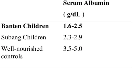
The children in Subang were clearly emaciated whereas the Banten children often
had a deceptively plump appearance with swollen abdomens and edematous limbs.
Ten percents children in Banten indicated vitamin A deficiencies (night blindness,
xeroftalmia, bitot spot) and in Subang only 1%, whereas vitamin A supplementation
were government program which routinely supplemented at POSYANDU each
month.
Many people in Subang have domestic fish-pond and as traditional tempe or tofu producer.
Table 2.1 Serum albumin concentration groups of children from each region
Serum Albumin
What is the basic biochemistry behind the Subang children’s emaciated appearance? Why do the Banten children have oedema?
How would you recommend the Banten relief workers modify their feeding procedures in the future?
Why are the children in Subang have lower percentage of vitamin A deficiencies?
Qualitative Tests for Ketone Bodies in Urine (Gerhardt’s and Rothera’s Tests):
Introduction:
The most important consequence of the increased metabolism of fat in starvation or diabetic patients is the accumulation in blood of the following substances:
- acetoacetic acid (CH3.CO.CH2.COOH).
- -hydroxybutyric acid (CH3.CHOH.CH2.COOH).
- acetone (CH3.CO.CH3).
It is now held that the liver is the main site of formation of the two acids given above. Unless adequate glucose is available, the liver is compelled to use an increased amount of fat, so that these acids are formed in quantities too great for the tissues to metabolise them. Then they begin to accumulate in the blood and to be excreted in the urine. It has been shown that diabetics are able to metabolise them normally. It is the increased production which is significant. In addition to the ketosis of diabetes, starvation ketosis arises in the same way. Testing the urine for these substances is an important part of the routine examination of diabetic or starved patients.
Gerhardt’s Ferric Chloride Test:
Principle:
The definite reaction is not yet unknown, probably the oxidation of acetoacetic acid by FeCl3 produces a red brown colour substance.
Procedure:
Add more ferric chloride after filtering and again observe. Similar colour is given by salicylic acid and salycilates as well. This can be differentiated from acetoacetic acid by their different behaviour towards heat. On thoroughly boiling the urine, acetoaccetic acid loses carbon dioxide, and is converted into acetone, which no longer gives the test. If the heating is done after adding the ferric chloride, the colour already formed disappears. Salicylates are unaffected by boiling the urine and the colour persists.
This test is only given by acetoacetic acid, not by the other two keton bodies. However, it has now been superseded by a version of the Rothera test, usually in the convenient stick form.
Rothera’s Nitroprusside Test:
Principle:
Sodium nitroprusside (sodium nitroferricyanide) in a acid condition will split to Na4Fe(CN)6-NaNO2 and Fe(OH)3, a strong oxidating agent. Acetoacetic acid and acetone will be oxidized and produces a red purple colour mix of substances. To stabilize these substances, a buffer solution (NH4)2SO4 is needed.
Procedure:
Saturate about 5 ml urine with solid ammonium sulphate and add a little sodium nitroprusside, either about 0.5 ml 20 g/l solution or a small quantity of powdered solid. Mix and add about 0.5 ml concentrated ammonia. A purple color is given by acetoacetic acid and acetone which is maximal in 15 min. If the ammonia is layered on, a purple ring is obtained at the junction of the liquids. This test is not given by salicylates.
Rothera’s test is much more sensitive than Gerhardt’s, being positive at about one part in 40 000 compared with one in a few thousands for the Gerhardt test.
Blood Urea Nitrogen (BUN) Determination
analysis, urease catalyzes the hydrolysis of urea to NH3 and CO2. The ammonia is converted to indophenol blue in the presence of sodium nitroferricyanide-phenol and hypochlorite reagents. The color of the unknown is read against a blank at A625nm and calculations are made by comparison with absorbency values of a standard of known concentration. Follow the procedure shown in the following Table.
Procedure for Determination of Blood Urea Nitrogen (BUN)a Tube no.
Mix and incubate at room temperature for 40 min. Add 4 ml of H2O, mix, and read in a
Spectrophotometer against the blank % T625 nm
A625 nm a
Use 13 x 100-nm test tubes for this assay
Commentary - Malnutrition
Malnutrition, defined as an unintentional weight loss of more than 10% associated with a serum albumin level below 3.2 g/dL, is not infrequent in hospitalized patients in the modern era of medicine. Regardless of the etiology, malnutrition is associated with a suboptimal surgical outcome, increased rate of infection, longer hospital stay, impaired wound healing, frequent hospital readmission for elderly, more frequent postoperative complications, and increased risk death. Malnutrition results from alterations in the intake, digestion and/or absorption, metabolism, excretion, and/or metabolic requirements of dietary energy, protein, and other nutrients. Understanding the pathophysiology of the patient’s disease is essential to the likelihood of finding a component of malnutrition. Furthermore, the severity of clinical illness may be an independent variable of nutritional compromise. Bedside nutritional assessment requires integration of the medical history, the physical examination, selected laboratory data, estimation of nutritional requirements, and approaches to nutritional intervention and its outcome. Accurate bedside assessment allows the physician to determine which patients may benefit from nutritional intervention, and careful patient selection is essential to the cost-benefit of nutritional support.
As a result of the widespread famine, the Subang children are suffering from a severe dietary deficiency of protein and energy foodstuffs known as marasmus (from Greek, meaning ‘to waste’). Occurrence of marasmus is not related to climate and used to occur in industrialised areas of Europe
In the absence of adequate food intake, the body mobilises its fat stores. However fatty acid cannot be used to maintain blood glucose, which is vital for a variety of organs, particularly the brain, and erythrocytes. Liver glycogen stores are rapidly exhausted, and so muscle protein must be catabolised to provide amino acid carbon sources for gluconeogenesis.
reveals the effects of the malnutrition. Among the principals protein produced by the liver is serum albumin, a major osmotic buffer of extracellular fluid. The marked deficiency of serum albumin in the Subang children leads to water retention in the tissues. Another important biosynthetic role for the liver is to provide apolipoproteins for the formation of serum lipoprotein complexes. These are responsible for the transport of lipids, especially triglycerides, around the body. When lipoprotein synthesis is compromised, lipids accumulate in the liver.
Ingestion of large amounts of protein results in an excess of amino acids over the immediate requirements for protein synthesis. The surplus amino acids cannot be stored; instead they are catabolised. The unwanted amino groups give rise to ammonia which must be detoxified by conversion to urea. This take place largely in the liver via the urea cycle. Presumably the Banten children’s damage livers have only a limited ability to handle excess amino acids. Thus, feeding large amounts of protein supplements, far from saving the Banten children as intended, inadvertently overloaded their weakened livers and contributed to their eventual demise. Smaller amounts of the supplements should be given at more frequent intervals to spread the load on the failing liver.
and humid climate provides ideal conditions for the growth of moulds in foodstuffs, particularly when they are stored under rather primitive conditions in rural areas.
In both marasmus and kwashiorkor the malnutrition leads to a generalised debility and an inability to resist infections, so that death usually results from pneumonia or the consequences of chronic diarrhoea.
Further Questions
1. Malnutrition is not simply a matter of dietary insufficiency of protein and/or calories. Vitamins and minerals will also be limited. Vitamin A deficiency is frequently a complicating factor. What is vitamin A, what are its roles and what are the symptoms of its deficiency? From what you can discover about vitamin A transport in the body, can you explain why vitamin A deficiency is particularly noticeable in kwashiorkor?
2. Which hormones are involved in controlling energy metabolism in starvation? 3. Why are fatty acids not gluconeogenic?
4. In a hitherto well-nourished individual who starves, why does muscle metabolism take place at a much higher rate in the first few days than later?
5. Chronic diarrhoea can be fatal. Why is this and how would you treat it?
Case :
Chronic renal failure
Laboratory activities:
1. Electrophoresis of plasma protein 2. Identification of albumin in urine 3. Blood calcium assay
Topic for discussion
a. Hierarchy and classification of biomolecules
b. Structural related function of important biomolecules c. General principle of analysis method of biomolecules
d. Biomolecules alterations in chronic renal failure and impact on its function
Case :
Famine in West Java
Laboratory activities:
1. Blood Urea Nitrogen assay 2. Rothera reaction
3. Gerhardt reaction
Topic for discussion
a. Resource and utility of some biomolecules b. Biological function of important biomolecules c. Biomolecules as nutrition