Isolation and analysis of an aciclovir-resistant murine
cytomegalovirus mutant
Toshio Minematsu
a,*, Seiichiro Mori
b, Yoshito Eizuru
b,
Yoichi Minamishima
aaDepartment of Microbiology,Miyazaki Medical College,Kihara,Kiyotake,Miyazaki889-1692,Japan
bDi6ision of Persistent and Oncogenic Viruses,Center for Chronic Viral Diseases,Faculty of Medicine,Kagoshima Uni6ersity, 8-35-1Sakuragaoka,Kagoshima890-8520,Japan
Received 27 October 1999; accepted 29 September 2000
Abstract
An aciclovir (ACV)-resistant murine cytomegalovirus (MCMV) was isolated from the Smith strain and the mutant was analysed. Attempts were also made to identify directly the mutated gene. The 50% inhibitory concentration (IC50)
of ACV for the mutant strain was30 times higher than that for the wild-type strain. The mutant strain was equally
sensitive to ganciclovir (GCV), but slightly resistant to cidofovir (CDV) and foscarnet (PFA) when compared with the wild-type. Molecular analysis of the mutant strain revealed that a single base mutation of cytosine (C) to guanine (G) occurred at the 2476th nucleotide position in the DNA polymerase gene region, resulting in an amino acid substitution of proline (Pro) with alanine (Ala) at codon 826. The marker transfer experiment confirmed that this mutation conferred ACV resistance to MCMV. This mutation at codon 826 was easily identified by means ofHaeIII digestion of the selected PCR product and electrophoresis. © 2001 Elsevier Science B.V. All rights reserved.
Keywords:Murine cytomegalovirus; Aciclovir; Ganciclovir; Cidofovir; Foscarnet; DNA polymerase; Drug-resistance
www.elsevier.com/locate/antiviral
1. Introduction
Aciclovir (ACV) has been used to treat patients with herpes simplex virus (HSV) and varicella-zoster virus (VZV) infections. ACV is an acyclic nucleoside analogue and its antiviral activity de-pends on two virus-coded enzymes, thymidine kinase (TK) and DNA polymerase. The viral TK phosphorylates ACV more efficiently than the cellular TK (Fyfe et al., 1978). As a result,
HSV-and VZV- infected cells contain much more ACV triphosphate (ACV-TP) than do uninfected cells (Elion et al., 1977; St. Clair et al., 1980). The ACV-TP then serves as a substrate and inhibits herpesvirus DNA polymerase. ACV-TP inhibits viral DNA polymerases more effectively than cel-lular DNA polymerases (Martin et al., 1994). Activation by viral TK and selective inhibition of viral DNA polymerase are crucial for the antiviral actions of ACV.
The entire sequence of human cytomegalovirus (HCMV) has been reported (Bankier et al., 1991). * Corresponding author. Fax: +81-985-856475.
HCMV was shown not to encode for an enzyme analogous to HSV TK (Zavada et al., 1976). Therefore, ACV is not used for treatment of active HCMV diseases. Murine cytomegalovirus (MCMV) has been utilized as a model for HCMV. As HCMV, MCMV does not encode a viral-specific TK. (Rawlinson et al., 1996). MCMV does not induce TK activity in either TK+ or TK− cells resistant to 5-bromod-eoxyuridine (BUdR) (Muller and Hudson, 1977; Eizuru et al., 1978; Burns et al., 1981). MCMV does not stimulate TK− cells to incorporate ex-ogenous thymidine (Muller and Hudson, 1977). Furthermore, the replication of MCMV is not inhibited by BUdR in TK− cells (Muller and Hudson, 1977; Eizuru et al., 1978; Burns et al., 1981). Thus, MCMV neither induces viral TK activity nor enhances cellular TK activity. Yet, wild-type MCMV shows high susceptibility to ACV in vitro and in vivo (Burns et al., 1981; Wingard et al. 1981; Glasgow et al., 1982).
It is of great interest to know why MCMV is susceptible to ACV even in the absence of TK activity. Therefore, to elucidate the mechanism of this susceptibility, we isolated an ACV-resistant MCMV mutant from the Smith strain and deter-mined the susceptibility of this mutant strain to various antiviral agents. Then we attempted to identify mutation(s) responsible for the ACV re-sistance of MCMV.
2. Materials and methods
2.1. Anti6iral agents
ACV and ganciclovir (GCV) were gifts from Nippon Wellcome, Kobe, Japan. Cidofovir (CDV) was generously provided by Professor E. De Clercq, Leuven University, Belgium. Foscar-net (PFA) was purchased from Sigma, St. Louis, MO.
2.2. Cells and medium
Mouse embryonic fibroblasts (MEF) were pre-pared by trypsinization of 14 – 18-day-old mouse embryos and used at the second to fourth passage
level for isolation, propagation and titration of MCMV. Balb/c 3T3 cells were used for transfec-tion. MEF were grown in Eagle’s minimum essen-tial medium (MEM, Nissui Pharmaceutical, Tokyo, Japan) supplemented with 5% heat-inacti-vated calf serum (Flow Laboratories, Irvine, VA), 60 mg/ml of kanamycin and 0.12% of sodium bicarbonate. The medium for Balb/c 3T3 cells contained 10% fetal bovine serum instead of 5% calf serum.
2.3. Viruses and isolation of ACV-resistant mutant
The Smith strain of MCMV that was plaque-purified and serially passaged in MEF was used as the wild-type strain. An ACV-resistant MCMV mutant was obtained by the following processes. The plaque-purified wild-type strain of MCMV was passaged in the presence of various concen-trations (5, 10, 25 and 50 mM, respectively) of ACV. The MCMV that replicated in the presence of an ACV concentration which yielded B20% of the control virus was used for the next cycle of selection using a medium with a higher concentra-tion of ACV. This cycle was repeated up to a final concentration of 50 mM of ACV. The stock sam-ples of both the wild-type and ACV-resistant MCMV were plaque-purified and then filtrated through the membrane filter with a pore size of 450 nm to remove multicapsid or aggregated viri-ons. Finally, stock samples were again plaque-purified twice.
2.4. Titration of 6iruses
number of plaques was evaluated under an in-verted microscope.
2.5. Susceptibility test of MCMV strains against anti6iral agents
The susceptibility of MCMV strains to antiviral agents was determined by the 50% plaque reduc-tion assay. The monolayers of MEF in 35 mm plastic dishes were inoculated with 100 plaque-forming units (PFU) of each strain of MCMV. After virus adsorption at room temperature for 1 h, the monolayers were overlaid with Eagle’s MEM containing 5% heat-inactivated calf serum, 60 mg/ml of kanamycin, 0.12% of sodium bicar-bonate, 2.25% methylcellulose and different con-centrations of each antiviral agent. Duplicate dishes were used for each concentration of antivi-ral agent. The dishes were incubated at 37°C under a humidified atmosphere containing 5% carbon dioxide. The 50% inhibitory concentration (IC50) was defined as the concentration of the agent that resulted in 50% plaque reduction.
2.6. Virus growth cur6e determinations
The monolayers of MEF in culture tubes were inoculated with the Smith strain of MCMV or ACV-resistant strain at a multiplicity of infection (M.O.I.) of 0.2. After adsorption at room temper-ature for 1 h, the inocula were aspirated off, and 2 ml of Eagle’s MEM containing 5% calf serum was added to each culture tube. The infected cells and culture fluid in duplicate tubes were collected every 3 h and stored at −80°C until titration. Then, the viruses were released from the cells by freeze – thawing three times followed by low-speed centrifugation. The supernatant fluid was stored at −80°C until titration. The infectious viruses were plaque-titrated with MEF.
2.7. Preparation of DNA
Viral DNA was prepared according to the Hirt’s procedure (Hirt, 1967). The Hirt’s superna-tant was treated with proteinase K (50 mg/ml) at 56°C for 2 h, followed by phenol: chloroform extraction and ethanol precipitation. Finally,
DNA was dissolved in 10 mM Tris-HCl and 1 mM EDTA (pH 8.0: TE) buffer. For marker transfer experiment, wild-type virions of MCMV were prepared as described by Ihara et al. (1994). The viral DNA was treated as mentioned above.
2.8. Amplification of 6iral DNA fragments by polymerase chain reaction (PCR)
The primers were designed to amplify the four-teen overlapping fragments of viral DNA poly-merase coding region and ten overlapping fragments of m97 coding region. Each primer was labelled with biotin at 5%end for non-radioisotope
single strand conformation polymorphism (SSCP) analysis and sequencing. The sequences of the primers for amplifying MCMV DNA polymerase gene are listed in Table 1 and the schematic diagram is shown in Fig. 1. The PCR reaction mixture consisted of viral DNA as template, ap-propriate primer pairs (1mM each), deoxyribonu-cleoside triphosphates (200 mM each), and 1.25 unit of Tth polymerase (Toyobo, Osaka, Japan) in a total volume of 50 ml PCR buffer. The fragment was amplified by 30 cycles of denatura-tion at 94°C for 1 min, annealing at 54°C for 1 min, and extension at 72°C for 1 min.
2.9. SSCP analysis and sequencing
For SSCP analysis, PCR product was diluted to 1/10 or 1/100 with a loading buffer consisting of 95% formamide, 10 mM EDTA, 0.1% bromophe-nol blue and 0.1% xylene cyabromophe-nol. After heating at 90°C for 2 min, 1 ml of the mixture was subjected to gel electrophoresis in 5% polyacrylamide gel containing 5% glycerol as described by Orita et al. (1989). After electrophoretic separation, DNA was transferred to positively charged nylon mem-brane by capillary blotting and radiated by Imag-ing high-chemiluminescent detection kit, according to instructions of the Manufacturer (Toyobo). For sequencing, PCR product was purified from agarose gel using QIAX II Extrac-tion Kits (Qiagen GmbH and Qiagen, Hilden, Germany). Sequencing of PCR product was car-ried out by dideoxy termination method using
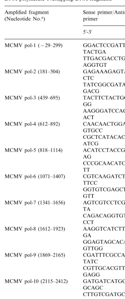
Table 1
The sequence of the primers defining the amplified MCMV DNA polymerase overlapping DNA fragments
Sense primer/Antisense Amplified fragment
(Nucleotide No.a) primer
5%-3%
MCMV pol-1 (−29–299) GGACTCCGATTTCGAG TACTGA
TTGACGACCTGGTCGT AGGTGT
MCMV pol-2 (181–504) GAGAAAGAGTACGTG CTC
MCMV pol-5 (818–1114) ACATCCTACCGATCGA AG
MCMV pol-7 (1341–1656) AGTCGTCCTCGACATG TA
CAGACAGGTGTAGAT CCT
MCMV pol-8 (1612–1923) AAGGTCATCTTCGACG GA
Table 1 (Continued)
Amplified fragment Sense primer/Antisense primer
(Nucleotide No.a)
5%-3%
aNucleotide numbers were counted from the start site of the RNA transcript from DNA polymerase gene
Kit (Toyobo). The cycling parameters were as follows: denaturation at 94°C for 1 min, anneal-ing at 54°C for 1 min, extension at 72°C for 1 min, and 30 cycles. The sequencing products were subjected to electrophoresis in 8% polyacrylamide gel containing 8.3 M urea. After electrophoretic separation, the DNA was transferred and irradi-ated as mentioned above.
2.10. Marker transfer experiments
To confirm the mutation related to ACV resis-tant, we constructed an ACV-resistant recombi-nant using marker transfer technique. The DNA fragment containing mutation was amplified by PCR and directly cloned into pCR ‘2.1 Vector using Original TA Cloning’ Kit according to the instructions of the Manufacturer (Invitrogen, Carlsbad, CA). Balb/c 3T3 cells were transfected simultaneously with plasmid DNA and viral DNA using LipofectAMINE PLUSa
Fig. 1. A schematic representation of the MCMV DNA polymerase region showing the location of the amplified fragments on an approximate amino acid scale. The numbers at intervals down length of PCR products show nucleotide range.
3.2. Molecular analysis of 6iral DNA polymerase and m97 regions
HCMV encodes a phosphotransferase which phosphorylates GCV. The gene responsible for the enzyme is UL97. MCMV also encodes similar enzyme, and the murine counterpart gene is m97. The viral DNA polymerase and m97 regions of both wild and mutant strains were compared by PCR-SSCP analysis. Only one fragment pair, nu-cleotide range from 2353 to 2649 in viral DNA polymerase cording region, showed a different mobility in SSCP analysis (Fig. 2). Then, the region of this fragment was sequenced to identify 2.11. Restriction enzyme clea6age analysis
PCR product was digested with Hae III (Nip-pon Gene, Toyama, Japan), and subjected to 2.5% agarose gel electrophoresis. The elec-trophoretic pattern was photographed under UV light.
3. Results
3.1. Isolation and susceptibility of ACV-resistant mutant
An ACV-resistant virus which can replicate in the presence of 50mM of ACV was derived from the Smith strain of MCMV. This virus was plaque-purified twice and used as the ACV-resis-tant strain throughout the experiments.
The susceptibility of wild and mutant strain to various antiviral agents was determined by 50% plaque reduction (Table 2). The 50% inhibitory concentration (IC50) of ACV was 90 mM for the mutant, which means that it was 30 times more resistant than the wild strain. However, the mu-tant strain was equally sensitive to GCV but slightly resistant to CDV and PFA as compared to the wild-type.
Table 2
Susceptibility of ACV-resistant MCMV to various antiviral agentsa
IC50(mM) Mutant
Antiviral agents
Wild Wild strain Mutant strain
ACV 3.2 90 28.1
GCV 16 16 1.0
2.3 0.9
CDV 0.4
400 4.7
PFA 85
Fig. 2. PCR-SSCP analysis. The DNA fragments amplified from wild-type MCMV (1) and two cloned ACV-resistant mutants (2 – 3) were subjected to SSCP analysis.
Table 3
Susceptibility of wild-type MCMV, mutant MCMV and re-combinant MCMV to various agentsa
Antiviral agents IC50(mM)
Wild-type Mutant Recombinant
80
2.8 82
ACV
8.6 9.8 9.6
GCV
CDV 0.3 0.8 0.8
340 320
80 PFA
aEach value presents the mean for duplicate determinations from two separate experiments.
the mutation(s). When the sequences were compared, the mutant had a single base mutation of cytosine (C) to guanine (G) at nucleotide position 2476, resulting in an amino acid change at codon 826 of proline to alanine (Fig. 3). To examine the possibility of any mutation in m97 region, ten overlapping fragments were also examined by PCR-SSCP analysis. However, there was no difference in the mobility between m97 regions of wild and mutant stains.
3.3. Drug resistance conferred by marker transfer of the mutant DNA to wild-type MCMV
The plasmid DNA containing the mutated viral DNA polymerase region was co-transfected in Balb/c 3T3 cells with the DNA of wild-type MCMV. Recombinant virus was selected twice in the presence of 50mM ACV. The susceptibility of the recombinant virus to the antiviral agents was the same as that of the ACV-resistant mutant
(Table 3). Median inhibitory concentration of ACV was 80 mM for the recombinant virus.
3.4. Replication of wild-type MCMV, mutant MCMV and recombinant MCMV in MEF
To investigate whether there was any biological difference among the three MCMV strains, the growth curves of these viruses in MEF were com-pared. Amounts of virus produced every 3 h during the virus life cycle were quantified by plaque titration. Overall, there was no marked difference in the time course of replication or in total virus yield among wild-type, mutant and recombinant MCMV strains (data not shown).
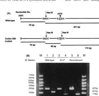
3.5. Identification of mutation site by PCR and restriction fragment length polymorphism (RFLP)
Identification of the mutation at codon 826 was accomplished by means of RFLP of the selected PCR product in the nucleotide range from 2353 to 2649. This region in wild strain contained one GGCC recognition site for Hae III. In the ACV-resistant strain, however, the sequence GCCC had changed to GGCC, which produced an additional recognition site for Hae III. Consequently, the codon 826 CCC (proline) had changed to GCC (alanine). The PCR products amplified from wild and mutant strains were digested with Hae III and subjected to 2.5% agarose gel electrophoresis. The PCR product from wild strain produced two bands, 76 and 221 bps. In contrast, the PCR product from the mutant strain produced three Fig. 3. Sequencing of wild-type, ACV-resistant and
bands, 76, 48 and 173 bps as shown in Fig. 4 (A – B). Therefore, the mutation was easily de-tected by digestion with Hae III followed by electrophoresis.
4. Discussion
Human cytomegalovirus (HCMV) causes seri-ous and often life-threatening diseases in fetuses, neonates and immunocompromised hosts, such as recipients of organ transplants or patients with malignancies or the acquired immune deficiency syndrome (AIDS) (Britt and Alford, 1996). Cur-rently, inhibitors of viral DNA synthesis are
avail-able as anti-CMV drugs. These include GCV, PFA and CDV.
Unfortunately, no animal model is available for studying antiviral agents against HCMV in vivo because of its strict species-specificity. MCMV has been well investigated as an animal model for evaluating anti-CMV compounds. MCMV is sus-ceptible to GCV, PFA and CDV (Smee et al., 1992, 1995; Okleberry et al., 1997; Duan et al. 1998); however, it is also sensitive to ACV (Burns et al., 1981; Wingard et al., 1981; Glasgow et al., 1982). The susceptibility of MCMV to ACV may reside in the DNA polymerase rather than the phosphotransferase, as mutants resistant to ACV also show less susceptibility to chemicals that
interact with DNA polymerase such as phospho-noacetic acid (PAA) (Burns et al., 1982; Sandford et al., 1985) and PFA (Elliott et al., 1991).
The analysis of ACV-resistant mutant may offer a clue to help elucidating the mechanism of the anti-MCMV effect of ACV. We isolated and char-acterized an ACV-resistant mutant of MCMV from the Smith strain. Drug susceptibility tests showed that the ACV-resistant mutant was equally sensitive to GCV but slightly resistant to CDV and PFA, when compared with the wild-type. As this ACV-resistant mutant showed cross-resistance to PFA, its DNA polymerase gene seemed to be the site of mutation. In fact, molec-ular analysis of the mutant revealed a novel muta-tion in the DNA polymerase gene, namely, a single base substitution (from C to G) at the 2476th nucleotide position in the DNA polymerase re-gion, with consequently an amino acid substitu-tion, from proline to alanine, at codon 826. It was confirmed by marker transfer experiment that this mutation conferred ACV resistance to MCMV.
The amplified fragment which contains the mu-tation was detectable by SSCP analysis. For rapid and direct identification of MCMV responsible for ACV-resistance, we employed RFLP analysis. SSCP followed by restriction enzyme cleavage provides a rapid sensitive detection of certain mutations in PCR-amplified DNA fragments. In our case, the mutation at codon 826 (CCC to GCC mutation) was detected byHaeIII digestion (recognition sequence GGCC) and electrophore-sis. However, this identification is specific only for CCC to GCC mutation. If the mutation were not CCC to GCC, of course, other endonuclease en-zymes would be required to recognize the muta-tion site.
Incidentally, no mutation could be found in the m97 gene that is homologous to HCMV UL97 (phosphotransferase). However, in this study both wild and mutant strains of MCMV were less susceptible to GCV than in previous reports (Smee et al., 1995; Okleberry et al., 1997).
Overall, there is much homology between MCMV and HCMV DNA polymerase gene. The amino acid sequences revealed that 576 of the 1097 amino acids of the MCMV DNA polymerase have an identically matched residue in the HCMV
DNA polymerase (Elliott et al., 1991). In addition, the amino acid residue Pro826 of MCMV DNA polymerase, shown to be responsible for ACV susceptibility of MCMV in this study, is conserved in the DNA polymerase of HCMV (Pro945) and rhesus monkey CMV (Pro792) (Swanson et al., 1998). This position of Pro in HCMV polymerase is located in the gap between I and VII conserva-tive regions. However, a mutation at the ho-mologous position to the Pro826 of MCMV has not been reported in the human or rhesus CMV DNA polymerase yet. In addition, there is no reduction in growth fitness of the MCMV mutant encoding the Pro826Ala mutation.
Antiviral effect of ACV requires initial phos-phorylation of ACV to form ACV monophos-phate and final inhibition of viral DNA synthesis by ACV-TP. This investigation showed that the DNA polymerase may at least be one of the target enzymes for the anti-MCMV action of ACV. This was evidenced by the fact that the ACV-resistant mutant had a mutation in the DNA polymerase gene. The results described in this paper are com-patible with those of a previous paper (Sandford et al., 1985). We have already reported on the role of MCMV DNA polymerase in the antiviral activity of ACV in vitro (Ochiai et al., 1992). In that study, we partially purified the DNA polymerase and determined its kinetic constants for ACV triphos-phate. We raised a possibility that MCMV DNA polymerase might be highly sensitive to ACV triphosphate. However we did not use an ACV-re-sistant mutant at that time.
pattern of ACV in MCMV-infected cells should be addressed in future studies.
References
Bankier, A.T., Beck, S., Bohni, R., Brown, C.M., Cerny, R., Chee, M.S., Hutchinson, C.A., Kouzarides, T., Mar-tignetti, J.A., Preddie, E., Satchwell, S.C., Tomlinson, P., Weston, K.M., Barrell, B.G., 1991. The DNA sequence of the human cytomegalovirus genome. DNA Seq. 2, 1 – 12. Britt, W.J., Alford, C.A., 1996. Cytomegalovirus. In: Fields,
B.N., Knipe, D.M., Howley, P.M. (Eds.), Fields Virology, third ed. Lippinocott-Raven, Philadelphia, pp. 2493 – 2523. Burns, W.H., Wingard, J.R., Bender, W.J., Saral, R., 1981. Thymidine kinase not required for antiviral activity of acyclovir against mouse cytomegalovirus. J. Virol. 39, 889 – 893.
Burns, W.H., Wingard, J.R., Sandford, G.R., Bender, W.J., Saral, R., 1982. Acyclovir in mouse cytomegalovirus infec-tions. Am. J. Med. 73, 118 – 124.
Duan, J., Paris, W., Kibler, P., Bousquet, C., Liuzzi, M., Cordingley, M.G., 1998. Dose and duration-dependent of ganciclovir treatment against murine cytomegalovirus in-fection in severe combined immunodeficient mice. Antiviral Res. 39, 189 – 197.
Eizuru, Y., Minamishima, Y., Hirano, A., Kurimura, T., 1978. Replication of mouse cytomegalovirus in thymidine kinase-deficient mouse cells. Microbiol. Immunol. 22, 755 – 764. Elion, G.B., Furman, P.A., Fyfe, J.A., De Miranda, P.,
Beauchamp, L., Schaeffer, H.J., 1977. Selectivity of action of an antiherpetic agent, 9-(2-hydroxyethoxymethyl)-guanine. Proc. Natl. Acad. Sci. USA 74, 5716 – 5720. Elliott, R., Clark, C., Jaquish, D., Spector, D.H., 1991.
Tran-scription analysis and sequence of the putative murine cytomegalovirus DNA polymerase gene. Virology 185, 169 – 186.
Fyfe, J.A., Keller, P.M., Furman, P.A., Miller, R.L., Elion, G.B., 1978. Thymidine kinase from herpes simplex virus phosphorylates the new antiviral compound, 9-(2-hydrox-yethoxymethyl)guanine. J. Biol. Chem. 253, 8721 – 8727. Glasgow, L.A., Richards, J.T., Kern, E.R., 1982. Effect of
acyclovir treatment on acute and chronic murine cy-tomegalovirus infection. Am. J. Med. 73, 132 – 137. Hirt, B., 1967. Selective extraction of polyoma DNA from
infected mouse cell culture. J. Mol. Biol. 26, 365 – 369. Ihara, S., Takekoshi, M., Mori, N., Sakuma, S., Hashimoto,
J., Watanabe, Y., 1994. Identification of mutation sites of a temperature-sensitive mutant of HCMV DNA poly-merase. Arch. Virol. 137, 263 – 275.
Martin, J.L., Brown, C.E., Matthews-Davis, N., Reardon, J.E., 1994. Effects of antiviral nucleoside analogs on hu-man DNA polymerases and mitochondrial DNA synthesis. Antimicrob. Agents Chemother. 38, 2743 – 2749.
Muller, M.T., Hudson, J.B., 1977. Thymidine kinase activity in mouse 3T3 cells infected by murine cytomegalovirus. Virology 80, 430 – 433.
Ochiai, H., Kumura, K., Minamishima, Y., 1992. Murine cytomegalovirus DNA polymerase: purification, character-ization and role in the antiviral activity of acyclovir. An-tiviral Res. 17, 1 – 16.
Okleberry, K.M., Warren, R.P., Smee, D.F., 1997. Metabolism of ganciclovir and cidofovir in cells infected with drug-resistant and wild-type strains of murine cy-tomegalovirus. Antiviral Res. 35, 83 – 90.
Orita, M., Iwahana, H., Kanazawa, H., Hayashi, K., Sekiya, T., 1989. Detection of polymorphism of human DNA by gel electrophoresis as single-strand conformation polymor-phism. Proc. Natl. Acad. Sci. USA 86, 2766 – 2770. Rawlinson, W.D., Farrell, H.E., Barrell, B.G., 1996. Analysis
of the complete DNA sequence of murine cytomegalovirus. J. Virol. 70, 8833 – 8849.
Rawlinson, W.D., Zeng, F., Farrell, H.E., Cunningham, A.L., Scalzo, A.A., Booth, T.W., Scott, G.M., 1997. The murine cytomegalovirus (MCMV) homolog of the HCMV phos-photransferase (UL97(pk)) gene. Virology 233, 358 – 363. Sandford, G.R., Wingard, J.R., Simons, J.W., Staal, S.P.,
Saral, R., Burns, W.H., 1985. Genetic analysis of the susceptibility of mouse cytomegalovirus to acyclovir. J. Virol. 54, 104 – 113.
Smee, D.F., Morris, J.L.B., Leonhardt, J.D., Mead, J.R., Holy, A., Sidwell, R.W., 1992. Treatment of murine cy-tomegalovirus infection in severe combined immunodefi-cient mice with ganciclovir, (S)-1-[3-hydroxy-2-(phos-phonylmethoxy)propyl]cytosine, interferon, and bropirim-ine. Antimicrob. Agents Chemother. 36, 1837 – 1842. Smee, D.F., Barnett, B.B., Sidwell, R.W., Reist, E.J., Holy´,
A., 1995. Antiviral activities of nucleosides and nucleotides against wild-type and drug-resistant strains of murine cy-tomegalovirus. Antiviral Res. 26, 1 – 9.
St. Clair, M.H., Furman, P.A., Lubbers, C.M., Elion, G.B., 1980. Inhibition of cellularaand virally induced deoxyri-bonucleic acid polymerases by the triphosphate of acy-clovir. Antimicrob. Agents Chemother. 18, 741 – 745. Swanson, R., Bergquam, E., Wong, S.W., 1998.
Characteriza-tion of rhesus cytomegalovirus genes associated with anti-viral susceptibility. Virology 240, 338 – 348.
Wingard, J.R., Bender, W.J., Saral, R., Burns, W.H., 1981. Efficacy of acyclovir against mouse cytomegalovirus in vivo. Antimicrob. Agents Chemother. 20, 275 – 278. Zavada, V., Erban, V., Rezacova, D., Vonka, V., 1976.
Thymidine-kinase in cytomegalovirus infected cells. Arch. Virol. 52, 333 – 339.
Zimmermann, A., Michel, D., Pavic´, I., Hample, W., Lu¨ske, A., Neyts, J., De Clercq, E., Mertens, T., 1997. Phosphory-lation of aciclovir, ganciclovir, penciclovir and S2242 by the cytomegalovirus UL97 protein: a quantitative analysis using recombinant vaccinia viruses. Antiviral Res. 36, 35 – 42.