Assessment of the application level of
radiation protection and awareness of
radiation safety regulations among the
radiographers at Yogyakarta Special Region,
Indonesia
Ahmad Rafiq Mohammad Abu Arrah1*, Arif Faisal2, Ahmad Hamim Sadewa3
1Faculty of Allied Medical Sciences, Arab American University, Palestine, 2Department of Radiology, 3Department of Biochemistry, Faculty of Medicine, Gadjah Mada University, Yogyakarta, Indonesia
ABSTRACT
A strong relation between cancers and radiation exposure has been reported. Radiation may damage DNA in the cell. Therefore, radiation protection program must be applied in the radiology department. Morover, the radiographer should have high level of awareness and risk assessment for radiation. Personal radiation monitoring is one of the main radiation protection, especially for pregnant worker and her fetus. This study was conducted to evaluate the application, awareness and risk assessment levels of radiation protection among radiographers at hospitals in Yogyakarta Special Region, Indonesia. This was a descriptive study, applying a cross sectional survey at hospitals in Yogyakarta. The subjects were radiographers of both governmental and private hospitals. There were 101 respondents from a total of 124 radiographers. The data obtained were tabulated and analyzed using Chi Square test. The study revealed that 69.3% of the respondents had low application level of radiation protection, 19.8% did not know the meaning of ALARA (As Low As Reasonably Achievable), 50.5% were not aware of Inverse Square Law. The study also reported that 36.6% of the respondents did not know the amount of radiation that entered their body last year, 61.4% of radiographers thought that the risk assessment of radiation was not enough, 18.8% of radiographers were never use any radiation monitoring device, and 90.1% stated that there was no additional protection or radiation monitoring to the pregnant radiographer. However, there were no significant differences between duration of working, type of imaging modality, academic level, and training course for radiation protection. In conclusion, there was no difference in the application, awareness, and risk assessment levels of radiation protection among the radiographers at hospital in Yogyakarta Special Region between duration of working, type of medical imaging modality, academic level, and training on radiation protection. In addition, the application, awareness, and risk assessment levels of radiation protection were not sufficient.
Key words:radiation protection – ALARA – radiographer – hospital – risk assessment
ABSTRAK
demikian, tidak terdapat perbedaan antara masa kerja, tipe modalitas pencitraan, tingkat pendidikan, dan pelatihan perlindungan radiasi. Dapat disimpulkan, tidak terdapat perbedaan dalam hal pelaksanaan, perhatian, tingkat penilaian risiko perlindungan radiasi diantara radiografer pada rumah sakit di DIY antara masa kerja, jenis modalitas pencitraan, tingkat pendidikan, dan pelatihan perlindugnan radiasi. Selain itu, pelaksanaan, perhatian dan tingkat penilaian risiko perlindungan radiasi dianggap tidak memadai.
Kata kunci: pelindung radiasi – ALARA – radiografer – rumah sakit – penilaian risiko
INTRODUCTION
The average annual radiation dose received by general public is 2.5mSv, and 15% of them are related to medical exposures.1,2 The use of radiation in medical
practices has evolved since its beginning and 30-50% of medical decisions are based on radiological examinations.3 However, the hazards of ionizing
radiation are irrefutable. For instance, according to recent studies in United Kingdom, 100-250 death per year occurs because of harmful effects of medical radiation exposures.1,4 Reducing radiographer and
patients radiation dose exposure through As Low As Reasonably Achievable (ALARA) is based on the recommendations of all radiation protection organizations such as International Commission on Radiological Protection (ICRP) and National Radiological Protection Board (NRPB).1-4
Awareness of medical practitioners on hazards of ionizing radiation has been reported to be one of the main factors for decreasing the radiographer’s and radiologist’s dose in medical practices.2,4 These studies
indicated that the improvement of information about radiation dose received in different diagnostic imaging procedures to radiographers and radiologist is important. The hazards of radiation exposure, therefore, help them to optimize the radiations protections during different medical imaging scanning. The first step of radiation protection can be obtained by conducting radiological investigations.4,5 If radiographers and radiologists are
aware of radiation dose received in different radiological investigations, they will avoid unnecessary examinations and performs counterpart examinations with low or without radiation risk.4-6 It seems important to instruct
MATERIALS AND METHODS
This was a descriptive study, applying a cross sectional survey. Radiographers were asked to give their responses on a number of questions related to the radiation protection among them. This study was carried out at hospitals in Yogyakarta Special Region, Indonesia during April 2011.
The studied population was all radiographers who worked in hospitals in Yogyakarta Special Region. Since the total numbers of radiographers in hospitals in Yogyakarta Special Region was small, most of them were included in the study, and therefore sampling was not performed. Data were collected from the studied population, which were a total of 124 radio-graphers from hospital in Yogyakarta Special Region. Data analysis was performed using a computer software package, which included univariate analysis, bivariate analysis and multivariate analysis. The protocol of the study has been approved by the Medical and Health Research Ethics Committee, Faculty of Medicine, Gadjah Mada University, Yogyakarta.
RESULTS
Characteristics of respondents
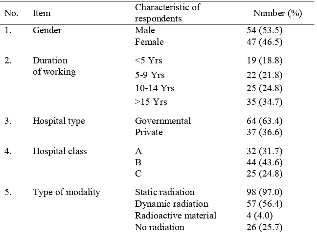
TABLE 1. The distribution of respondents according to gender, duration of working, hospital type, hospital class, and type of modality
No. Item Characteristic of
respondents Number (%)
1. Gender Male 54 (53.5)
Female 47 (46.5)
2. Duration
of working
5 Yrs 19 (18.8)
5-9 Yrs 22 (21.8)
10-14 Yrs 25 (24.8)
15 Yrs 35 (34.7)
3. Hospital type Governmental 64 (63.4)
Private 37 (36.6)
4. Hospital class A 32 (31.7)
B 44 (43.6)
C 25 (24.8)
5. Type of modality Static radiation 98 (97.0)
Dynamic radiation 57 (56.4)
Radioactive material 4 (4.0)
No radiation 26 (25.7)
Note: all the values were valid; static radiation includes conventional X ray, CT scan
and radiotherapy; dynamic radiation includes fluoroscopy and cath. Lab.; radioactive material includes nuclear medicine; no radiation includes MRI and ultrasonography
The hospitals were divided into three classes. Class A which provides all basic and specialty medical services with the minimal capacity about 400 beds. Class B which provides basic and some specialty medical services with minimal capacity about 200 beds, and class C which provides basic medical services with minimal capacity about 100 beds. Basic medical services include internal medicine, surgery, pediatrics and gynecology.
Application level of radiation protection
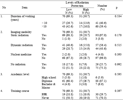
TABLE 2 showed the application level of radiation protection related to the duration of working, type of medical imaging modality, academic level, and training on radiation protection. The rate
of radiographers who did not apply the required level of radiation protection was 69.3% while 30.7% of the radiographers did not apply radiation protection. There was no protection difference between the duration of working, type of imaging modality, academic level, training course for radiation protection and application level of radiation protection.
TABLE 2. Application level of radiation protection in relation to the duration of working, type of imaging modality, academic level and training course for radiation protection.
No
Item
Levels of Radiation
Protection Number
(%) p
Low N (%)
High N (%)
1. Duration of working
(years)
70 (69.3) 31 (30.7) 0.534
10 27 (26.7) 14 (13.9) 41 (40.6)
10 43 (42.6) 17 (16.8) 60 (59.4)
2. Imaging modality 70 (69.3) 31 (30.7)
Static radiation Yes 69 (68.3) 29 (28.7) 98 (97.0) 0.170
No 1 (1.0) 2 (2.0) 3 (3.0)
Dynamic radiation Yes 41 (40.6) 16 (15.8) 57 (56.4) 0.515
No 29 (28.7) 15 (14.9) 44 (43.6)
Nuclear medicine Yes 2 (2.0) 2 (2.0) 4 (4.0) 0.393
No 68 (67.3) 29 (28.7) 97 (96.0)
No radiation Yes 18 (17.8) 8 (7.9) 26 (25.7) 0.992
No 52 (51.5) 23 (22.8) 75 (75.3)
3. Academic level 70 (69.3) 31 (30.7) 0.595
High school 5 (5.0) 1 (1.0) 6 (5.9)
Diploma 61 (60.4) 27 (26.7) 88 (87.1)
Bachelor& Master
4 (4.0) 3 (3.0) 7 (6.9)
4. Training course 70 (69.3) 31 (30.7) 0.397
PPR 19 (18.8) 11 (10.9) 30 (29.7)
Never 51 (50.5) 20 (19.8) 71 (70.3)
Awareness level of radiation protection
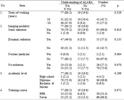
TABLE 3 showed the understanding of ALARA concept related to the duration of working, type of medical imaging modality, academic level, and training on radiation protection. The rate of radiographers who know the ALARA concept was
TABLE 3. Understanding ALARA concept in relation to duration of working, type of imaging modality, academic level and training course for radiation protection
No Item
Understanding of ALARA Number
(%) p
Yes N (%)
No N (%)
1. Years of working
(years)
77 (80.2) 19 (19.8) 0.329
10 31 (32.3) 10 (10.4) 41 (42.7)
10 46 (47.9) 9 (9.4) 55 (57.3)
2. Imaging modality 77 (80.2) 19 (19.8)
Static radiation Yes 76 (79.2) 19 (19.8) 95 (99.0) 0.618
No 1 (1.0) 0 (0.0) 1 (1.0)
Dynamic radiation Yes 47 (49.0) 8 (8.3) 55 (57.3) 0.135
No 30 (31.3) 11 (11.5) 41 (42.7)
Nuclear medicine Yes 0 (0.0) 2 (2.1) 2 (2.1) 0.004
No 77 (80.2) 17 (17.7) 94 (97.9)
No radiation Yes 24 (25.0) 2 (2.1) 26 (27.1) 0.070
No 53 (55.2) 17 (17.7) 70 (72.9)
3. Academic level 77 (80.2) 19 (19.8) 0.299
High school 2 (2.1) 2 (2.1) 4 (4.2)
Diploma 70 (72.9) 16 (16.7) 86 (89.6)
Bachelor & Master
5 (5.2) 1 (1.0) 6 (6.3)
4. Training course 77 (80.2) 19 (19.8) 0.972
PPR 24 (25.0) 6 (6.3) 30 (31.3)
Never 53 (55.2) 13 (13.5) 66 (68.8)
TABLE 4 showed the frequency of some answers related to the application and awareness levels of radiation protection, such the use of badge film which showed that only 33 (32.7%) of the radiographers who always used it during their working. Moreover, there were 37 (36.6%)
TABLE 4.Frequency table of some answers which in relation to the application and awareness levels of radiation protection
Item
Answer MissingNumber (%)
Understanding of ALARA concept Yes 77 (80.2) 0
No 19 (19.8)
Applying ALARA concept Yes 65 (64.4) 3
No 33 (32.7)
If RSO visiting the department Yes 85 (84.2) 0
No 16 (15.8)
Radiation amount for radiographer during last year
<5 rem 57 (56.4) 0
5 rem 4 (4.0)
>5 rem 3 (3.0)
Don’t know 37 (36.6)
If radiographer have badge film Yes 87 (86.1) 0
No 14 (13.9)
If the radiographer use badge film Never 19 (18.8) 0
Rarely 28 (27.7)
Sometimes 3 (3.0)
Mostly 18 (17.8)
Always 33 (32.7)
Instructions for pregnant worker Yes 58 (57.4) 0
No 43 (42.6)
Demonstrating embryo dose (<50 mrem)
Yes 10 (9.9) 0
No 91 (90.1)
Risk assessment level of radiation protection
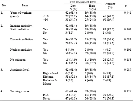
TABLE 5 showed the risk assessment level of radiation protection in relation to the years of working, type of medical imaging modality, academic level, and training on radiation protection. TABLE 5 also showed that there were 61.4% of
No Item
Risk assessment level
Number
(%) p
Low N (%)
High N (%)
1. Years of working
(years)
62 (61.4) 39 (38.6) 0.446
10 27 (26.7) 14 (13.9) 41 (40.6)
10 35 (34.7) 25 (24.8) 60 (59.4)
2. Imaging modality 62 (61.4) 39 (38.6)
Static radiation Yes 59 (58.4) 39 (38.6) 98 (97.0) 0.163
No 3 (3.0) 0 (0.0) 3 (3.0)
Dynamic radiation Yes 34 (33.7) 23 (22.8) 57 (56.4) 0.683
No 28 (27.7) 16 (15.8) 44 (43.6)
Nuclear medicine Yes 4 (4.0) 0 (0.0) 4 (4.0) 0.106
No 58 (57.4) 39 (38.6) 97 (96.0)
No radiation Yes 15 (14.9) 11 (10.9) 26 (25.7) 0.653
No 47 (46.5) 28 (27.7) 75 (74.3)
3. Academic level 62 (61.4) 39 (38.6) 0.089
High school 6 (5.9) 0 (0.0) 6 (5.9)
Diploma 53 (52.5) 35 (34.7) 88 (87.1)
Bachelor & Master
3 (3.0) 4 (4.0) 7 (6.9)
4. Training course 62 (61.4) 39 (38.6) 0.127
PPR 15 (14.9) 15 (14.9) 30 (29.7)
Never 47 (46.5) 24 (23.8) 71 (70.3)
TABLE 5. Risk assessment level of radiation protection in relation to duration of working, type of imaging modality, academic level and training course for radiation protection
Other issues
TABLE 6 showed the application level of radiation protection related to type of hospital and
hospital class. There was no difference between type and class of hospitals on application level of radiation protection.
TABLE 6. Application level of radiation protection in relation to the type and class of hospital
No
Item
Levels of radiation protection Number
(%) p
Low N (%)
High N (%)
1. Type of hospital 31 70 (69.3) (30.7) 0.544
Governmental 21 43 (42.6) 64 (20.8) (63.4)
Private 10 27 (26.7) 37 (9.9) (36.6)
2. Hospital class 31 70 (69.3) (30.7) 0.060
TABLE 7 showed the risk assessment level of radiation protection in relation to the type of hospital.
There was no difference between type of hospital an risk assessment level
TABLE 7. Risk assessment level of radiation protection in relation to the type of hospital
Item
Risk assessment level
Number
(%) p
Low N (%)
High N (%)
Type of Hospital 39 62 (61.4) (38.6) 0.467
- Governmental 23 41 (40.6) 64 (22.8) (63.4)
- Private 16 21 (20.8) 37 (15.8) (36.6)
TABLE 8 showed the application level of radiation protection at nuclear medicine department. It demonstrated that half of radiographers at nuclear medicine department did not use protective gloves and syringe to prepare radioactive materials. In
addition, they thought that the cautions posters and lights were not enough. But all of them reported that there was a protective container for radioactive wastes.
TABLE 8. Application level of radiation protection at Nuclear Medicine Department
DISCUSSION
Application Level of Radiation Protection
Cancer induction is the most important somatic effect of low-dose ionizing radiation. There is a long history of the association between radiation exposure and elevated incidence of cancer.7 The respondents
of this study were from different hospitals either governmental or private, and from different classes of hospital, A, B or C. The number of respondents was 101 of a total 124 radiographers at hospital in
Education plays an important role in ensuring compliance with protective standards and practices.8
Training is required for all persons involved in the use of X-rays on humans for diagnostic purposes.9
The result showed that there was low application level of radiation protection in all groups (69.3%) such as in those who have worked more or less than 10 years, working on different medical imaging modality, have different academic levels and whether already have training courses on radiation protection or not. Item
Answer N (%)
Whether there is a protective gloves for radioactive material 2 Yes (50.0)
No
2 (50.0)
Whether there is a protective syringe for radioactive material 2 Yes (50.0)
No
2 (50.0)
Whether there are enough cautions (danger) posters or lights in the unit
Yes
2 (50.0)
No
2 (50.0)
Whether there is a protective container for radioactive wastes 4 Yes (100.0)
No
radiographers did not have any device for monitor-ing the amount of radiation, and most of them were new radiographers. There was 32.7% of the radio-graphers who used badge film and the rest did not use it during working, and 18.8% of them were never use badge film.
The occupational dose limit to the fetus is 50 mrem/month during the 9 months of gestation. For monitoring purposes, radiation workers must declare their pregnancy in writing.11 The study revealed that
female radiographers who are employed in the radiology department during the pregnan-cy period, even when they worked at first trimester and with all types of imaging modalities, worked without any radiation monitoring device for their fetus. Some of them did not use personal badge film, and there was no program to protect the pregnant radiographers or control program on the amount of radiation exposed to the fetus. There were more than 90% of radiographers supported this result.
As a result, the hypothesis which stated that there was a difference in the application level of radiation protection among the radiographers at hospital in Yogyakarta Special Region between duration of working, type of medical imaging modality, academic level, and training on radiation protection was rejected. More importantly, the application level of radiation protection is not enough and needs significant improvements.
Awareness level of radiation protection
Increasing the awareness towards the hazards associated with ionizing radiation and its consequent disorders and diseases requires more attention as a part of a comprehensive radiation safety program.5
Professionals radiologists have a duty to understand the concepts behind radiation protection so they can be fully equipped to protect themselves and their patients. The goal of ALARA is to provide a balance between producing a quality image during a diagnostic or interventional procedure and protecting the patient by using the possible lowest dose of radiation.8 There was 80.2% of radiographers who
understand the purpose of ALARA concept. It was indicated by the fact that there was no significant difference between the radiographers in relation to
The practice guidelines and technical standards state that safe and effective use of diagnostic and therapeutic radiology requires specific training, skills and techniques. Several professional skills and knowledge of the radiological technologist are 1) basic understanding of radiation biology; 2) basic understanding of patient protection practices; 3) continuing education with regular radiation protection modules.8 This study showed that 36.6%
of the respondents did not know the amount of radiation exposure to their body during last year. Therefore, they did not aware about the hazard of radiation on the cell and how it can cause many diseases especially cancer.
The result of this study showed that the radiographers participating in this study still did not have information about these issues. This may due to 1) they did not have any training course about radiation protection; 2) most of them have three years study (diploma) which may not be enough, so the responsible authority should improve their performance through training course; seminars and workshop and 3) medical imaging departments did not have an effective and detail radiation safety program to ensure adequate safety of patients and radiation workers.
As a result, the hypothesis which states that there was a difference in the awareness level of radiation protection (safety) among the radio-graphers at hospital in Yogyakarta Special Region between duration of working, type of medical imaging modality, academic level, and training on radiation protection was rejected. Generally, the awareness level of radiation protection is not adequate.
Risk assessment level of radiation protection
Any system of verification includes record-keeping. The requirements for recording occupatio-nal exposures usually were determined by the regulatory.12 There was 61.4% of radiographers who
thought that the risk assessment authorized of radiation was not enough and this percentage was high in all of the variables.
difference between duration of working, type of imaging modality, academic level and training course on radiation protection (p> 0.05). Therefore, the hypothesis that stated that there was a difference in the risk assessment in level of radiation protection (safety) among the radiographers at hospital in Yogyakarta Special Region between duration of working, type of medical imaging modality, academic level, and training on radiation protection was rejected. The risk assessment needs more applicat-ion in the radiology departments.
Other issues
Radiological technologists should be encouraged to use protective equipment and procedures.8 In relation
to nuclear medicine unit, it seems there were a lot of risk for the radiographers because the radio-graphers did not use protective gloves to prepare the radioactive materials. There was no protective syringe to give the radioactive material to the patient. We found that half of the answers were answered by yes, but during the discussion it was found that syringe was not fit for use. The container wastes of radioactive material remained open all the time, and the radiation leakage from these wastes can be harmful to everyone in that area. The RSO is responsible for placing radiation warning signs on locations prescribed by the levels of radioactivity, certain exposure rates, and
special working conditions.11 There was no
adequate warning signs to indicate it‘s a danger area for public.
CONCLUSION
It could be concluded that :
1. The application level of radiological protection was not sufficient. There wass no difference in the application level of radiation protection among the radiographers at hospital in Yogyakarta Special Region between duration of working, type of medical imaging modality, academic level, and training on radiation protection.
ing, type of medical imaging modality, academic level, and training on radiation protection. 3. The risk assessment level for radiation
protec-tion was not enough. There was no difference in the risk assessment in level of radiation pro-tection (safety) among the radiographers at hos-pital in Yogyakarta Special Region between duration of working, type of medical imaging modality, academic level, and training on ra-diation protection.
ACKNOWLEDGMENT
We thank all radiographers from hospitals in Yogyakarta Special Region who participated in this study. We also thank all of Director of hospitals in Yogyakarta Special Region for their permission to perform this study.
REFERENCES
1. Quinn AD, Taylor CG, Sabharwal T, Sikdar T. Radiation
protection awareness in non-radiologists. Br J Radio 1997;70: 102–6.
2. Rahman N, Dhakam S, Shafqut A, Qadir S, Tipoo FA.
Knowledge and practice of radiation safety among invasive cardiologists. J Pak Med Assoc 2008; 58: 119-22.
3. Ghazi-khanlou Sani K, Momennezhad M, Zakavi SR,
Sabzevari S. Effects of lead aprons on decreasing the dose received by personnel in nuclear medicine departments. J Med Sci 2008; 10: 30-4.
4. Shiralkar S, Rennie A, Snow M, Galland RB, Lewis MH,
Gower-Thomas K. Doctors’ knowledge of radiation exposure: questionnaire study. BMJ 2003; 327: 371-2.
5. Tavakoli M, Seilanian Toosi F, Saadatjou S. Knowledge
of medical students on hazards of ionizing radiation. J Med Educ Spring 2003; 3:3-6.
6. Victoria Marx M. The Radiation dose in interventional
radiology study: knowledge brings responsibility. J Vasc Interv Radiol 2003; 14: 947–51.
7. Hall EJ, Giaccia AJ. Radiobiology for the radiologist 6th
ed. Philadelphia: Lippincott Wilkins & Williams, 2006.
8. Colangelo JE, Johanston J, Killion JB, Wright DL. Radiation
biology and protection. J Rad Tech 2009; 5: 436-40.
9. Metivier H, Arranz L, Gallego E, Sugier A editors. Current