www.elsevier.com/locate/jinsphys
The importance of the spermathecal duct in bumblebees
E. Schoeters
*, J. Billen
Zoological Institute, University of Leuven, Naamsestraat 59, B-3000 Leuven, Belgium
Received 13 December 1999; received in revised form 11 February 2000; accepted 11 February 2000
Abstract
The elongated spermathecal duct of bumblebees has been studied in hibernating queens, queens shortly after hibernation, mature egg-laying queens and uninseminated queens captured during summer, and workers. Only rather small size differences are found when comparing spermathecae of queens and workers. Clear differences between bumblebee queens and workers are found when comparing the histochemistry of the spermathecal ducts. Adult queens, regardless of age and reproductive status have spermathecal ducts that contain PAS positive material, whereas workers do not. It is suggested that the polysaccharides in the spermathecal ducts of queens are necessary as a source of energy for the rapid activation of spermatozoa passing through the duct prior to oocyte fertilization. An ultrastructural investigation revealed the presence of high glycogen content in the cells lining the duct of queens. Assuming that sperm cells are kept in a rather inactive state in the reservoir, the carbohydrate (glycogen) probably serves as an energy source for the sperm. The comparatively increased spermathecal duct length of bumblebees may increase the retention time of sperm inside the lumen. 2000 Elsevier Science Ltd. All rights reserved.
Keywords: Bombus; Spermatheca; Ultrastructure; Glycogen; Histochemistry
1. Introduction
Queen and worker bumblebees in general contain few ovaries, each consisting of several ovarioles. Usually four ovarioles on each lateral oviduct are found. A sper-matheca with a very long spersper-mathecal duct opens into the dorsal wall of the median oviduct. The duct is also commonly found as part of the sperm storage system of other insects, but in bumblebees the duct is much longer than in representatives of other hymenopteran groups. In order to determine whether or not a female has been inseminated successfully, dissections can reveal a dense opaque mass of sperm if the female has mated success-fully or can reveal a transparent lumen if insemination has not taken place. Concerning spermathecal func-tioning, a well-documented case is that of the honey bee with queens that live two to four years (Ruttner and Koeniger, 1971; Dallai 1972, 1975; Gessner and Gessner, 1976). Maintenance of sperm viability is a
criti-* Corresponding author. Tel.: +32-16-32-45-86; fax: + 32-16-32-45-75.
E-mail address: [email protected] (E. Schoeters).
0022-1910/00/$ - see front matter2000 Elsevier Science Ltd. All rights reserved. PII: S 0 0 2 2 - 1 9 1 0 ( 0 0 ) 0 0 0 5 2 - 4
cal aspect of the reproductive success of the queen (Hitchcock, 1956).
Recently, the reproductive biology of the bumblebee has received attention, with emphasis on haploid and diploid males (Duchateau and Marie¨n, 1995; Duchateau et al., 1994) and on female mating frequencies under natural conditions (Schmid-Hempel and Schmid-Hem-pel, 2000). However, almost no attention has been paid to the spermathecal duct, so one of our aims is to fill this gap and to suggest a mechanism for the contribution of each subunit to the functioning of the spermatheca in bumblebees, including the spermathecal duct.
insects, the number of ultrastructural studies is very lim-ited, with the studies of Dallai (1972, 1975) on the Apis mellifera spermatheca and the study of Wheeler and Krutzsch (1994) on the ant Crematogaster opuntiae as the two most representative studies. The references men-tioned here mainly deal with structure. Among the prob-lems encountered have been elucidation of the nature of the secretion produced by the spermathecal gland (Juberthie-Jupeau and Cazals, 1985; Villavaso, 1975), the role of the epithelium of the reservoir (Poole, 1970; Rojas-Rousse and Palevody, 1981; King and Ratcliffe, 1969), the functioning of the spermathecal muscle(s)/pump(s) (Rodriguez, 1994), the transfer of spermatozoa into the reservoir (Gessner and Ruttner, 1977), the number of sperm cells reaching the reservoir lumen (Ro¨seler, 1973) and the release of sperm out of it (and sex control) (Gerber and Klostermeyer, 1970). Consideration of spermathecal anatomy and physiology becomes even more complex when considering social insects with their distinct queen-worker differences in various degrees and consequently spermatheca size dif-ferences (Ito and Ohkawara, 1994; Peeters et al., 1999). We here present a general, ultrastructural and histoch-emical characterization of the spermatheca reservoir, its accessory gland and the spermathecal duct in bumblebee queens and workers. For queens, this study has been done on individuals belonging to distinct phases of their reproductive cycle.
2. Material and methods
2.1. Material
Bumblebee workers of various species (Bombus hor-torum,B. pratorum,B. ruderatusandB. terrestris) were collected in the field near Willebroek and Wijgmaal (Belgium). Bumblebee queens (B. ruderatus, B. terrestris) were collected in Leuven (Belgium).Bombus terrestris queens with known reproduction state were provided by Hombio (St–Katelijne–Waver, Belgium). These include hibernating queens, queens shortly after hibernation, and mature egg-laying queens. Also unin-seminated queens captured during summer were studied.
2.2. Methods
Bumblebees of the species investigated were dissected in Insect Ringer solution (Joly: 7.05 g NaCl, 0.35 g KCl, 0.21 g CaCl2 and 0.25 g NaHCO3, all added to 1 liter of distilled water) and then fixed in 2% glutaraldehyde. A first group of samples was dehydrated in a graded ethanol series. After dehydration, samples were put into LRWhite resin (three times rinsed). Consequently embedding in LRWhite was performed (at 50°C).
A second group of samples was also dissected in 2%
glutaraldehyde. After postfixation in 2% osmium tetrox-ide in cacodylate buffer, the glands were block-stained in uranyl acetate and dehydrated in a graded acetone series, followed by embedding in Araldite. Serial semi-thin sec-tions were stained with methylene blue and thionin.
PAS and glycogen: The standard PAS method as found in Kiernan (1981) was used for detection of sugars. An amylase test with human saliva was perfor-med as control to see whether glucosidic linkages of gly-cogen were hydrolysed, resulting in loss of histochem-ical staining.
Thin sections, made with a Reichert Ultracut E micro-tome were stained with uranyl acetate and lead citrate in an LKB 2168 Ultrostainer, and examined in a Zeiss EM 900 electron microscope.
Samples for SEM analysis were critical point dried in a Balzers CPD 030 critical point drying device, after complete dehydration (in methylal), and examined in a Philips XL 30 ESEM.
3. Results
3.1. General structure of the spermatheca
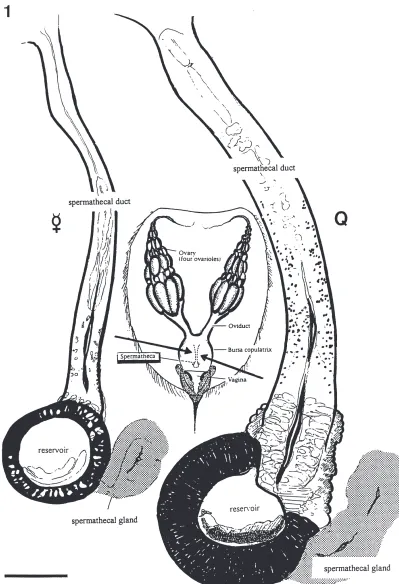
The spermatheca of bumblebees consists of a rela-tively small reservoir for sperm storage, a paired sperma-thecal gland visible as two rather elongated structures on this reservoir and a long straight spermathecal duct between receptacle and the common oviduct (Figs. 1 and 2). Spermathecal morphology does not show significant gross morphological differences between queens and workers (Fig. 1).
The sperm duct, which is extremely long in bumble-bees, is 600 to 700µm long in workers and 900 µm to 1 mm in queens. Its diameter varies from 30 to 50 µm in workers and from 60 to 100µm in queens. The sperm-athecal gland is 150 µm long (diameter 60–70 µm) in workers and measures up to 250µm in length (diameter 70–100 µm) in queens (Figs. 1 and 2).
Fig. 2. Scanning picture of the reservoir with the spermathecal gland (*) attached and the position of the muscular pump (arrow). Ovd= ovid-uct; res=reservoir; spd=spermathecal duct. Scale bar=100µm. Fig. 3. Longitudinal semi-thin section of the spermatheca duct and reservoir of a Bombus terrestris queen. The lumen of the spermathecal duct (spd) is clearly showing its cuticular lining. The position of the muscu-lar pump (pu) is indicated. ep=columnar epithelium of the reservoir; res=reservoir. Scale bar=100 µm. Fig. 4. Sample of spermatheca of
Bombus pratorum(queen after hibernation) stained for carbohydrates. The positively stained material is located in the duct (arrows). The spermathecal gland (*) can be distinguished on top of the reservoir. Scale bar=100µm. Fig. 5. Same sample as in Fig. 4 but stained with a methylene–blue thionin mixture to show stored sperm in the sperma-theca reservoir and spermasperma-thecal gland (*). Scale bar=50µm. Fig. 6. Longitudinal semi-thin section of the spermathecal duct in an artifici-ally hibernating queen ofBombus terrestris. PAS-positive material is widely distributed along the course of the duct, except in its very proxi-mal (near reservoir, on the right in figure) and distal (near oviduct) regions. Scale bar=100µm.
cal reservoir itself and its epithelium. In bumblebees this reservoir is relatively small and this of course emphas-izes the relative importance of the spermathecal duct.
3.2. Histology and histochemistry with special attention to the spermathecal duct
Different regions can be distinguished within the sper-mathecal duct. The proximal region or neck near the res-ervoir is provided with muscles (region of the sperm pump). The rest of the spermathecal duct is lined with a columnar epithelium and its cuticle (Fig. 3). The chemical nature of the substances present in the sperma-theca and in the spermasperma-thecal duct and its cells was examined by using routine histochemical methods (PAS).
Most parts of the bumblebee spermatheca show very low reactivity to the histochemical test performed. How-ever, the most prominent histochemical feature of the cells lining the spermathecal duct is their very strong positive PAS-reaction (Figs. 4 and 6). In addition to this, the intensity of staining was zero when the samples had been treated with human saliva (amylase test). This (together with the ultrastructural results) indicates that the polysaccharide component present in the secretion is glycogen.
Glycogen is only present in large quantities in the cells lining the spermathecal duct of queens. Further experiments have confirmed this presence in queens before insemination, during hibernation, shortly after hibernation, in mature egg-laying queens and in unin-seminated queens captured during summer. The same staining of worker spermathecal ducts, however, revealed only small quantities of this material.
3.3. Ultrastructure
3.3.1. The spermathecal duct
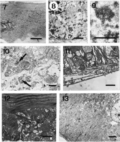
The spermathecal duct is formed by an epithelium with variable characteristics according to the queen’s reproductive state. Glycogen is found at different locations in the cells (Fig. 7). In spermatheca of bumble-bees that are not actively laying, glycogen is present at the basal side of the cells in dense aggregations. In virgin females, large accumulations can be seen. When queens start egg-laying, glycogen stores are gradually mobilized and are gradually found more near the apical side of the cells, suggesting intracellular migration (Figs. 8–10). The observation of glycogen is especially clear in the proximal and medial region of the spermathecal duct (Fig. 1). The part of the spermathecal duct near the ovi-duct does not show the specializations mentioned here. In workers, glycogen content of the duct is far lower.
3.3.2. Reservoir epithelium
Bumblebees have a well developed spermathecal wall (columnar epithelium) lined with a thin cuticular layer. Nuclei of these cells usually are large. Mitochondria are dominant cytoplasmatic organelles. Sometimes, mito-chondria are large and are situated in between the numerous invaginations of the basal plasma membrane of the cells (Fig. 11). Apically, mitochondria are also well represented underneath the long microvillar special-isations of the apical plasma membrane (Fig. 12). Microtubular structures are also found (Fig. 13).
3.3.3. The spermathecal gland
The spermathecal gland consists of two elongated branches that join adjacent to the long spermathecal duct. Its lumen opens into the reservoir at the point where duct and reservoir meet. At this junction, a mus-cular pump occurs. Two cell types occur in the gland: the duct cells, usually with a more electron dense cyto-plasm, and the actual glandular cells, with a less electron dense cytoplasm.
Fig. 7. Virgin queen ofB. terrestris. PAS-staining/TEM. Distribution of sugars in spermathecal duct epithelium. Scale bar=1µm. Fig. 8. Insemi-nated, egg-laying queen of B. terrestris. Basal side of the epithelium with numerous dense inclusions corresponding with sugar material. Scale bar=1µm. Fig. 9. Detail of glycogen particles. Scale bar=0.5µm. Fig. 10. Central part of spermathecal duct of inseminated, egg-laying queen of
B. terrestris. Dense spherical inclusions near the spermathecal duct lumen (arrows). Scale bar=2µm. Fig. 11. Ultrastructural aspects of the columnar epithelium of the reservoir. Basal region of the reservoir cell in aB. ruderatusworker bumblebee. Note strongly infolded membrane. Scale bar=1
µm. Fig. 12. Apical part of the same type of cell with long and slender microvilli. Scale bar=1µm. Figure 13. Basel part of the reservoir cell in aB. terrestrisqueen bumblebee. Note strongly infolded membrane and mitochodria. Scale bar=2µm.
4. Discussion
4.1. Assumed contribution of each subunit of the spermatheca to its overall functioning
The spermathecal duct is rich in glycogen. Between the duct and the glands, a pump mechanism consisting
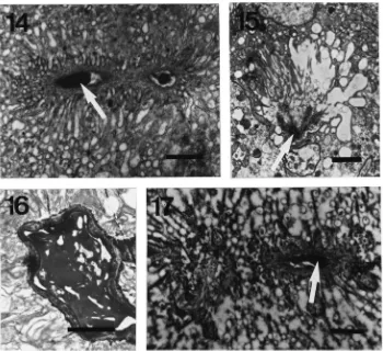
Fig. 14. The spermathecal gland in queens of different reproductive stages ofB. terrestris. Artificially hibernating queen. Part of a secretory cell of the spermathecal gland, showing similar aspect to that of the worker, except the dense secretory material (arrow) in the lumen. Scale bar=1µm. Fig. 15. Inseminated queen before hibernation. The end apparatus shows an intermediate degree of microvillar distortion. Dense material (arrow) is present in the lumen (arrow). Scale bar=2µm. Fig. 16. Inseminated queen before hibernation. A lot of secretory material can be observed inside the larger ducts. Scale bar=1µm. Fig. 17. Egg-laying queen. Microvillar distortion is commonly found in the secretory cells of the spermathecal gland. The lumen again shows the presence of dense material (arrow). Scale bar=1µm.
The epithelial wall of the reservoir clearly is a trans-porting rather than a secretory epithelium. The combi-nation of some structural adaptations such as apical microvilli, numerous basal infoldings with elongated and numerous mitochondria, the absence of secretory ves-icles, the presence of microtubuli, make it a typical trans-port epithelium. In ants, such asCrematogaster opuntiae pinocytotic vesicles were observed in this region (Wheeler and Krutzsch, 1994), but not for the honey bee (Dallai, 1975). In bumblebees we also failed to detect pinocytotic vesicles that would be involved in the uptake of small protein molecules (Berridge and Oschman, 1972; Wheeler and Krutzsch, 1994).
The spermathecal gland has a secretory function. Each secretory cell has an end apparatus with microvilli. Microvillar distortion in active glands could be related to solvent transport together with an array of solutes, as is observed for insect goblet cells as described by Berridge and Oschman (1972). From available literature data and our own observations we conclude that the solute(s) probably correspond with a (glyco)protein. It is interesting that the gland in workers does not show
signs of secretory activity near the end apparatus in each gland cell, and that no distorted extracellular spaces are detectable.
4.2. Biochemical capacities of sperm cells in social insects
Blum et al. (1962) have demonstrated that the sperma-tozoa of the honey bee rapidly oxidize free sugars that occur in the seminal plasma. These authors pointed out that 40 minutes after ejaculation, the concentration of fructose is reduced by 80 per cent. Oxidizing free sugars is a feature these spermatozoa share with those of ver-tebrates (Yanagimachi, 1988). Apparently, the use of glycolysable material is limited to animals in which internal fertilization occurs. In this regard, Mohri (1957) and Humphrey (1950) have demonstrated for sea-urchins and oysters respectively that spermatozoa are not cap-able of metabolizing glycolyscap-able material which was added to suspensions of spermatozoa.
plasma, Blum et al. (1967) have pointed out that also phospholipids serve as a source of oxidative energy for endogenous respiration after aerobic incubation. The use of such substrates is most likely responsible for the fact that, under aerobic conditions, the spermatozoa are highly motile for at least 3 h after ejaculation (Blum et al., 1962). Data of Gessner and Gessner (1976) suggest that generation of the spermathecal fluid of the queen honey bee involves active energy dependent secretion of K+ and of some other ions by the epithelium lining the spermathecal wall.
Linking the physiology of the spermatheca to its struc-ture is challenging. The present study offers data to con-sider the spermathecal duct as a fundamental and important part of the spermatheca, both because of its position in between the receptacle and the oviduct, its length and the positive staining for carbohydrates. Dallai (1975) is, to our knowledge, the only author who has closely examined the ultrastructure of the spermathecal duct (for Apis mellifera).
4.3. The function of the polysaccharides in the spermathecal duct
Our structural and histochemical observations are indicative for a hitherto mostly neglected role for the spermathecal duct. It appears not to be a simple con-ducting channel for transport of spermatozoa to and from the reservoir, but it exhibits an important physiological role with an additional difference between the apparently anatomically similar spermatheca of queens and work-ers. We assume that the mechanism involving glucose is most likely to be activated when spermatozoa need to receive energy when necessary for their metabolism shortly before fertilization, because sugars are always present in the spermathecal duct of queens and the sugar material is supposed to migrate towards the lumen of the spermathecal duct, a process which probably is under hormonal control. Among animals in general, several examples can be encountered involving polysaccharide containing metabolites of the female genital tract with an effect on the metabolism of the sperm. In viviparous fish for example, secretions of the female genital tract provide the sperm with essential nutrients, because apart from acquiring its motility, sperm needs to get energy-rich nutrients for further metabolism (Yanagimachi, 1988). In mammals, similar data are known from rats and humans. For the latter it is known that the spermato-zoa can readily use sugars (and other substances) present in the cervical mucosa. Hormonal changes can strongly affect the composition and hence the viscosity of the mucosa, resulting in facilitating or inhibiting sperm pass-age (Yanagimachi, 1988).
Integration of the ultrastructural data available in literature on spermathecal reservoir epithelia and our his-tochemistry data on the spermathecal duct of
bumble-bees, makes us conclude that the reservoir region is important for osmoregulation and for maintenance of ion levels, whereas the cells lining the spermathecal duct are very important for delivery of energy-rich material for sperm metabolism. In this regard, the impact of the female on the sperm cells is important, probably also a physiological example of cryptic female choice (Eberhard, 1996).
Examples of such cryptic female choice are found at the level of extreme spermathecal duct elongation in some spider species (Eberhard, 1997). Cryptic choice by the female is usually considered to be advantageous to the female by increasing the difficulty of the sperm or other components of semen to penetrate, or sometimes limiting the entrance of male genitalia (Eberhard, 1997). Flanders (1962) and Hitchcock (1956) mention that non-hatchability can be phenotypic because they found that a single queen can either deposit hatchable eggs ing the first half of her life and non-hatchable eggs dur-ing the last half, or deposits durdur-ing her lifetime a series of eggs which gradually decrease in hatchability. Queens also lay mostly non-hatching eggs after being chilled or starved (Hitchcock, 1956). With the results presented here in mind, the latter assumption may be understood if we assume that starvation does not provide extra sugars needed for the spermathecal duct, resulting in less efficient egg-laying. The impact on spermatozoa of the polysaccharide material provided by the spermathecal duct is expected to be considerable, since Duvoisin et al. (1999) have reported that the time required to fill the spermatheca ranged from 30 to 80 min.
Initially in the present study we did not exclude the possibility that the polysaccharide material could corre-spond with reabsorbed material from semen after sperm passage through the duct, since literature data exist men-tioning such regions near the spermathecae of Dolicho-poda, Gryllus and Gryllotalpa (Martoja, 1977). How-ever, it became evident that for bumblebees this is not the case since uninseminated queens also have the poly-saccharides in their spermathecal duct.
Acknowledgements
Eric Schoeters is Postdoctoral Fellow of the Fund for Scientific Research — Flanders (Belgium) (F.W.O.). We are also indebted to Hombio for kindly providing the bumblebee queens and to Koen Collart for section prep-aration for ultrastructural examination.
References
Ahmed, L., Gillot, C., 1982. The spermatheca ofMelanoplus sanguin-ipes (Fabr.). I. Morphology, histology, and histochemistry. Inter-national Journal of Invertebrate Reproduction 4, 281–295. Berridge, M.J., Oschman, J.L., 1972. Transporting Epithelia. Academic
Press, New York.
Blum, M.S., Glowska, Z., Taber, S., 1962. Chemistry of the drone honey bee reproductive system — II. Carbohydrates in the repro-ductive organs and semen. Annals of the Entomological Society of America 55, 135–139.
Blum, M.S., Bumgarner, J.E., Taber, S., 1967. Composition and poss-ible significance of fatty acids in the lipid classes in honey bee semen. Journal of Insect Physiology 13, 1301–1308.
Clements, A.N., Potter, S.A., 1967. The fine structure of the sperma-thecae and their ducts in the mosquito Aedes aegypti. Journal of Insect Physiology 13, 1825–1836.
Dallai, R., 1972. Fine structure of the spermatheca ofApis mellifera. Redia 53, 413–425.
Dallai, R., 1975. Fine structure of the spermathecal gland ofApis melli-fera. Journal of Insect Physiology 21, 89–109.
Davey, K.G., Webster, G.F., 1967. The structure and secretion of the spermatheca of Rhodnius prolixus Sta˚l: A histochemical study. Canadian Journal of Zoology 45, 653–657.
Duchateau, M.J., Marie¨n, J., 1995. Sexual biology of haploid and dip-loid males in the bumble beeBombus terrestris. Insectes Sociaux 42, 255–266.
Duchateau, M.J., Hoshiba, H., Velthuis, H.H.W., 1994. Diploid males in the bumble beeBombus terrestris: sex determination, sex alleles and viability. Entomologia Experimentalis et Applicata 71, 263– 269.
Duvoisin, N., Baer, B., Schmid-Hempel, P., 1999. Sperm transfer and male competition in a bumblebee. Animal Behaviour 58, 743–749. Eberhard, W.G., 1996. Female Control: Sexual Selection by Cryptic
Female Choice. Princeton University Press, Princeton (NJ). Eberhard, W.G., 1997. Sexual selection by cryptic female choice in
insects and arachnids. In: Choe, J.C., Crespi, B.J. (Eds.), The Evol-ution of Mating Systems in Insects and Arachnids. Cambridge Uni-versity Press, pp. 32–57.
Flanders, S.E., 1962. Physiological prerequisites of social reproduction in the Hymenoptera. Insectes Sociaux 9, 375–388.
Gerber, H.S., Klostermeyer, E.C., 1970. Sex control by bees: a volun-tary act of egg fertilisation during oviposition. Science 167, 82–84. Gessner, B., Gessner, K., 1976. Inorganic ions in the spermathecal fluid and their transport across the spermathecal membrane of the queen bee, Apis mellifera. Journal of Insect Physiology 22, 1469–1474.
Gessner, B., Ruttner, F., 1977. Transfer der Spermatozoen in die Sper-matheka der Bienenko¨nigin. Apidologie 8, 1–18.
Grodner, M.L., 1979. Fine structure of the spermathecal gland of the cotton boll weevil, Anthonomus grandis Boheman. Coleoptera: Curculionidae. International Journal of Insect Morphology and Embryology 8, 51–58.
Gupta, B.L., Smith, D.S., 1969. Fine structural organization of the spermatheca in the cockroach,Periplaneta americana. Tissue and Cell 1, 295–324.
Happ, G.M., Happ, C.M., 1970. Fine structure and histochemistry of the spermathecal gland in the mealworm beetle,Tenebrio molitor. Tissue and Cell 2, 443–466.
Happ, G.M., Happ, C.M., 1975. Fine structure of the spermatheca of the mealworm beetle (Tenebrio molitor L.). Cell and Tissue Research 162, 253–269.
Hitchcock, J.D., 1956. Honey bee queens whose eggs all fail to hatch. Journal of Economical Entomology 49, 11–14.
Humphrey, G.F., 1950. The metabolism of oyster spermatozoa. Aus-tralian Journal of Experimental Medical Sciences 28, 1–13. Ito, F., Ohkawara, K., 1994. Spermatheca size differentiation between
queens and workers in primitive ants, relationships with repro-ductive structure of colonies. Naturwissenschaften 81, 138–140. Juberthie-Jupeau, L., Cazals, M., 1985. Ultrastructure et maturation de
la glande accessoire de la spermatheque chezSpeonomus delarou-zeei Fairm. (Coleoptera: Catopidae) du milieu souterrain. Inter-national Journal of Insect Morphology and Embryology 14, 75–86. Kiernan, J.A., 1981. Histological and Histochemical Methods.
Perga-mon Press.
King, P.E., Ratcliffe, N.A., 1969. The structure and possible mode of functioning of the female reproductive system inNasonia vitrip-ennis(Hymenoptera; Pteromalidae). Journal of Zoology (London) 157, 319–344.
Kurzrok, R., Birnberg, C., 1958. A study of semen mucus penetration and its relation to a test of ovulation. International Journal of Fer-tility 3, 134–138.
Martoja, R., 1977. Organes ge´nitaux femelles: Voies ge´nitales et glandes annexes. In: Grasse´, P.-P. (Ed.), Traite´ de Zoologie, Anato-mie, Syste´matique, Biologie: Insectes: Game´togene`ses, Fe´cond-ation, Me´tamorphoses, 8. Fascicule V-A, Masson, Paris, pp. 1–123. Mohri, H., 1957. Endogenous substrates of respiration in sea-urchin spermatozoa. Journal of the Faculty of Sciences of Tokyo Univer-sity 8, 51–63.
Noirot, C., Quennedey, A., 1991. Glands, gland cells, glandular units: some comments on terminology and classification. Annales de la Socie´te´ Entomologique de France 27, 123–128.
Pabalan, N., Davey, K.G., Packer, L., 1996. Comparative morphology of spermathecae in solitary and primitively eusocial bees (Hymenoptera; Apoidea). Canadian Journal of Zoology 74, 802– 808.
Peeters, C., Ito, F., Gobin, B., Fannes, W., Billen, J., 1999. Queen-worker divergence in the spermathecae of ponerine and myrmeciine ants. Proceedings of the XIIIth IUSSI Congress, Adelaide, Aus-tralia 1999, p. 369.
Poole, H.K., 1970. The wall structure of the honey bee spermatheca with comments about its function. Annals of the Entomological Society of America 63, 1625–1628.
Quennedey, A., 1998. Insect epidermal gland cells: ultrastructure and morphogenesis. In: Harrison, W.H. (Ed.), Microscopic Anatomy of Invertebrates, vol. 11a, Wiley-Liss, New York. pp. 177–207. Rodriguez, V., 1994. Function of the spermathecal muscle in
Chely-morpha alternans Boheman (Coleoptera: Chrysomelidae: Cassidinae). Physiological Entomology 19, 198–202.
Rojas-Rousse, D., Palevody, C., 1981. Structure et fonctionnement de la spermatheque chez l’endoparasite solitaireDiadromus pulchellus
Wesmael (Hymenoptera: Ichneumonidae). International Journal of Insect Morphology and Embryology 10, 309–320.
Ro¨seler, P.-F., 1973. Die Anzahl Spermien im Receptaculum seminis von Hummelko¨niginnen (Hymenoptera: Apidae, Bombinae). Api-dologie 4, 267–274.
Ruttner, F., Koeniger, G., 1971. Die Fu¨llung der Spermatheka der Bienenko¨nigin: active Wanderung oder passiver Transport der Spermatozoen? Zeitschrift fu¨r Vergleichende Physiologie 72, 411–422.
Suzzoni, J.P., 1972. Ultrastructure de la glande de la spermatheque chez Phosphuga atrataL. (Coleoptera Silphidae). Zeitschrift fu¨r Zellforschung 128, 426–437.
Tombes, A.S., Roppel, R.M., 1972. Ultrastructure of the spermatheca of the granary weevil Sitophilus granarius L. (Coleoptera: Curculionidae). International Journal of Insect Morphology and Embryology 1, 141–152.
Villavaso, E.J., 1975. The role of the spermathecal gland of the boll
weevil, Anthonomus grandis. Journal of Insect Physiology 21, 1457–1462.
Wheeler, D.E., Krutzsch, P.H., 1994. Ultrastructure of the spermatheca and its associated gland in the ant Crematogaster opuntiae
(Hymenoptera: Formicidae). Zoomorphology 114, 203–212. Yanagimachi, R., 1988. Mammalian fertilization. In: Knobil, E., Neil,