* corresponding author:[email protected]
Low CD4
+
T cell counts are not risk factor
for Malassezia species infection in HIV/AIDS
patients
Epi Panjaitan1*, Satiti Retno Pudjiati2, Agnes Sri Siswati2
1Sangata District Hospital, East Kutai District, East Kalimantan Province, Department of
Dermatology and Venereology, Faculty of Medicine Universitas Gadjah Mada/Dr Sardjito General Hospital, Yogyakarta
ABSTRACT
Human immunodefiiency virus (HIV) infection and aquired immunodeficiency syndrome (AIDS) cause a progressive depletion of CD4+ T cell populations accompanied by progressive impairment
of cellular immunity and increasing susceptibility to opportunistic infections. Seborrheic dermatitis is one of the most common skin opportunistic infections on HIV/AIDS patients. Malassezia species is bilieved as the causative of seborrheic dermatitis. The aim of the study was to evaluate low CD4+ T cell counts as risk factor for Malassezia sp. infection in HIV/AIDS patients. This was
an observational study with cross-sectional design conducted on HIV/AIDS patients who attended in Department of Dermatology and Venereology, Faculty of Medicine Universitas Gadjah Mada/ Dr Sardjito General Hospital, Yogyakarta and met the inclusion and exclusion criteria. Culture of Malassezia sp. was conducted in Department of Microbiology and classified as high (>100 CFU/ tape) and low (<100 CFU/tape) density colonies. CD4+ T cell counts were measured in Department
of Clinical Pathology and classified as high (>200 cells/mm3) and low (<200 cells/mm3) CD4+ T
cell counts. A total of 83 subjects with HIV/AIDS comprising 54 (65.1%) males and 29 (34.9%) females aged 20 - >60 years were involved in the study. The number of Malassezia sp. colony on subjects with high and low CD4+ T cell counts were 31.55 ± 26.21 and 25.2 ± 33.89 CFU/
tape, respectively. No significantly relationship between between CD4+ T cell count and Malassezia
sp. colony number was observed in the study (p=0.607; 95%CI=0.04-5.19; RP=0.452). In conclusion, low CD4+ T cell counts is not risk factor for Malassezia sp. infection in HIV/AIDS
patients.
ABSTRAK
Infeksi human immunodefiiency virus (HIV) dan aquired immunodeficiency syndrome (AIDS) menyebabkan penurunan progresif jumlah sel T CD4+ yang diikuti dengan gagguan imunitas
seluler dan meningkatnya risiko infeksi oportunitis. Dermatitis seboroik merupakan salah satu infeksi oportunitis kulit yang umum terjadi pada penderita HIV/AIDS. Malassezia sp. dipercaya penyebab dermatitis seboroik. Tujuan penelitian ini untuk mengkaji kadar sel T CD4+ yang
rendah sebagai risiko terjadinya infeksi Malassezia sp. pada penderita HIV/AIDS. Penelitian ini merupakan penelitian observasional dengan rancangan potong lintang yang dilakukan terhadap penderita HIV/AIDS yang berkunjung ke Bagian Kulit dan Kelamin, Fakultas Kedokteran, Universitas Gadjah Mada/RSUP Dr. Sardjito, Yogyakarta dan memenuhi kriteria pemasukan dan pengeluaran. Kultur Malassezia sp. dilakukan di Bagian Mikrobiologi dan dikelompokkan sebagai koloni densitas tinggi ((>100 CFU/tape) dan rendah (<100 CFU/tape). Kadar sel T CD4+ ditetapkan di Bagian Patologi Klinik dan dikelompokkan sebagai sel T CD4+ tinggi (>200 sel/mm3) dan rendah (<200
sel/mm3). Total 83 penderita HIV/AIDS terdiri 54 (65.7%) pria dan 29 (34.9%) wanita berumur
CFU/tape. Tidak ada hubungan bermakna adalah kadar sel T CD4+ dengan jumlah koloni Malassezia
sp dalam penelitian ini (p=0,607; 95%CI=0,04-5,19; RP=0,452). Dapat disimpulkan, kadar sel T CD4+ yang rendah bukan merupakan faktor terjadinya infeksi Malassezia sp. pada penderita
HIV/AIDS.
Keywords: Malassezia species - colony - CD4+ T cell – risk factor – opportunitic infection
INTRODUCTION
The number of people infected with human immunodeficiency virus (HIV) continues to rise significantly in most parts of the world in the last decade. It was estimated that between 30 and 36 million people in the world are living with HIV in 2012. Approximately 1.8 to 2.3 million of them develop to be acquired immunodeficiency syndrome (AIDS).1 In Indonesia, approximately 150.200 cases of HIV and 55.700 cases of AIDS with 9.700 deaths from AIDS have been reported in 2014. In Yogyakarta Special Region, 2.611 cases of HIV and 916 cases of AIDS have been reported in 2014.2
Human immunodefiiency virus infection causes a progressive depletion of CD4+ T cell populations accompanied by progressive impairment of cellular immunity and increasing susceptibility to opportunistic infections.3 Seborrheic dermatitis is one of the most common skin opportunistic infections on HIV/AIDS patients. Seborrheic dermatitis is a chronic inflammatory skin disorder that particularly affects the sebaceous-land-rich areas of skin. In general population, the prevalence of seborrheic dermatitis varies between 3 and 5%, while in patients with HIV its prevalence increases between 20 and 30% and up to 80% in patients with AIDS.1,4,5
body with varied density depending on age, body site, geographic area, and the presence of normal or diseased skin.6 The highest density is found in the seborrhoic areas, namely the scalp, face, chest and upper trunk, whereas the lowest density is found on the hands.7 The pathogenesis of Malassezia sp in seborrheic dermatitis is multifactorial involving external factors such as climate, drug use, lifestyle and internal factors such as immune deficiency, amount of sebum, hyperhidrosis, pregnancy.8
The role of Malassezia sp. in seborrheic dermatitis in patients of HIV/AIDS has been investigated by some authors with varied results. The associtation of seborrheic dermatitis and AIDS first reported by Eisenstat in 1984.9 Further studies showed tha the prevalence of seborrheic dermatitis in patients with HIV differs according to various authors. 10-12 It appears that the subtype of Malassezia sp.
and level of CD4+ T cell are found to be risk factors of seborrheic dermatitis in patients of HIV/AIDS. This study was conducted to evaluate level of CD4+ T-cell in HIV/AIDS patients in correlation with density of Malassezia sp. colony.
MATERIALS AND METHODS
Subjects
Sardjito General Hospital between June and September 2012 and fulfulled the inclusion and exclusion criteria were involved in this study. The inclusion criteria were male or female HIV/ AIDS patients aged over 18 years and agreed to participate in this study by signed an informed consent. The exclusion criteria were pregnant women, currently use of oral contraceptives, use of topical or systemic antifungal, corticosteroids, antibiotics and immunosupresive agents during two last months, and patients with hyper-hidrosis. Ethical approval for the study was obtained from the Medical and Health Research Ethics Committee, Faculty of Medicine, Universitas Gadjah Mada, Yogyakarta.
Protocol of study
Subjects who welling to participate in the study were given a questionnaire to be filled. The questionnaire consisted of interview-administrative questions regarding age, education, occupation, marrital status and duration of illness. Body weight and body height of subjects were then measured using digital weight completed with microtoise statur meter. Culture of Malassezia sp. was conducted in Department of Microbiology, Faculty of Medicine, Universitas Gadjah Mada, Yogya-karta. Sample was taken from forehead skin of subjects. The Malassezia sp. were then isolated from the skin with contact plates containing a modified Leeming-Notman’s agar medium. The culture plates were incubated at 32 oC for 14 days. The relative humidity in the incubator was 85% and all culture plates were incubated in
plastic bags. Followed after 14 days incubation, the Malassezia sp. were identified and the number of colonies was counted, giving a semi-quantitative number of organisms. The number of colonies was then classified into two groups i.e. subjects with high colonies density (≥ 100 CFU/tape) and those with low colonies density (<100 CFU/tape).
Blood sample for CD4+ T cell measuremnet was taken from veins within fossa cubiti. The measurement of CD4+ T cell was conducted in Department of Clinical Pathology, Faculty of Medicine, Universitas Gadjah Mada, Yogyakarta. CD4+ T cell counts were then classified into two groups i.e. subjects with high CD4+ T cell counts (
≥ 200 cells/mm3) and those with low CD4+ T cell counts (<200 cells/mm3).
Statistical analysis
Descriptive statistics were used to analyze the characteristics of subjects. Data were presented as percentage. Chi Square test was used to evaluate the correlation between the CD4+ T cell count and the density of Malassezia sp. colonies. p value <0.05 was considered significant.
RESULTS
Characteristics of subjects
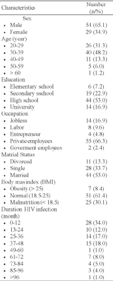
TABLE 1. Characteristics of subjects
Risk factor of HIV infections/AIDS
Multi partner (64 or 77.1% subjects) was found to be risk factors of HIV infection,
TABLE 2. Risk factor of HIV infections/AIDS
Relationship between sex and CD4+ T cell
count
The relationship between sex and CD4+ T cell count is presented in TABEL 3. The CD4+ T cell count of male patients with HIV/AIDS (283.66 ± 274.08 cells/mm3) was higher than female patients (258.81 ± 200.56 cells//mm3). However, it was not significantly different (p=0.637).
TABLE 3. Relationship between sex and CD4+T
cell count
Relationship between sex, age, and Malassezia sp. colony
TABLE 4. Relationship between sex, age, and colony of Malassezia sp colony
Relationship between CD4+ T cell count
and Malassezia sp. colony
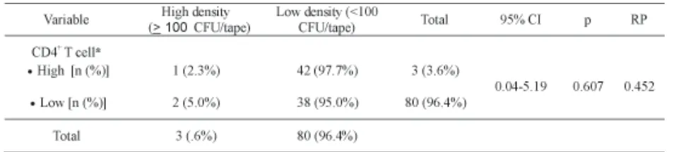
The relationship between CD4+ T cell count and Malassezia sp. is presented in TABEL 5. The number of Malassezia sp. colony on subjects with high and low CD4+ T cell counts were 31.55 ± 26.21 and 25.2 ± 33.89 CFU/ tape, respectively. Low density colony (<100 CFU/tape) was observed on most subjects both who had CD4+ T cell count > 200 cells/mm3
(42 or 97.9% subjects) and < 200 cells/mm3 (38 or 95.0% subjects). High density colony was observed only on one subject on subjects who had CD4+ T cell count > 200 cells/mm3 and two subjects on subjects who had CD4+ T cell count < 200 cells/mm3. No significantly relationship between between CD4+ T cell count and Malassezia sp. colony number was observed in the study (p=0.607).
TABLE 5. Relationship between CD4+ T cell counts and Malassezia sp colony
*High: CD4+ Tcell counts > 200 cells/mm3; Low: CD4+ Tcell counts > 200 cells/mm3
DISCUSSION
This study showed that the prevalence of HIV/AIDS on males was higher than on females. Moreover, the highest prevalence of HIV/AIDS was observed on patients aged between 30 and 39 years. These results were not in accordance with a report that released
occupational of HIV/AIDS patients in this study was similar to that reported by the Directorate General of Disease Control and Environmental Health.
Multi partner was found to be risk factor for HIV infection in this study. It was similar with the report that released by the Directorate General of Disease Control and Environmental Health. It was reported that unsafe sex (77%) was risk factor for HIV infection, followed by the use unsterile syringe by injecting drug users (8.5%), mother to child transmission (5.1%), male to male sex (2.7%).13 This study proved that unsafe intercourse was the largest risk factor for HIV infection.
The CD4+ T cell counts in female patients tended to be higher than in male patients in this study although it was not significantly different. A study conducted in Netherlands showed that the mean CD4+ T cell counts were significantly higher in female HIV/AIDS patients than male patients.14 Similar results were obtained in the studies that conducted in India and in Nigeria.15,16 It has been suggested that a sex hormone effect could be one possible explanation for the reported higher in CD4+ T cell counts in felamle HIV/AIDS patients.17
The number of Malassezia sp. colony in male patients was not signidicantly different than in female patients. A study conducte in Japan showed that the density of Malassezia sp. colony in male patients was higher than those females and the highest density was observed on patients aged from 30 to 40 years.18 However, a study conducted in Canada showed that no significantly different in number of Malassezia sp. colony between male and female patients was observed.19
evalute the relationship between Malassezia sp. colonization and the immune status of HIV/AIDS patients have been performed with different results. Hakansson et al.20 reported that there was no correlation between the number of Pityrosporum ovale (Malassezia sp.) and the immune status of the HIV-seropositive patients. Moreover, Munoz-Perez et al.21 reported that Malassezia sp. was not assocoated with the onset of seborrheic dermatitis lesions formation on HIV/AIDS patients. In contrast, Farrokh et al.22 demonstrated that low CD4+ T cell counts was associated with the incidence of seborrheic dermatitis in HIV-positive patients. Furthermore, Schechtman et al.23 demonstrated that there was a trend between numbers of Malassezia yeasts present on lesional skin, severity of seborrheic dermatitis and CD4+ T cell counts in HIV-positive patients. Nnoruko et al.24 showed that seborrheic dermatitis had occurred at CD4+ T cell counts of > 200 cells/ mm3 in HIV/AIDS patients.
The variability of data obtained in several studies could be explained by the differences in sampling sampling techniques, media culture race or region. Several sampling techniques are available for Malassezia culture including adhesive tapes stripping, swabbing, agar contact plate and scrub method. The sampling techniques could influence the quality of sample obtained. Dixon media or Leeming and Notman agar are often used for Malassezia cultures. The media used for the cultures could influence Malassezia recovery from sample.18,25
CONCLUSIONS
ACKNOWLEDGEMENTS
Authors would like to thank all subjek who have participated in the study.
REFERENCES
1. Ramdial PK and Grayson W. Human
immunodeficiency virus (HIV) and acquired immunodeficiency syndrome (AIDS)-associated cutaneous diseases. In: Calonje E, Brenn T, Lazar A, McKee PH, editors. McKee’s pathology of the skin. 4th eds. Philadelphia: Elsevier, 2001: 895-6.
2. Directorate General of Communicable Diseases and Enviromental Health. Cases of HIV/AIDS in Indonesia. Jakarta: Ministry of Health, Republic of Indonesia, 2014.
3. Okoye AA and Picker LJ. CD4+ T cell depletion
in HIV infection: mechanisms of immunological failure. Immunol Rev 2013; 254(1):54-64. 4. Naldi L and Rebora A. Seborrheic dermatitis. N
Engl J Med 2009; 360: 381-96.
5. Chatzikokkinou P, Sotiropoulos K, Katoulis A, Luzzati R, Trevisan G. Seborrheic dermatitis – an early and common skin manifestation in HIV patients. Acta Dermatovenerol Croat 2008; 16(4):226-30.
6. Ashbee, HR. Recent developments in the
immunology and biology of Malassezia species. FEMS Immunol Med Microbiol 2006; 47:14-23. 7. Aspres N and Anderson C. Malassezia yeasts in the pathogenesis of atopic dermatitis. Aust J Dermatol 2004; 45(4):199-207.
8. Lee YW, Yim SM, Lim SH, Choe YB, Ahn, KJ. Quantitative investigation on the distribution of Malassezia species on healthy human skin in Korea. Mycoses 2006; 49(5): 405–410. 9. Eisenstat BA, Wormser GP. Seborrheic dermatitis
and butterfly rash in AIDS. N Engl J Med 1984;311(3):189.
10. Blanes M, Belinchón I, Merino E, Portilla J, Sánchez-Payá J, Betlloch I. Current prevalence and characteristics of dermatoses associated with human immunodeficiency virus infection. Actas Dermosifiliogr 2010;101(8):702-9.
11. Schechtman RC, Midley G, and Hay RJ. HIV disease and Malassezia yeasts: a quantitative study of patiens presenting with seborrheic dermatitis. Br J Dermatotol 1995; 133(5): 694-8.
12. Berger RS, Stoner MF, Hobbs ER, Hayes TJ, Boswell RN. Cutaneous manifestations of early human immunodeficiency virus exposure. J Am Acad Dermatol 1988;19(2 Pt1):298-303. 13. Ditjen Pengendalian Penyakit dan Penyehatan
Lingkungan, Kementiran Kesehatan, Republik Indonesia. Statistik Kasus HIV/AIDS di Indonesia Dilapor s/d Desember 2012. [cited 2013 January 11]. Avaible from: www.spiritia.or.id/stats/Stat Curr.pdf
14. Prins M, Brettle RP, Robertson JR, Aguado IH, Broers B, Carre N, et al. Geographical variation in disease progression in HIV-1 seroconverted injecting drug users in Europe. Int J Epidemiol 1999; 28: 541–549.
15. Thakar MR, Abraham, PR, Arora S, Balakrishnan P, Bandyopadhyay B, Joshi AA. Establishment of reference CD4+ T cell values for adult Indian population. AIDS Res Ther 2011; 8:35. doi:10.1186/1742-6405-8-35
16. Akinbami A, Dosunmu A, Adediran A, Ajibola S, Oshinaike O, Wright K, et al. CD4 count pattern and demographic distribution of treatment-na¿ve HIV patients in Lagos, Nigeria. AIDS Res Treat 2012; 2012:352753. doi:10.1155/2012/352753 17. Maini MK, Gilson RJ, Chavda N, Gill S, Fakoya A, Ross EJ, et al. Reference ranges and sources of variability of CD4 counts in HIV-seronegative women and men. Genitourin Med 1996, 72(1):27-31.
18. Sugita T, Boekhout T, Velegraki A, Guillot J, Hadina S, Cabanes FJ. Epidemiology of Malassezia-Related Skin Disease. In: Teun B, Eveline G, Peter M, Aristea V editors. Malassezia and the skin: science and clinical practice, 1st ed.
Heidelberg: Springer, 2010: 65-119.
19. Gupta AK and Bluhm R. Seborrheic dermatitis. J Eur Acad Dermatol Venereol 2004; 18(1): 13-26. 20. Håkansson C, Faergemann J, Löwhagen GB. Studies on the lipophilic yeast Pityrosporum ovale in HIV-seropositive and HIV-seronegative homosexual men. Acta Derm Venerol 1988; 68(5):422-6.
22. Farrokh R, Ghaderi E, Moradi G, Mafakheri L. The relationship between skin manifestations and CD4 counts among HIV-positive patients. Park J Med Sci 2008; 24(1):114-7.
23. Schechtman RC, Midgley G, Hay RJ. HIV disease and Malassezia yeasts: a quantitative study of patients presenting with seborrhoeic dermatitis. Br J Dermatol 1995; 133(5):694-8.
24. Nnoruka EN, Chukwuka JC, Anisuiba B. Correlation of mucocutaneous manifestations of
HIV/AIDS infection with CD4 counts and disease progression. Int J Dermatol 2007; 46(Suppl2):14-8.
25. Jagielski T, Rup E, Zió³kowska A, Roeske K, Macura AB, Bielecki J. Distribution of