Zoologischer Anzeiger 248 (2010) 299–312
Description of the male reproductive system of
Paguristes eremita
(Anomura, Diogenidae) and its placement in a phylogeny of diogenid
species based on spermatozoal and spermatophore ultrastructure
Tina Tirelli
a,, Daniele Silvestro
b, Daniela Pessani
a, Christopher C. Tudge
c,da
Dipartimento di Biologia Animale e dell’Uomo, Via Accademia Albertina, 13, 10123 Torino, Italy
b
Senckenberg Research Institute, Frankfurt/M, Germany and Biodiversity and Climate Research Center, Frankfurt/M, Germany
c
Department of Invertebrate Zoology, National Museum of Natural History, Smithsonian Institution, Washington, DC 20013-7012, USA
d
Biology Department, American University, Washington, DC 20016-8007, USA
Received 28 July 2009; received in revised form 7 January 2010; accepted 9 January 2010
Corresponding Editor: S. De Grave
Abstract
The male gonopores, male reproductive apparatus, spermatophore and spermatozoa of the Mediterranean hermit crab Paguristes eremita are described, using interference phase microscopy, scanning electron microscopy and transmission electron microscopy. A correlation is made between the gonopore morphology and the different kinds of setae accompanying them, and the reproductive biology of these crabs. Each testes merges into a tubular duct made up of four zones: (1) the collecting tubule with free spermatozoa; (2) the proximal zone, where the ampulla of the spermatophores starts to be formed; (3) the medial zone, where the ampulla is completed, the stalk lengthens and the pedestal is formed; (4) the distal zone, where the mature spermatophores are stored. The sizes of the different parts of the spermatophore and of the sperm are given and their exterior morphology and ultrastructure described and compared to congeners. The morphology of the gonopore, male reproductive system, spermatophore and spermatozoa ofP. eremitaare species-specific, clearly distinguishing the species from the other members of the family. The available spermatozoal and spermatophore data is used to placeP. eremitawithin a sperm phylogeny of the hermit crab family Diogenidae.
&2010 Elsevier GmbH. All rights reserved.
Keywords:Hermit crab; Reproductive tract; Morphology; Ultrastructure; Phylogenetic analysis
1. Introduction
Recently, comparisons of the functional morphology of genitalia and subsequent sperm transfer and storage
mechanisms, and the structure of spermatozoa and spermatophores have been studied extensively in the Decapoda and provide useful information on phyloge-netic relationships and evolutionary divergence (see
Bauer 1986, 1991; Kronenberger et al. 2004). In particular, the reproductive apparatus of hermit crabs, made up of gonopores (sometimes sexual tubes), testes and vasa deferentia containing spermatophores and
www.elsevier.de/jcz
0044-5231/$ - see front matter&2010 Elsevier GmbH. All rights reserved. doi:10.1016/j.jcz.2010.01.001
Corresponding author. Tel.:þ39 011 6704538;
fax:þ39 011 6704508.
spermatozoa, show a species-specific morphology, that has been successfully used in recent phylogenetic studies (Tudge 1997;Tirelli et al. 2008).
The hermit crab family Diogenidae is comprised of 20 genera and approximately 1200 species (McLaughlin, 2003; Lemaitre. pers. comm.) and for 13 genera (Allodardanus Haig & Provenzano, 1965, Aniculus
Dana, 1852, Bathynarius Forest, 1989, Cancellus
H. Milne Edwards, 1836, Ciliopagurus Forest, 1995,
IsochelesStimpson, 1858, PaguropsisHenderson, 1888,
Petrochirus Stimpson, 1858, Pseudopaguristes
McLaughlin, 2002, Pseudopagurus Forest, 1952, Stra-tiotesThomson, 1899,TiseaMorgan & Forest, 1991 and
Trizopagurus Forest, 1952) the reproductive apparatus morphology is still unknown or incompletely known (Mantelatto et al. 2009) and could be very important for comparative studies.
Paguristes eremita (Linnaeus, 1767) is a common Mediterranean hermit crab belonging to the family Diogenidae. It lives on sandy bottoms of the infralittoral zone. Its larval distribution (Thiriot 1974), shell use, and epibiotic relationships (Tirelli et al. 2006a) are well known, but no complete description of the male reproductive apparatus is available for this species.
Therefore the aim of this paper is to expand the knowledge of the reproductive morphology ofP. eremita
by analyzing and describing the male gonopores, the reproductive tract, the mature spermatophores and their stages of maturation, studying the sperm ultrastructure, and comparing them with similar structures in other members of the family Diogenidae. A second phyloge-netic analysis of selected Diogenidae is performed, adding to a previous one (Tirelli et al. 2008) characters concerning P. eremita sperm and spermatophore ultra-structure.
This paper is part of a series that aims to describe the male reproductive apparatus of four of the most common Mediterranean hermit crab species: Calcinus tubularis Linneus, 1967 (see Tirelli et al. 2006b),
Clibanarius erythropus Latreille, 1818 (see Tirelli et al. 2007), Diogenes pugilator Roux, 1829 (see Tirelli et al. 2008) and Paguristes eremita (present study). In particular, forP. eremitawe present new research that, together with the limited data already published (Mouchet 1931), contributes to the knowledge of the reproductive apparatus of this species.
2. Materials and methods
2.1. Specimens
Ten adult specimens (indicated by 410.00 mm carapace length and gonopores present) of P. eremita
were collected on a sandy bottom at 9 m depth from
Caorle, Veneto, Mediterranean Sea (45134’5800
N, 12154’1500
E) during summer 1998. After collection, the specimens were immediately preserved in 4% formalin in seawater or in 2.5% glutaraldehyde.
For each individual the cephalothorax length (CL) was measured from the tip of the rostrum to the V-shaped groove at the posterior edge of the cepha-lothorax, with a stereomicroscope fitted with a grad-uated eyepiece.
The use of animals was approved by the appropriate animal care review committee at the Dipartimento di Biologia Animale e dell’Uomo dell’Universita`di Torino
(Italy), where the study was carried out.
2.2. Microscopy and morphometry
Observations were made under interference phase-contrast (IPM) light microscopy, scanning electron microscopy (SEM), and transmission electron micro-scopy (TEM). Five specimens were prepared for light microscopy observations, three specimens for SEM observations, and two specimens for TEM observations according to standard protocols described inTirelli et al. (2006b, 2007).
The distal part of the vas deferens was used for the spermatophore analysis. A regression analysis was performed to compare the width of the last portion of the distal part of the vas deferens (VDW) and the size (CL) of the crabs.
Twenty spermatophores from each of 5 specimens used for IPM analysis were measured. The following measure-ments were taken: ampulla length; ampulla width; stalk length; stalk width; pedestal length; pedestal width. A regression analysis was performed to compare the ampulla dimensions and the width of the distal vas deferens.
Measurement of the different layers making up the spermatophore wall were performed.
Lastly, the different regions of the spermatozoa for each one of the 2 specimens observed under TEM were measured.
2.3. Phylogenetic analysis
The diogenid taxa included in this analysis are taken fromTudge (1997), except for the following four hermit crabs (listed with their source):Calcinus tubularis(from
Tirelli et al. 2006b),Calcinus tibicenHerbst, 1791 (from
Amadio and Mantelatto 2009), Clibanarius vittatus
Bosc, 1802 (from Matos et al. 1993; Hess and Bauer 2002), Diogenes pugilator (from Manjo´n-Cabeza and
absence of the peripheral acrosome zone (0=absent, 1=present).
The outgroup taxon, Pagurus bernhardus Linneus 1758 (family Paguridae) is chosen because of its sister-group relationship to the diogenid taxa (plus the coenobitids) in the phylogeny ofTudge (1997).
The matrix is available, upon request, from the first author (TT).
The parsimony analysis was performed using PAUP version 4.0b10 (Swofford 2002) on a data matrix established as a Nexus file by MacClade 4.08 (Maddison and Maddison 2005). Heuristic searches found the most parsimonious trees, with the following options: the starting trees were obtained by stepwise addition (random addition sequence, RAS, 100 replicates) and then rearranged by the branch swapping algorithm tree-bisection-reconnection (TBR).
All characters were treated as unordered and initially equally weighted, but then reweighted according to their own rescaled consistency index (RC), using the iterative protocol ofFarris (1969, 1988).
Support for tree topology was evaluated by perform-ing a bootstrap analysis (500 replicates, 10 RAS, TBR).
3. Results
3.1. Gonopores
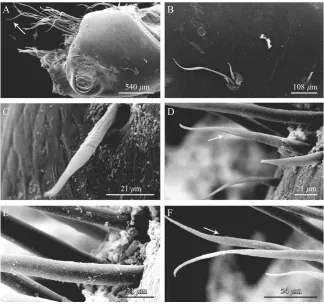
The gonopores ofP. eremitaare paired structures, on both the left and right coxa of P5, and are flush with each coxal surface. Each gonopore is a circular pore covered by a membranous operculum, possibly hinged mesially. A ventral view of a single coxa with gonopore is shown in Fig. 1A. At a short distance from the gonopore there are two kinds of setae: annulate without setules and annulate with setules. In particular the setae are assembled into four isolated groups of three setae each. The three setae making up each group (Fig. 1B) differ from each other: one is short, annulate without setules (stl=45.0470.37mm, stw=5.0070.20mm; mean71 SD,n=6) (Fig. 1C), one is medium, annulate with a setule in the distal third (stl=100.0871.25mm, stw=9.0070.24mm; mean71SD, n=6) (Fig. 1D) and one is long, annulate with setules along the apical portion (stl=210.0871.69mm, stw=9.8870.60mm; mean71SD, n=6) (Fig. 1E and F). The longest setae are annulate without setules (stl=300.5472.45mm,
540 m 108 m
21 m 21 m
21 21 m
21 m 54 54 54 mm
stw=9.9670.24mm; mean71SD,n=6) and constitute a thick row, along the coxal edge (Fig. 1A).
All four types of setae have a central axis, which is slightly curved and flattened (almost spatula shaped) in the distal portion, and a distinct annulus dividing them into a basal portion showing slight longitudinal grooves and a distal smooth portion (Fig. 1BE).
3.2. Testes, vasa deferentia and stages of
spermatophore maturation
The male reproductive tract is composed of paired gonads, consisting of testes and vasa deferentia, located dorsally in the pleon. The testes lie dorsal to the gut, on the large hepatopancreas, while the vasa deferentia descend vertically into the hepatopancreas and release the spermatophores externally through the paired gonopores.
The paired, lobe-like testes are composed of cystic structures. Each testis merges into a tubular duct. Throughout their length the vasa deferentia are com-posed of two main layers: an inner secretory epithelium (Fig. 2A) and an outer muscular layer (Fig. 2B). The
epithelium is generally composed of cells containing cytoplasmic organelles, such as cisternae of the rough endoplasmic reticulum filled with secretory products (Fig. 2C) occupying a large fraction of the cell, some Golgi bodies, and many small vesicles. The secretory material produced by the epithelium is evident in the vasa deferentia lumen (Fig. 2D).
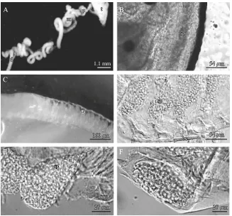
The vas deferens is composed of four zones: (1) highly coiled narrow collecting tubule, (2) proximal, (3) medial, and (4) distal zones (Fig. 3A). The proximal and the medial zones are characterized by a very high number of coils.
The amorphous sperm mass flows embedded in the seminal fluid from the testes to the adjacent collecting tubule (Fig. 3B). In the proximal zone, the sperm mass is subdivided by the dense primary spermatophore layer, which envelops it. In between the proximal and the medial zones, the aggregated spermatozoa are separated and compartmentalized to form the ampullae (Fig. 3C). In the medial zone, the stalk and foot are built and the stalk lengthens (Fig. 3D). The mature spermatophores (Fig. 3E and F) then are stored in the distal zone.
CL was 11.2071.16 mm and VDW was 292.0070.05. VDW was positively correlated with the size of the crab (r2=0.437;F=11.658;po0.05).
0.54 m
1.3 m
2.1 m
0.8 m
1.1 m
gl
gl edp se
l
ml se
rer sm
3.3. Spermatophore
The spermatophore ofP. eremita (Fig. 3DF) has a sperm-filled ampulla elevated on a stalk, which is connected to a pedestal or foot. The ampulla is generally cylindrical in shape but its basal end (the one towards the stalk) is narrower (Fig. 3E), therefore the whole ampulla has an upside down pear shape. The ampulla is 45.277.3mm (n=100) wide and 92.677.2mm (n=100) long. The stalk is 49.276.5mm (n=6) in length, and 6.571.4mm (n=6) in width, and the width is constant for most of the stalk length. The stalk gradually thickens in its distal portion to envelop the ampulla. The pedestal has a characteristic shape: it resembles a half-moon with the convexity towards the stalk (Fig. 3D). It is 2.570.0mm (n=6) long and 10.871.3mm (n=6) wide. The stalk inserts at around half of the pedestal width. The mature spermatophores in the reproductive duct are enveloped by protective layers and they are connected by a basal cord (Fig. 3D).
As the only spermatophore portion measured in the spermatophores coming from all the specimens was the
ampulla, we performed a regression analysis to compare the ampulla length (AL) and the distal vas deferens width (VDW). No correlation between AL and VDW was found.
An accessory ampulla, associated with the main ampulla, is present only in spermatophores, which have not yet ended the maturation process, therefore have not yet fully developed the stalk.
Observations of the spermatophore wall ultrastruc-ture (Fig. 2E) show an inner, slightly granular and moderately electron-dense layer, showing in its middle some quite electron-dense patches, followed by a thin outer fibrillar layer. The fibrillar layer is just 1/10 as thick as the granular one, being 0.12970.002mm (n=2) while the granular one is 1.01270.107mm (n=2).
3.4. Spermatozoa
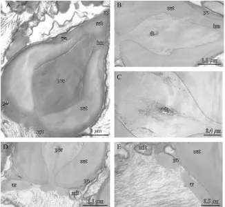
Spermatozoa (Fig. 4A) are composed of an almost conical acrosomal vesicle capped by an operculum. At the base of the acrosomal vesicle, three arms are
1.1 mm
160 160 m
54 54 m
54 54 m
27 27 m 27
27 m
160 m
54 m
54 m
27 m
27 m
sp sp t
c p
m
d
a
s
f bcbc
a
sp sp
s
s s
sp sp
a sp sp
t c p
m
d
a
s
f bc
a
sp
sp
a sp
sp
a
a a
positioned to form a 1201 angle between each other.
P. eremita total sperm length is 4.7mm, total sperm width is 3.6mm, acrosomal length is 4.4mm, and acrosomal width is 3.5mm (more detailed sperm cell dimensions are shown inTable 1).
The acrosomal vesicle is posteriorly penetrated by the perforatorial chamber (Fig. 4A–D), which in longitudinal section is conical in shape. This is an invaginated column, constricted at its base, which swells to form a kind of bulb
and then tapers anteriorly. Inside the chamber dense tubules (Fig. 4B and C), with a tendency to be arranged longitudinally, occur in the middle portion. The outer acrosome zone (Fig. 4A and B) is less electron-dense than the chamber wall and slightly more granular in appear-ance. In the basal portion of the perforatorial chamber, the outer acrosome zone is close to the chamber itself, then, from approximately half the sperm length, it extends anteriorly separated from the perforatorial
1 m
Fig. 4. Paguristes eremita: TEM of spermatozoa. (A) acrosomal vesicle of spermatozoon capped by the operculum (op), subopercular zone (sub), perforatorial chamber (per) surrounded by the inner acrosome zone (inn), the outer acrosome zone (out) and the peripheral acrosome zone (pa); (B) perforatorial chamber, showing dense tubules (dt), surrounded by the inner (inn), outer (out) and peripheral (pa) acrosome zones; (C) detail of the perforatoral chamber clearly showing the dense tubules (dt); (D) basal portion of the spermatozoa showing the connection between the cytoplasm and the perforatorial chamber (per), the outer (out) and peripheral (pa) acrosome zones and the cytoplasmic region with degenerating mitochondria (mit) and base of the microtubular arms (ar); (E) detail of the basal portion of the sperm with mitochondria (mit) and base of the microtubular arms (ar) and acrosome with peripheral (pa) and outer (out) acrosome zones.
Table 1. Various dimensions of the spermatozoa ofPaguristes eremita.
Maximum width (mm) Length (mm)
pa out inn per cyt pce acr per acr Total
n 4 4 4 4 4 4 4 4 4 4
mean 0.188 0.904 0.269 1.596 3.593 0.237 3.535 3.360 4.395 4.744 s.d. 0.019 0.072 0.050 0.197 0.291 0.025 0.354 0.285 0.139 0.136
chamber by the inner acrosome zone (Fig. 4A). This last zone is slightly granular and more electron-dense than the outer acrosome zone. Exterior to the outer acrosome zone there is the peripheral acrosome zone (Fig. 4A), extending around the periphery of the acrosome vescicle, from the operculum to the base of the perforatorial chamber. This is a homogeneous, finely granular and moderately electron-dense zone, barely distinguishable in the basal portion of the spermatozoa. The peripheral acrosome zone progressively widens anteriorly, filling the gap left by the outer acrosome zone, which, in turn, gets narrower. The operculum is a highly electron-dense cap that anteriorly delimits the acrosomal vesicle (Fig. 4A); the area beneath the operculum makes up a granular moderately electron-dense subopercular zone (Fig. 4A). It is of similar appearance to the inner acrosome zone but is slightly more electron-dense. The cytoplasm envelops the base of the acrosomal vesicle and is directly connected with the perforatorial chamber contents (Fig. 4A and D). In the cytoplasm there are spherical mitochondria, both cristate and degenerate (Figs. 4D and E), membrane systems and the characteristic ‘‘triad’’ pattern of the base of the three microtubular arms (Fig. 4D and E). The nucleus is homogeneous and granular with visible fibrillar chromatin.
3.5. Phylogenetic analysis
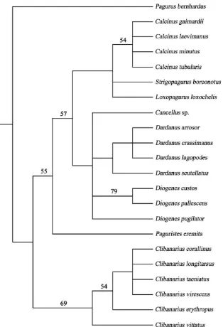
The heuristic analysis of the 21 diogenid taxa using 19 spermatozoal and spermatophore characters gave 43 equally parsimonious trees after the reweighting proce-dure, rooted using the outgroup method. The cladogram shown inFig. 5is the strict consensus of the 43 MP trees; the bootstrap values are reported above the branches. The pagurid hermit crab,P. bernhardus, is designated as the outgroup and the tree length=20 steps; consistency index (CI)=0.75; retention index=0.91.
4. Discussion
4.1. Gonopores
In Paguristes eremita the male gonopores are not raised on any type of external prolongation of the coxae (such as genital papillae) or of the ejaculatory ducts (such as elongate sexual tubes). Therefore it seems very likely that during copulation the males of P. eremita
ejaculate spermatophores from the flush gonopores onto, or close to, the gonopores on the ventral face of the coxae of pereopod 3 of the female, as reported by
Hess and Bauer (2002) for Clibanarius vittatus and by
Tirelli et al. (2007) for Clibanarius erythropus. So, as generally accepted for all hermit crabs (see Tudge and Lemaitre 2004, 2006), fertilization is external.
The male gonopores are closed by a thin, possibly non- or weakly calcified membrane termed an opercu-lum. Although indistinct, a hinge line is possibly visible on the mesial side of the membranous operculum. A similar hinge has been reported in male hermit crabs of C. vittatus (see Hess and Bauer 2002), Diogenes pugilator (see Manjo´n-Cabeza and Garcia Raso 2000)
and C. erythropus (see Tirelli et al. 2007) and in some carideans of the genera HeptacarpusHolmes, 1900 (see
Bauer 1976),ProcarisChace & Manning, 1972,Lysmata
Risso, 1816 (seeFelgenhauer et al. 1988), andChorismus
Bate, 1888 (see Mascetti et al. 1997). Probably, as already noted forC. erythropus(Tirelli et al. 2007), the operculum is used as protection from dehydration during emersion periods. When copulation occurs, spermatophores are extruded due to the internal pressure exerted on the operculum, which then opens outwards with a door-like mechanism. Nevertheless, as no direct observations have ever been made on opercular opening, chemical dissolution, muscular action or mechanical breakage cannot be excluded.
Among the decapods, the gonopore opercula of female Brachyura had been more extensively studied than in the Anomura. In particular, gonopores of female brachyurans close under several mechanisms (Hartnoll 1968, 2006;
Thompson and McLay 2005): (1) simple closure by muscle action, like inAcanthocyclusLucas, 1844,JonasHombron & Jacquinot, 1846, Bellia H. Milne Edwards, 1848,
Erimacrus Benedict, 1892 and TelmessusWhite, 1846 (see
Thompson and McLay 2005); (2) closure by soft oper-culum, like inHeteroziusA. Milne Edwards, 1867,Peltarion
Hombron & Jacquinot, 1846, Pteropeltarion Dell, 1972,
TrichopeltarionA. Milne Edwards, 1880,PseudocorystesH. Milne Edwards, 1837 (see Thompson and McLay 2005),
Uca vocans (see Nakasone et al. 1983;Salmon 1984) and
Uca lactea (see Murai et al. 1987; Goshima and Murai 1988); or (3) closure by calcified operculum, like in
Corystoides Lucas, 1844 and Corystes Bosc, 1802 (see
Thompson and McLay 2005). When the operculum is calcified, it locally decalcifies at certain times permitting internal fertilization, as it happens in the families Dorippi-dae and GrapsiDorippi-dae (seeBrockerhoff and McLay 2005a, b, c) and in Uca pugnax Smith, 1870 (see Greenspan 1982),
Ilyoplax pusilla De Haan, 1835 (see Henmi and Murai 1999), and Macrophthalmus hirtipes Heller, 1862 (see
Jennings et al. 2000).
In contrast with other hermit crab species, in particular D. pugilator (Manjo´n-Cabeza and Garcia
Raso 2000) and C. erythropus (Tirelli et al. 2007),
Isocheles sawayai Forest & de Saint Laurent, 1968 (Mantelatto et al. 2009),P. eremitaessentially lacks any setae immediately surrounding the male genital aperture of the gonopore and, instead, has little groups of setae located at a distance, as reported for Calcinus tibicen
cephalothorax and genital openings during spermato-phore transfer, an essential role during reproduction as underlined by Tirelli et al. (2007) for the ones surrounding the C. erythropus gonopore. Similar sen-sory roles have been previously inferred for the pleopodal (gonopodal) setae in some male brachyurans (Beninger et al. 1991; Elner and Beninger 1992, 1995;
Tsuchida and Fujikura 2000).
Even though there is no insertion of any appendage associated with the gonopores in hermit crabs, an important sensory role for the male coxal setae is expected during mating and spermatophore placement, but direct evidence
for it is currently lacking. A sensorial function has also been suggested for some of the setae found on the extensive sexual tube of the Australian endemic hermit crab,
Micropagurus acantholepis (Stimpson, 1858) (see Tudge and Lemaitre 2004). Although the presence of these setae is evident, their sensorial function and real role during reproduction may only be proven through careful ablation experiments.
The annulate setae without setules and annulate setae with setules (Watling 1989) present on the coxae of male
P. eremita show pronounced size differences, more so than distinct morphological ones. The shallow
itudinal grooves that extend from their base to the conspicuous annulus are unlikely to be preferential flowing channels to convey spermatophores close to the female gonopore, as the setae do not immediately surround the genital aperture. This is in contrast to a similarly suggested function of longitudinal grooves present along the ejaculatory duct of some crab gonopods (Beninger et al. 1988, 1991; Tsuchida and Fujikura 2000;Moriyasu et al. 2002). The shallow setal grooves may instead correspond to insertions by muscle groups, as suggested by Felgenhauer (1992), because these setae probably need to move in all directions during mating. This muscle insertion hypothesis, as already reported for C. erythropus (Tirelli et al. 2007) seems to be supported by the fact that grooves are present only on the basal portion of the setae. However, this muscle insertion hypothesis still needs to be confirmed by histological and TEM investigations. An alternative hypothesis could be that the longitudinal grooves may be artifacts of the progressive extrusion (lengthening) of setae through the cuticle during their growth (Tirelli et al. 2007).
The medium and long, annulate, setae show various setules in their terminal portion that may serve as mechanical functions during spermatophore transfer. While, in contrast with what is reported forC. erythropus
(Tirelli et al. 2007), they do not serve in connecting setae to each other to form a setal tube, as there are too few of them and they are not close enough to each other.
These speculations about the possible functional differences between the two types of setae (sensu
Watling 1989) undoubtely need further investigation at the histological and TEM level in order to be confirmed.
4.2. Testes, vasa deferentia and stages of
spermatophore maturation
Male specimens ofP. eremitahave their reproductive organs in the pleon. The internal reproductive apparatus is dorsal to the hepatopancreas and midgut. Its external morphology is similar to that of other hermit crabs, in terms of components and position in the abdomen (McLaughlin 1980). They are long and highly coiled as already reported for other hermit crabs (Mouchet 1931;
Matthews 1953, 1956;Greenwood 1972;Manjo´n-Cabeza
and Garcia Raso 2000; Scelzo et al. 2004;Tirelli et al. 2006b, 2007, 2008; Amadio and Mantelatto, 2009;
Mantelatto et al. 2009).
As intact testes and vasa deferentia were dissected a greater number of coils, were observed, compared to
Mouchet (1931), probably because the technique used here preserved the spatial orientation of the entire reproductive tract (Tirelli et al. 2006b, 2007).
Each one of the paired, lobe-like testes, is composed of cystic structures, and continues into a tubular duct, the vas deferens. Throughout its length the vas deferens has an inner secretory epithelium composed of cells containing cisternae of rough endoplasmic reticulum and an outer muscular layer, in agreement with the description given by Kronenberger et al. (2004)for the anomuran Galathea intermedia Liljeborg, 1851 and by
Manjo´n-Cabeza and Garcia Raso (2000) for the
diogenidD. pugilator.
The vas deferens is made up of four zones, as seen in other decapods such as the shrimp Pleoticus muelleri
(Bate, 1888) (see Diaz et al. 2002), the crayfish Cherax
Erichson, 1846 (see Talbot and Beach 1989) and the diogenid hermit crabsD. pugilator(seeManjo´n-Cabeza
and Garcia Raso 2000), Calcinus tubularis (see Tirelli et al. 2006b), andClibanarius erythropus(seeTirelli et al. 2007). As in these other described diogenids, there is a narrow collecting tubule producing the secretion that envelops the spermatic mass, and in the same portion spermatozoa are carried towards the proximal zone, as stated previously by Kooda-Cisco and Talbot (1986),
Ro et al. (1990), and Tirelli et al. (2006b, 2007). The spermatophore ampulla starts to be built in the proximal zone, where the sperm mass becomes sub-divided into successive portions, as already noted for
Enoplometopus A. Milne Edwards, 1862 sp. (see Haley 1984),HomarusWeber, 1795 sp. (seeKooda-Cisco and Talbot 1986), Galathea intermedia Liljeborg, 1851 (see
Kronenberger et al. 2004),C. tubularis(seeTirelli et al. 2006b) andC. erythropus(seeTirelli et al. 2007). In the medial zone, the spermatophore is completed, showing the definitive ampulla shape, a moderately short stalk and a foot. In the distal zone the mature spermato-phores are simply stored, waiting to be transferred to the female during copulation (Dudenhausen and Talbot 1983;Kooda-Cisco and Talbot 1986;Talbot and Beach 1989;Ro et al. 1990;Tirelli et al. 2006b, 2007).
4.3. Spermatophore
The present study supports the hypothesis that light microscope observations of spermatophores can be used successfully to distinguish hermit crab families of the Paguroidea, and even within the family Diogenidae, in agreement with Tudge (1991) and Tirelli and Pessani (2007).
P. eremita exhibits spermatophores with the classic tripartite structure typical of anomurans and their general morphology described here is in agreement with the description of the spermatophores of P. eremita
pagurids, parapagurids, and lithodids (Tudge 1991, 1999a; Tudge et al. 1998a), taxa in which the sperma-tophores do not undergo a significant stalk elongation. In agreement with Tudge (1999a), the accessory ampullae are byproducts of the process of main spermatophore production within the vas deferens. During spermatophore formation, a continuous sperm column is periodically constricted by the vas deferens musculature. The joining sperm column is retained throughout the remainder of spermatophore formation and sometimes only detaches from one of the adjacent spermatophores. If stalk elongation follows ampulla formation (as in the majority of diogenids), the sperm column material is stretched, and becomes progressively thinner until the two ampullae of adjacent spermato-phores are completely separate. If stalk elongation is absent, as in pagurids, parapagurids, and lithodids, the joining sperm column is incorporated into the short stalk, appearing as the accessory ampullae.
In the present study the pedestal, although partially covered by the protective structures which envelop the spermatophore, was observed, whileMouchet (1931)did not describe this portion of the spermatophore. The absence of observation and description of the pedestal by Mouchet (1931) is probably due to the fact that Mouchet did not succeed in obtaining spermatophores completely free from the mucilage which envelopes them, and simply reported a cord like structure connecting the spermatophores one to the others, while our observations place the cord like structure at the base of the pedestal.
The spermatophore ofP. eremitasuperficially resem-bles the general morphology reported in some pagurids, including Pagurus bernhardus (see Tudge, 1999a),
Pagurus cuanensis Bell, 1845 (see Mouchet 1931),
Pagurus excavatus (Herbst, 1791) (see Mouchet 1931;
Tudge 1999a),Pagurus longicarpusSay, 1817 (seeTudge 1999a), Pagurus pollicarisSay, 1817 (seeTudge 1999a), andPagurus prideauxLeach, 1815 (seeMouchet 1931), but the spermatophores of P. eremita, even if looking generally like the spermatophores of some pagurids when mature, lack the accessory ampulla. This latter structure is present in immature spermatophores of
P. eremitathough.
Moreover, the total length of the spermatophore of
P. eremita is shorter than that of P. longicarpus and
P. pollicaris, half the length of the spermatophores of P. bernhardus, and 2/5’s of spermatophore length of
P. excavatus (P. eremita=144.3mm, P. longicarpus=158 mm, P. pollicaris=152mm, P. bernhardus=250mm and
P. excavatus=315mm). As expected, the ampulla length has an analogous trend (P. eremita=92.6mm, P. long-icarpus=97mm,P. pollicaris=113mm,P. bernhardus=220 mm, andP. excavatus=284mm). The ampulla width though is similar in all the above-mentioned species, except for
P. excavatus(P. eremita=45.2mm,P. longicarpus=43mm,
P. pollicaris=52mm, P. bernhardus=50mm, and
P. excavatus=79mm).
The ultrastructure of the ampulla wall ofP. eremitais homogeneously granular, therefore generally in agree-ment with those described forPagurus hirtimanusWhite, 1847, P. prideaux, and Porcellanopagurus Filhol, 1885 (Tudge 1999b). Moreover, P. eremita ampulla walls show also a thin external fibrillar layer.
In conclusion, it seems possible to recognize sperma-tophores produced by P. eremita from the ones produced by other hermit crabs on the basis of their morphometry, in agreement withScelzo et al. (2004). It seems also possible to recognize them from spermato-phores produced by other members of the same family, as already shown by Tirelli and Pessani (2007) for the diogenids C. tubularis, C. erythropus, and D. pugilator. But it should be noted that Amadio and Mantelatto (2009)found differences in spermatophore morphology due to hermit crab size and the different vas deferens regions considered.
4.4. Spermatozoa
The spermatozoa of P. eremita show the typical morphology of anomurans sperm: (1) an ovoid to elongate acrosomal vesicle organized into concentric zones; (2) an electron-dense operculum capping the acrosomal vesicle; (3) a perforatorial chamber partially or wholly penetrating the acrosomal vesicle; (4) cyto-plasm with degenerate mitochondria and membrane systems; (5) a generally diffuse posterior nucleus; and (6) three or more microtubular arms emanating from the cytoplasm (seeJamieson 1991andJamieson and Tudge 2000for reviews;Tudge et al. 2001, Tirelli et al. 2006b, 2007).
The typical acrosome vescicle shape recorded for the Anomura, varies from spherical to cylindrical, with a length:width ratio of approximately 1 or more, and has been reported for all anomurans studied to date, except for the symmetrical hermit crab Pylocheles A. Milne Edwards, 1880 (Tudge et al. 2001). P. eremita, has a length:width ratio of 1.3 and a slightly ovoid acrosome shape, and is in total agreeement with data reported in the literature (Tudge 1995b, 1997;Scelzo et al. 2006).
Apart from these general anomuran characteristics, the sperm cells ofP. eremitashow some similarities with the spermatozoa described for the genus Clibanarius
(Tudge 1992, 1995a, b;Tudge and Justine 1994). In fact,
P. eremita show a quite large and ovoid acrosome, which constitutes almost the entire sperm volume, and a perforatorial chamber, which resembles the one de-scribed forClibanarius. The spermatozoa of P. eremita
are also smaller than Clibanarius species, being 3.6mm wide by 4.7mm long, while the smallest Clibanarius
3.7mm5.4mm.P. eremitaspermatozoa lack the dense perforatorial ring at the base of the acrosomal vesicle (found in Clibanarius spermatozoa), and do not show any microvillar projections on the interior wall of the perforatorial chamber. This last characteristic has been described in all the other investigated diogenids, except a member of the genusCancellusH. Milne Edwards, 1836 (Tudge 1995a, b) and Loxopagurus loxochelis (Scelzo et al. 2006). Moreover, compared to all the other diogenids investigated, except for L. loxochelis (Scelzo et al. 2006), the spermatozoa of P. eremita have one more exteriormost layer constituting the acrosome: the peripheral acrosome zone. This external zone has been previously described, among the anomurans for the hippids Emerita talpoida Say, 1817 and Hippa pacifica
Dana, 1852 (see Tudge et al. 1999), and among the brachyurans for Cancer pagurus Linnaeus, 1758 (see
Tudge et al. 1994), Segonzacia mesatlantica (Williams, 1988) and Austinograea alayseae Guinot, 1990 (see
Tudge et al. 1998b), for example. Furthermore,
P. eremita spermatozoa show, inside the perforatorial chamber, some structures resembling the perforatorial tubules already reported for some brachyurans like
C. pagurus (Tudge et al. 1994), S. mesatlantica, and
A. alayseae(Tudge et al. 1998b).
In conclusion, the spermatophore morphology is most similar to that reported for some pagurids, except for the fact that when mature P. eremita spermatophores lack the accessory ampulla, but the spermatozoal ultrastructure does not support this pagurid relation-ship. P. eremita spermatozoa, instead, show a certain number of characters typical of diogenids including the apomorphy of theClibanariusspermatozoon – the bulb shape of the perforatorial chamber (Tudge 1992, 1995a, b, 1997; Tudge and Justine 1994). Also the absence of microvillar projections in the perforatorial chamber has been similarly reported for the diogenid L. loxochelis
(Scelzo et al. 2006). Therefore,P. eremitaspermatozoal ultrastructure does not provide any clear apomorphies, that separate it from the other diogenid genera already described but shares some characters with select diogenid genera.
As this research is the last of a series regarding the description of the male reproductive apparatus of
C. tubularis(seeTirelli et al. 2006b),C. erythropus (see
Tirelli et al. 2007), D. pugilator(see Tirelli et al. 2008), andP. eremita(present study), it is possible to underline the similarities and the differences among the reproduc-tive apparatus of these species.
The male gonopores of C. erythropus (Tirelli et al. 2007), D. pugilator (Manjo´n-Cabeza and Garcia Raso
2000), and P. eremita are not raised on any type of external prolongation of the coxae or ejaculatory ducts and show a thin, possibly non- or weakly calcified membrane closing it and numerous setae around it (C. erythropus andD. pugilator) or at a short distance
from it. The setae seem to have two functions: assisting in conveying spermatophores to the female gonopores and/or working as active sensors of the female cephalothorax and genital openings.
In all four species, testes merge into a tubular duct made up of four zones, where spermatophores undergo different maturation phases. The spermatophore am-pulla is constituted of two halves meeting at the lateral ridge, which, together with the ampulla wall, shows a mostly fibrillar ultrastructure inC. tubularis(Tirelli et al. 2006b), C. erythropus (Tudge 1999b), and D. pugilator
(Tirelli et al. 2008) and a homogenously granular one in
P. eremita(present paper). The ampulla shape varies in the four species from round/ovoidal to quite long and narrow (P. eremita); in C. erythropus lateral processes are present, as in the other members of the same genus described up to now. The stalk is generally long and thin (except for C. tubularis) and the foot quite large.
Spermatozoa have a large acrosomal vesicle, whose shape varies among the four species, posteriorly penetrated by the perforatorial chamber (Tudge and Justine 1994; Tirelli et al. 2006b, 2007, 2008). The perforatorial chamber shows microvillus-like projec-tions, extending radially into the lumen, except for
P. eremita. The acrosome shows an inner and an outer acrosome zone in all four species. Only in D. pugilator
there is also a thick ray zone (Tirelli et al. 2008). Moreover, inD. pugilator, the various acrosome zones, in transverse section, have a circular profile at the perforatorial chamber level and a trilobed profile immediately beneath the subopercular zone (Tirelli et al. 2008). In C. tubularis, instead, there is an area that looks like the typical acrosome ray zone but the homology between this area and the acrosome ray zone seems uncertain (Tudge 1992, 1995a, b, 1997;Jamieson and Tudge 2000,Tirelli et al. 2006b). Only inP. eremita
is a peripheral acrosome zone present. All four species show an acrosome anteriorly delimited by a highly electron-dense operculum, beneath which there is a subopercular zone; a cytoplasmic region enveloping the base of the acrosomal vesicle, with only a small portion connected with the perforatorial chamber; spherical mitochondria, cristate and degenerate membrane sys-tems and the characteristic ‘‘triad’’ pattern of the base of the three microtubular arms; and finally a homogeneous and granular nucleus.
4.5. Phylogenetic analysis
The tree presented here (Fig. 5) shows similarity (as expected) to the Bayesian consensus tree and Quartet Puzzling parsimony tree (both in their figure 6) ofTirelli et al. (2008).P. eremitawas simply inserted between the monophyletic Clibanarius clade and remainder of the diogenid taxa. Fig. 5 shows (and Tirelli et al. 2008), though the low support values, a basal position of the genus Clibanarius and its monophyly, and the weaker substructure of this clade probably reflects their biogeography; Pacific versus Atlantic (see Tirelli et al. 2008for comments).
Such a basal position of the genusClibanariusis also supported by the molecular analyses performed by
Morrison et al. (2002) and Mantelatto et al. (2006). In the current phylogenetic reconstruction, as previously mentioned, the position ofP. eremitais uncertain between
Clibanariusand all the other diogenid genera. This fluidity of placement is also demonstrated in the molecular phylogeny of Mantelatto et al. (2006), where their monophyletic Paguristes clade variously allied with the other diogenid taxa according to which cladistic metho-dology (NJ, MP or Bayesian) was applied. From a somatic morphological perspective the generaClibanarius
andPaguristesare quite similar to one another (e.g. equal or subequal chelae compared to the majority of the diogenids which have noticeably larger left chelae) and so their adjacent placement in this tree of reproductive characters is not so surprising. Inherent in this analysis, like many using solely reproductive characters, are the problems of low clade support and within-genus poly-tomies (e.g. the genusCalcinusinFig. 5) associated with small numbers of characters, limited taxonomic sampling, and biased biogeographic sampling. Extending the analysis to more species within each genus, taxa from other geographic regions (this analysis is biased towards the Indo-West Pacific by two-thirds), other morphological structures and characters, and even including molecular sequence data for these genera/species, would make an interesting comparison for the phylogeny generated here from reproductive characters alone.
Acknowledgements
The authors would like to thank Professor Ezio Campantico for assistance with transmission electron microscopy and the anonymous reviewers for improve-ments to the manuscript.
References
Amadio, L.M., Mantelatto, F.L., 2009. Description of the male reproductive system of the hermit crab Calcinus tibicen(Decapoda: Anomura: Diogenidae). J. Crust. Biol. 29, 466–475.
Bauer, R.T., 1976. Mating behaviour and spermatophore transfer in the shrimp Heptacarpus pictus (Stimpson) (Decapoda: Caridea: Hippolytidae). J. Nat. Hist. 10, 415– 440.
Bauer, R.T., 1986. Phylogenetic trends in sperm transfer and storage complexity in decapod crustaceans. J. Crust. Biol. 6, 313–325.
Bauer, R.T., 1991. Sperm transfer and storage structures in penaeoid shrimps. A functional and phylogenetic perspec-tive. In: Bauer, R.T., Martin, J.W. (Eds.), Crustacean Sexual Biology. Columbia University Press, New York, pp. 183–207.
Beninger, P.G., Elner, R.W., Poussart, Y., 1991. Gonopods of the majid crabChionoecetes opilio(O. Fabricius) (Decapo-da: Majidae) and a hypothesis for fertilization. J. Crust. Biol. 11, 217–228.
Beninger, P.G., Elner, R.W., Foyle, T., Odense, P., 1988. Functional anatomy of the male reproductive system and the female spermatheca in the snow crabChionoecetes opilio
(O. Fabricius) (Decapoda: Majidae) and a hypothesis for fertilization. J. Crust. Biol. 8, 322–332.
Brockerhoff, A.M., McLay, C.L., 2005a. Mating behaviour, female receptivity and male–male competition in the intertidal crabHemigrapsus sexdentatus(Brachyura: Grap-sidae). Mar. Ecol. Prog. Ser. 290, 179–191.
Brockerhoff, A.M., McLay, C.L., 2005b. Comparative analy-sis of the mating strategies in grapsid crabs with special references to the intertidal crabsCyclograpsus lavauxiand
Helice crassa(Decapoda: Grapsidae) from New Zealand. J. Crust. Biol. 25, 507–520.
Brockerhoff, A.M., McLay, C.L., 2005c. Factors influencing the onset and duration of receptivity of female purple rock crabs,Hemigrapsus sexdentatus(Brachyura: Grapsidae). J. Exp. Mar. Biol. Ecol. 314, 123–135.
Diaz, A.C., Fernandez Gimenez, A.V., Petriella, A.M., Fenucci, J.L., 2002. Morphological and functional study of the male reproductive tract in the shrimp Pleoticus muelleriBate (Decapoda, Penaeoidea). Inv. Repr. Dev. 42, 69–74.
Dudehausen, E.E., Talbot, P., 1983. An ultrastructural comparison of soft and hardened spermatophores from the crayfishPacifastacus leniusculusDana. Can. J. Zool. 61, 182–194.
Elner, R.W., Beninger, P.G., 1992. The reproductive biology of snow crab, Chionoecetes opilio: a synthesis of recent contributions. Am. Zool. 32, 524–533.
Elner, R.W., Beninger, P.G., 1995. Multiple reproductive strategies in snow crab, Chionoecetes opilio: physiological pathways and behavioral plasticity. J. Exp. Mar. Biol. Ecol. 193, 93–112.
Farris, J.S., 1969. A successive approximations approach to character weighting. Syst. Zool. 18, 374–385.
Farris, J.S., 1988. Hennig86 version 1.5 manual; software and MSDOS program.
Felgenhauer, B.E., Abele, L.G., Kim, W., 1988. Reproductive morphology of the anchialine shrimpProcaris ascensionis
(Decapoda: Procarididae). J. Crust. Biol. 8, 333–339. Goshima, S., Murai, M., 1988. Mating investment of male
fiddler crabs,Uca lactea. Anim. Behav. 36, 1249–1251. Greenspan, B.N., 1982. Semi-monthly reproductive cycles in
male and female fiddler crabs,Uca pugnax. Anim. Behav. 30, 1084–1092.
Greenwood, J.G., 1972. The male reproductive system and spermatophore formation in Pagurus novae-zealandiae
(Dana) (Anomura: Paguridae). J. Nat. Hist. 6, 561–574. Haley, S.R., 1984. Spermatogenesis and spermatophore
production in the Hawaiian red lobster Enoplometopus occidentalis (Randall) (Crustacea: Nephropidae). J. Mor-phol. 180, 181–193.
Hartnoll, R.G., 1968. Morphology of the genital ducts in female crabs. J. Linn. Soc. 47, 279–300.
Hartnoll, R.G., 2006. Reproductive investment in Brachyura. Hydrobiologia 557, 31–40.
Henmi, Y., Murai, M., 1999. Decalcification of vulvar operculum and mating in the ocypodid crab Ilyoplax pusilla. J. Zool. (London) 247, 133–137.
Hess, G.S., Bauer, R.T., 2002. Spermatophore transfer in the hermit crab Clibanarius vittatus (Crustacea, Anomura, Diogenidae). J. Morphol. 253, 166–175.
Jamieson, B.G.M., 1991. Ultrastructure and phylogeny of crustacean spermatozoa. Mem. Queens. Mus. 31, 109–142. Jamieson, B.G.M., Tudge, C.C., 2000. Crustacea–Decapoda. In: Jamieson, B.G.M. (Ed.), Reproductive Biology of Invertebrates. Progress in Male Gamete Ultrastructure and Phylogeny, vol. IX, part c. John Wiley & Sons, Chichester, pp. 1–95.
Jennings, A.C., McLay, C.L., Brockerhoff, A.M., 2000. Mating behaviour ofMacrophthalmus hirtipes(Brachyura: Ocypodidae). Mar. Biol. 137, 267–278.
Kooda-Cisco, M., Talbot, P., 1986. Ultrastructure and role of the lobster vas deferens in spermatophore formation: the proximal segment. J. Morphol. 188, 91–103.
Kronenberger, K., Brandis, D., Tu¨rkay, M., Storch, V., 2004.
Functional morphology of the reproductive system ofGalathea intermedia(Decapoda: Anomura). J. Morphol. 262, 500–516. Maddison, W.P., Maddison, D.R., 2005. In: MacClade:
Analysis of Phylogeny and Character Evolution, version 4.08. Sinauer Associates, Sunderland, MA, USA.
Manjo´n-Cabeza, M.E., Garcia Raso, J.E., 2000. Morphologi-cal reproductive aspects of males of Diogenes pugilator
(Roux, 1829) (Crustacea, Decapoda, Anomura) from southern Spain. Sarsia 85, 195–202.
Mantelatto, F.L., Robles, R., Biagi, R., Felder, D.L., 2006. Molecular analysis of the taxonomic and distributional status for the hermit crab generaLoxopagurusForest, 1964 and Isocheles Stimpson, 1858 (Decapoda, Anomura, Diogenidae). Zoosystema 28, 495–506.
Mantelatto, F.L., Scelzo, M.A., Tudge, C.C., 2009. Morpho-logical and morphometric appraisal of the spermatophore of the southern hermit crabIsocheles sawayaiForest and de Saint Laurent, 1968 (Anomura: Diogenidae), with com-ments on gonopores in both sexes. Zool. Anz. 248, 1–8. Mascetti, P., Fernandez de la Reguera, R., Albornoz, L.,
Oyarzo´n, S., Gorny, M., Wehrtmann, I., 1997. Gonopore
development and sex change in the Antarctic shrimp
Chorismus antarcticus(Caridea: Hippolytidae). Polar Biol. 17, 384–488.
Matos, E., Matos, P., Oliveira, E., Azevedo, C., 1993. Aspectos morfolo´gicos e ultraestruturais do espermatozo´ide de ermita˜o Clibanarius vittatus Bosc, 1802 (Crustacea, Decapoda) do litoral norte do Brasil. Rev. Brasiliera Cieˆncias Morfolo´g. 10, 126–131.
Matthews, D.C., 1953. The development of the pedunculate spermatophore of a hermit crab, Dardanus asper (De Haan). Pacif. Sci. 7, 255–266.
Matthews, D.C., 1956. The probable method of fertilization in terrestrial hermit crabs based on a comparative study of spermatophores. Pacif. Sci. 10, 303–309.
McLaughlin, P.A., 1980. In: Comparative Morphology of the Recent Crustacea. W.H. Freeman & Company, San Francisco.
McLaughlin, P.A., 2003. Illustrated keys to families and genera of the superfamily Paguroidea (Crustacea: Decapo-da: Anomura), with diagnoses of genera of Paguridae. Mem. Mus. Victoria 60, 111–144.
Moriyasu, M., Benhalima, K., Duggan, D., Lawton, P., Robichaud, D., 2002. Reproductive biology of male Jonah crab, Cancer borealis Stimpson, 1859 (Decapoda, Cancri-dae) on the Scotian shelf, Northwestern Atlantic. Crusta-ceana 75, 891–913.
Morrison, C.L., Harvey, A.W., Lavery, S., Tieu, K., Huang, y, Cinningham, C.W., 2002. Mitochondrial gene rearrange-ments confirm the parallel evolution of the crab-like form. Proc. R. Soc. London Part B Biol. Sci. 269 (1489), 345–350. Mouchet, S., 1931. Spermatophores des Crustace´s De´capodes
Anomoures et Brachyoures et castration parasitaire chez quelques Pagures. Ann. Stat. Oce´anogr. Salammbo 6, 1–203.
Murai, M., Goshima, S., Henmi, Y., 1987. Analysis of the mating system of the fiddler crab,Uca lactea. Anim. Behav. 35, 1334–1342.
Nakasone, Y., Okamine, H., Asato, K., 1983. Ecology of the fiddler crab Uca vocans vocans (Linnaeus) (Decapoda: Ocypodidae). II. Relation between mating system and the drove. Galaxea 2, 119–133.
Ro, S., Talbot, P., Leung-Trujillo, J., Lawrence, A.L., 1990. Structure and function of the vas deferens in the shrimp
Penaeus setiferus: segments 1–3. J. Crust. Biol. 10, 455–468. Salmon, M., 1984. The courtship, aggression and mating system of a ‘‘primitive’’ fiddler crab (Uca vocans: Ocypo-didae). Trans. Zool. Soc. London 37, 1–50.
Scelzo, M.A., Mantelatto, F.L., Tudge, C.C., 2004. Sperma-tophore morphology of the endemic hermit crab Loxopa-gurus loxochelis (Anomura, Diogenidae) from the southwestern Atlantic – Brazil and Argentina. Inv. Repr. Dev. 46, 1–9.
Scelzo, M.A., Medina, A., Tudge, C.C., 2006. Spermatozoal ultrastructure of the hermit crab Loxopagurus loxochelis
(Moreira, 1901) (Anomura, Diogenidae) from the south-western Atlantic. In: Asakura, A. (Ed.), Biology of Anomura II, Crus. Res., Special Number 6; 2006, pp. 1–11. Swofford, D.L., 2002. In: PAUPn
Phylogenetic Analysis Using Parsimony (n
Talbot, P., Beach, D., 1989. The role of the vas deferens in the formation of the spermatophore of the crayfishCherax. J. Crust. Biol. 9, 9–24.
Thiriot, A., 1974. Larves de De´capodes Macrura et Anomura,
espe`ces europe´ennes; caracte`res morphologiques et obser-vationse´cologiques. Thalassia Jugoslavica 10, 341–378. Thompson, G.A., McLay, C.L., 2005. Mating behaviour of
Heterozius rotundifrons(Crustacea: Brachyura: Bellidae): is it a hard or soft shell matter?. Mar. Fresh. Res. 56, 1107–1116. Tirelli, T., Pessani, D., 2007. Multivariate analysis: a useful tool in separating spermatophores produced by diogenids,
Clibanarius erythropus, Diogenes pugilator, and Calcinus tubularis(Decapoda, Anomura, Diogenidae). Crustaceana 80, 999–1011.
Tirelli, T., Guarneri, D., Pessani, D., 2006a. Studio prelimi-nare degli epibionti presenti su conchiglie occupate dal paguroPaguristes eremita. Biol. Mar. Medit. 13, 656–658. Tirelli, T., Campantico, E., Pessani, D., Tudge, C.C., 2006b. Description of the male reproductive apparatus of the hermit crabCalcinus tubularis(Anomura, Diogenidae). In: Asakura, A. (Ed.), Biology of Anomura II, Crus. Res., Special Number 6. Kokusai-Bunken Printing Inc, Tokyo, pp. 13–21.
Tirelli, T., Campantico, E., Pessani, D., Tudge, C.C., 2007. Reproductive biology of the Mediterranean hermit crabs: male reproductive apparatus of Clibanarius erythropus
(Decapoda Anomura). J. Crust. Biol. 27, 404–410. Tirelli, T., Pessani, D., Silvestro, D., Tudge, C.C., 2008.
Reproductive biology of the Mediterranean hermit crabs: fine structure of spermatophores and spermatozoa of
Diogenes pugilator (Decapoda, Anomura) and its bearing on a sperm phylogeny of the Diogenidae. J. Crust. Biol. 28, 534–542.
Tsuchida, S., Fujikura, K., 2000. Heterochely, relative growth, and gonopod morphology in the bythograeid crab,
Austinograea williamsi (Decapoda, Brachyura). J. Crust. Biol. 20, 407–414.
Tudge, C.C., 1991. Spermatophore diversity within and among the hermit crab families, Coenobitidae, Diogenidae and Paguridae (Paguroidea, Anomura, Decapoda). Biol. Bull. 181, 238–247.
Tudge, C.C., 1992. Comparative ultrastructure of hermit crab spermatozoa (Decapoda: Anomura: Paguroidea). J. Crust. Biol. 12 (3), 397–409.
Tudge,C.C.,1995a. Ultrastructure and phylogeny of anomuran crab spermatozoa. Ph.D. Thesis, Zoology Department, The University of Queensland, Australia, pp. 1–346.
Tudge, C.C., 1995b. Ultrastrucure and phylogeny of the spermatozoa of the infraorders Thalassinidea and Anom-ura (Decapoda, Crustacea). In: Jamieson, B.G.M., Ausio, J., Justine, J.L. (Eds.), Advances in Spermatozoal
Phylogeny and Taxonomy. Me´m. Mus. Natl. Hist. Nat., Paris, vol. 166, pp. 251–263.
Tudge, C.C., 1997. Phylogeny of the Anomura (Decapoda, Crustacea): spermatozoa and spermatophore morphologi-cal evidence. Contr. Zool. 67, 125–141.
Tudge, C.C., 1999a. Spermatophore morphology in the hermit crab families Paguridae and Parapaguridae (Paguroidea, Anomura, Decapoda). Inv. Repr. Dev. 35, 203–214. Tudge, C.C., 1999b. Ultrastructure of the spermatophore
lateral ridge in hermit crabs (Decapoda, Anomura, Paguroidea). Crustaceana 72, 77–84.
Tudge, C.C., Justine, J.L., 1994. The cytoskeletal proteins actin and tubulin in the spermatozoa of four decapod crabs (Crustacea, Decapoda). Acta Zool. 75, 277–285.
Tudge, C.C., Lemaitre, R., 2004. Studies of male sexual tubes in hermit crabs (Crustacea, Decapoda, Anomura, Pagur-oidea). I. Morphology of the sexual tube inMicropagurus acantholepis(Stimpson, 1858), with comments on function and evolution. J. Morphol. 259, 106–118.
Tudge, C.C., Lemaitre, R., 2006. Studies of male sexual tubes in hermit crabs (Crustacea, Decapoda, Anomura, Pagur-oidea). I. Morphology of the sexual tube in the land hermit crabs,Coenobita perlatusandC. clypeatus(Coenobitidae). In: Asakura, A. (Ed), Biology of Anomura II, Crus. Res., Special Number 6, pp. 121–131.
Tudge, C.C., Grellier, P., Justine, J.L., 1994. Actin in the acrosome of the spermatozoa of the crab.Cancer pagurus
L. (Decapoda, Crustacea). Mol. Repr. Dev. 38, 178–186. Tudge, C.C., Scheltinga, D.M., Jamieson, B.G.M., 1999.
Spermatozoal ultrastructure in the Hippoidea (Anomura, Decapoda). J. Submicr. Cyt. Pathol. 31, 1–13.
Tudge, C.C., Scheltinga, D.M., Jamieson, B.G.M., 2001. Spermatozoal morphology in the symmetrical hermit crab,
Pylocheles(Bathycheles) sp. (Crustacea, Decapoda, Anom-ura, Paguroidea, Pylochelidae). Zoosystema 23 (1), 117– 130.
Tudge, C.C., Jamieson, B.G.M., Sandberg, L., Erse`us, C., 1998a. Ultrastructure of the mature spermatozoon of the king crab Lithodes maja(Lithodidae, Anomura, Decapo-da): further confirmation of a lithodid–pagurid relation-ship. Inv. Biol. 117, 57–66.
Tudge, C.C., Jamieson, B.G.M., Segonzac, M., Guinot, D., 1998b. Spermatozoal ultrastructure in the three species of hydrothermal vent crab, in the generaBythograea, Austino-graea andSegonzacia(Decapoda, Brachyura, Bythograei-dae). Inv. Repr., 13–23.