Characteristics of Cerebrospinal Fluid in Tuberculous Meningitis
Patients with Hydrocephalus
Abstract Objective: To describe cerebrospinal fluid (CSF) characteristics in tuberculous meningitis (TBM) with hydrocephalus patients as diagnostic criteria of TBM.
Methods: A cross-sectional using retrospective method was applied in this study by obtaining medical records of TBM with hydrocephalus patients that were treated at Department of Neurosurgery Dr. Hasan Sadikin General Hospital, Bandung from January 2014–September 2016.
Results: Sixty one records were included in the study. Patient characteristics such as age, gender, and CSF laboratory features such as color and clarity, cells and differential count, protein, and glucose were recorded and descriptively analyzed. The majority of CSF macroscopic appearance was seen to be clear (88%) and colorless (88%). Median for CSF cell count was 25 cells/mm3 with
lymphocyte predominance (median: 76%). Increased protein from normal value range was seen in CSF (median: 50 mg/dL) while the mean for CSF glucose in this study remained in its normal value range (mean: 58.9±26.68 mg/dL) with lower CSF to blood glucose ratio (mean: 0.41±0.27).
Conclusions: Clear CSF with colorless appearance, lymphocyte pleocytosis, high protein level, and low CSF glucose as well as blood glucose ratio remain typical CSF characteristics of TBM patients found at the Department of Neurosurgery, Dr. Hasan Sadikin General Hospital, Bandung.
Keywords: Cerebrospinal fluid, hydrocephalus, tuberculous meningitis
pISSN: 2302-1381; eISSN: 2338-4506; http://doi.org/10.15850/ijihs.v6n2.1129 IJIHS. 2018;6(2):57–62
Received: November 2, 2017
Revised:
May 15, 2018
Accepted:
September 5, 2018
Correspondence:
Astrid Tamara Maajid Budiman, Faculty of Medicine, Universitas Padjadjaran
Jl. Raya Bandung-Sumedang KM 21 Jatinangor, Indonesia e-mail: [email protected]
Introduction
Tuberculosis (TB) currently remains a major global health issue. Indonesia is currently in the top three in terms of TB cases where 10% of the total TB population in the world is found
in this country.1 One of extrapulmonary TB
with high mortality and morbidity, esepcially in children, is tuberculosis meningitis (TBM).2
Hydrocephalus is the most frequently found complication of TB and one of the predictors of mortality and morbidity.2
Early diagnosis and proper management
of TBM is an important factor that influences mortality and morbidity. Cerebrospinal fluid (CSF) examination is an important parameter in the diagnosis of TBM . This CSF examinations
includes macroscopic, microscopic, chemical, and microbiological examinations. Bacterial
culture or acid-fast bacilli (AFB) observation in CSF are used to establish TBM definitive
diagnosis.2,3 A previous study has reported
that the ratio of positive culture results were
13% among TBM patients.4 In several cases,
low results of bacterial culture and AFB on CSF may be seen. Hence it is important to
observe the macroscopic, microscopic, and chemical characteristic as a reference for the laboratory-based diagnosis of TBM.4
Astrid Tamara Maajid Budiman,1 Nida Suraya,2 Ahmad Faried,3 Ida Parwati2
1Faculty of Medicine, Universitas Padjadjaran
2Department of Clinical Pathology, Faculty of Medicine, Universitas Padjadjaran-Dr. Hasan Sadikin General
Hospital, Bandung
3Department of Neurosurgery, Faculty of Medicine, Universitas Padjadjaran-Dr. Hasan Sadikin General Hospital,
This study aimed to observe the chemical, macroscopic, and microscopic characteristics
of CSF in TBM patients with hydrocephalus at
the Department of Neurosurgery Dr. Hasan Sadikin General Hospital, Bandung.
Methods
This design used in this study was retrospective cross-sectional descriptive study. TBM patients with hydrocephalus treated at the Department of Neurosurgery, Dr. Hasan Sadikin General Hospital, Bandung who met the inclusion and exclusion criteria were included. The inclusion criteria in this study were inpatients during the period of January 2014–September 2016,
with a computerized tomography (CT) scan
results that suggested hydrocephalus who
had undergo CSF examination with specimens derived from ventricular drainage at the Clinical Pathology Laboratory of Dr. Hasan Sadikin
General Hospital, Bandung, with macroscopic, microscopic, and chemical characteristic result, including protein and glucose levels. Exclusion criteria included congenital hydrocephalus, meningitis caused by organisms other than Mycobacterium tuberculosis, positive Human
Immunodeficiency Virus (HIV) status, and
incomplete medical record. Samples were selected using the total sampling method.
Approval from the Ethics Committee of the Faculty of Medicine, Universitas Padjadjaran under the ethical clearance No. 102/UN6. C1.3.2/KEPK/PN/2016 was gained. This study
was conducted from August to December 2016 by collecting data from the medical records of TBM inpatients with hydrocephalus i who were treated at the Department of Neurosurgery, Dr. Hasan Sadikin General Hospital, Bandung.
CSF of these patients were examined at the Laboratory of Clinical Pathology of the same
hospital.
Variables used in this study were age, CSF
color, clarity, leukocyte cell numbers, cell count percentage, protein level, and glucose level. Data were then analyzed descriptively by counting the number and percentage for the results of macroscopic examination while for microscopic and chemical examination results, mean and standard deviations or median and range were used based on the distribution of the data.
Results
There were 477 patients with TBM in Dr. Hasan Sadikin General Hospital Bandung from 2014
to 2016. A total of 170 (35.6%) patients were
treated at the Department of Neurosurgery Dr. Hasan Sadikin General Hospital, Bandung
with 149 cases (31.2%) were diagnosed as
TBM with hydrocephalus. Sixty six cases
(44.3) met the inclusion criteria but five of
them must be excluded, resulting a total of
61 (40.9%) cases. All were included in the study. All CSF samples collected from these 61
patients were taken from ventricular drainage
such as extra ventricular drainage (EVD) or ventriculoperitoneal (VP) shunt.
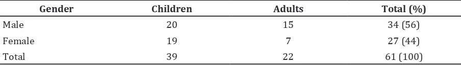
The number of male patients was higher than the number of female patients (Table 1). Age categorization of children and adults in this study referred to the existing consensus in Dr. Hasan Sadikin General Hospital, Bandung with 0–14 years categorized as children, and
>14 years classified as adults. The number of
pediatric patients was higher when compared to adults patients. Median age was 6 years old
with a range of 6 months–58 years.
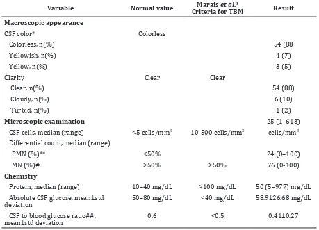
Examination of CSF was performed on TBM
patients with hydrocephalus and the results were compared to normal values and criteria from Marais et al.3 (Table 2). Cerebrospinal fluid color in TBM patients with hydrocephalus
Gender Children Adults Total (%)
Male 20 15 34 (56)
Female 19 7 27 (44)
Total 39 22 61 (100)
in this study was generally colorless. Similarly,
the clarity level of CSF in this study generally
showed a clear result.
The median of the CSF leukocyte cells was 25 cells/mm3 (range was 1-613 cells/mm3).
The differential cell count of the cerebrospinal
fluid was presented in the form of percentage
of mononuclear (MN) and polymorphonuclear cells (PMN). Mononuclear cells were shown to
be the dominant cell in the CSF. The scope of
percentage for both PMN and MN was 0% to 100%. There were two cases which showed
the percentage of PMN of 100%, with a CSF leukocyte cell count of 1 cell/mm3 in both.
The median CSF protein was higher than
the normal value but not as high as the one in Marais et al.3 criteria. The mean CSF glucose was 58.9±26.68 mg/dL which was still in the normal limit. The CSF to blood glucose ratio
could only be calculated in 11 patients (mean:
0.41±0.27).
Discussion
This study reveals that the prevalence of TBM with hydrocephalus is predominated by males.
This finding is in agreement with the results
of previous studies.4–6 A study found that the
prevalence of pediatric patient with TBM with hydrocephalus as complication is 71% while the same prevalence for adults is 12%.7 This
study also shows that more children has TBM with hydrocephalus compared to adult.
Macroscopic examination on the color and
clarity of CSF was performed. The macroscopic appearance of CSF in this study was generally
clear and colorless. This is in line with the
Variable Normal value Marais et al.3
Criteria for TBM Result
Macroscopic appearance
CSF color* Colorless
Colorless, n(%) 54 (88
Yellowish, n(%) 4 (7)
Yellow, n(%) 3 (5)
Clarity Clear Clear
Clear, n(%) 54 (88)
Cloudy, n(%) 6 (10)
Turbid, n(%) 1 (2)
Microscopic examination 25 (1–613)
CSF cells, median (range) <5 cells/mm3 10-500 cells/mm3 cells/mm3
Differential count, median (range)
PMN (%)** <50% 24 (0–100)
MN (%)# >50% >50% 76 (0-100)
Chemistry
Protein, median (range) 10–40 mg/dL >100 mg/dL 50 (5–977) mg/dL
Absolute CSF glucose, mean±std
deviation 50–80 mg/dL <40 mg/dL 58.9±26.68 mg/dL
CSF to blood glucose ratio##,
mean±std deviation 0.6 <0.5 0.41±0.27
Note: *CSF = Cerebrospinal fluid; **PMN = polymorphonuclear; #MN = mononuclear; ##Could only be done in 11 patients
diagnosis criteria of TBM according to Marais
et al.3 Tuberculous meningitis is a chronic
disease characterized by clear CSF with not
really high leukocyte count and is dominated by lymphocytes.3,8,9 The inflammation response of T cell as TBM pathogenesis is demonstrated through the predominance of lymphocytes
in microscopic examination of CSF, as seen in
previous studies and this study.5,10–13
Marais et al.3 stated that a diagnosis criteria of chemical examination of CSF includes an increase in CSF protein (>100 mg/dL). This study shows the amount of CSF protein was not greater than 100 mg/dL as stated in the
criteria diagnosis by Marais et al.3 However, the median CSF protein was in accordance with previous studies stating that the mean CSF protein was not more than 100 mg/dL despite the increase of the CSF protein value when
compared to the normal value.6 As the top
referral hospital in West Java, Dr. Hasan Sadikin General Hospital, Bandung, in particular the
hospital’s Clinical Pathology Laboratory, has their own normal CSF protein range standard of 10–40 mg/dL. The CSF protein in TBM
which is supposed to be increased according to the pathogenesis of TBM is considered as the result from the increased permeability of blood brain barrier or decreased reabsorption in the arachnoidalis villi.8,13
The normal value of glucose in CSF is 2/3 of the plasma glucose, or approximately 50 ti=o 80 mg/dL.8,13 As already explained in the criteria for TBM diagnosis, the CSF glucose should be less than 40 mg/dL (2.2 mmol/L) or the ratio between CSF glucose and plasma glucose is lower than 50%.3 This decrease in
glucose is resulted from anaerobic glycolysis caused by bacteria or disturbance in glucose
transport mechanism.8,13 The mean glucose
level in this study was >40 mg/dL and wa still in the normal range of CSF glucose. This
is in contrast with the diagnostic criteria by Marais et al.3 and some previous studies.6,9 A
previous study presented that the number of
patients with glucose level <40 mg/dL was
48% or less than a half of all patients and that the glucose level had a wide range between
12.1–156.7 mg/dL as also shown in this study (2–135 mg/dL).11 There are only 11 patients whose CSF glucose to blood glucose ratio can be measured (median: 0.41±0.27). This result
is in line with the diagnostic criteria by Marais
et al.3 which stated that CSF glucose to blood
glucose ratio is <50%.
Most previous studies that have discussed
the CSF characteristics in TBM use CSF to blood glucose ratio <0.5 as one ofdiagnostic criteria
for TBM.4,14 The previous studies showed that the sensivity of the low absolute CSF glucose was low, that is 68% and 69,5%.12,15 On the
other hand, previous studies also showed that
the sensitivity of low CSF to blood glucose ratio was 90%.15 This explains why the normal absolute CSF glucose level cannot rule out the
possibility of infection and it had also been suggested that considerations should be given
to use CSF to blood glucose ratio as additional
information for diagnosis of TBM.15
The difference of CSF content in this study
compared to Marais et al.3 criteria might be caused by the CSF source itself.16 Previous study had shown difference CSF composition between ventricular and spinal CSF that may
be caused by the disease’s pathophysiology.16
The impaired blood-brain barrier leads to a higher protein composition which may slow
down the circulation of CSF and may impair CSF absorption or obstruct the CSF pathway
that causes hydrocephalus after aggregation
of CSF protein and leukocytes caudally.16
The median of age in this study (median:
6 years) reflects a higher distribution among
children below 6 years old. Several previous studies have shown age-related variations in
CSF protein content.8,17 It is shown here that there is a lower protein CSF value in children
when compared to adults and this might result
in lower protein CSF in this study compared to
the Marais et al.3 criteria.8,17
Furthermore, the use of antimicrobial agent before ventricular drainage may also affect CSF
protein and glucose content.18 Time of interval
median between the occurence of tuberculosis to hydrocephalus development was 21 days (range: 3–180 days). In between, patients consumed antituberculous drugs. A previous
study has shown that CSF glucose will increase and CSF protein will decrease significantly
after the use of antibiotic, especially in the period of >12 hours postantibiotics.18
There are several limitations in the study including the small sample size, i.e. 61 samples. This is due to the limited time and rejection from some TBM patients with hydrocephalus to undergo ventricular drainage that includes
both EVD and VP shunts. In addition, incomplete
patient records and archives creates another
limitation, especially when the result of CSF in
patient who underwent ventricular drainage is missing. Another reason is that blood glucose
measurement done 15–30 minutes prior to the
ventricular drainage was not done in almost all
samples. This makes the calculation of CSF to
References
1. World Health Organization. Global tuberculosis report 2015. 20th ed. Geneva: World Health
Organization, 2015.
2. Rock RB, Olin M, Baker CA, Molitor TW, Peterson PK. Central nervous system tuberculosis: pathogenesis and clinical aspects. Clin Microbiol Rev. 2008;21(2):243–61.
findings in 185 children with tuberculous meningitis at a single centre and relationship with the stage of the disease. Ital J Pediatr [serial on the internet]. 2015 Oct [cited 2017 Aug 12];41(75):[about 6p.]. Available from: https://www.ncbi.nlm.nih.gov/pmc/articles/ PMC4606503/.
5. Pinto VL, Lima MA, Rolla VC, Rebelo MC, Boia MN. Atypical cerebrospinal fluid profile in tuberculous meningitis. Trop Doct. 2009;39(2):76–8.
6. Shaikh MA, Shah M, Channa F. Criteria indicating morbidity in tuberculous meningitis. J Pak Med Assoc. 2012;62(11):1137–9.
7. Rajshekhar V. Management of hydrocephalus in patients with tuberculous meningitis. Neurol India. 2009;57(4):368–74.
8. Karcher DS, McPherson RA. Cerebrospinal, synovial, serous body fluids, and alternative specimens. In: McPherson RA, Pincus MR, editors. Henry’s clinical diagnosis and management by laboratory methods. 22nd ed.
Philadelphia: Elsevier Saunders; 2011. p. 480– 92.
9. Qamar FN, Rahman AJ, Iqbal S, Humayun K. Comparison of clinical and CSF profiles in children with tuberculous and pyogenic
meningitis; role of CSF protein: glucose ratio as diagnostic marker of tuberculous meningitis. J Pak Med Assoc. 2013;63(2):206–10.
10. Salekeen S, Mahmood K, Naqvi IH, Baig MY, Akhter ST, Abbasi A. Clinical course, complications and predictors of mortality in patients with tuberculous meningitis--an experience of fifty two cases at Civil Hospital Karachi, Pakistan. J Pak Med Assoc. 2013;63(5):563–7.
of cerebrospinal fluid parameters for the diagnosis of tuberculous meningitis. Int J Infect Dis [serial on the internet]. 2013 Dec [cited 2016 Nov 18];17(12):[about 5p.]. Available from:https://www.ijidonline.com/article/ S1201-9712(13)00214-2/fulltext.
13. Cerebrospinal fluid. In: Strasinger SK, Di Lorenzo MS, editors. Urinalysis and body fluids. 6th ed. Philadelphia: F.A. Davis Company; 2008.
p.177–95.
14. Thwaites G, Fisher M, Hemingway C, Scott G, Solomon T, Innes J. British Infection Society guidelines for the diagnosis and treatment of tuberculosis of the central nervous system in adults and children. J Infect. 2009;59(3):167– 87.
15. Solomons RS, Visser DH, Donald PR, Marais BJ, Schoeman JF, van Furth AM. The diagnostic value of cerebrospinal fluid chemistry results in childhood tuberculous meningitis. Childs Nerv Syst. 2015;31(8):1335–40.
16. Alfayate-Miguelez S, Martinez-Lage-Azorin L, Marin-Vives L, Garcia-Martinez S, Almagro MJ, Martinez-Lage JF. Normal ventricular-CSF may comfound the diagnosis of tuberculous
ventricular drainage that may also affect the results compared to those of previous studies
that mostly used lumbar fluid as specimens.
In conclusion, the diagnosis of tuberculous meningitis is still problematic, especially with the high number of the hydrocephalus seen.
CSF presentations such as clear and colorless CSF, lymphocyte pleocytosis, increased protein level, and lower CSF to blood glucose ratio
are still the typical characteristics found in TBM with hydrocephalus in Dr. Hasan Sadikin
General Hospital, Bandung. Examination of CSF
in TBM may be done in every level of health facilities as it is the easiest way to diagnose
this disease. This study suggests the use of CSF
meningitis hydrocephalus. Neurocirugia (Astur). 2011;22(2):157–61.
17. Miftode EG, Dorneanu OS, Leca DA, Juganariu G, Teodor A, Hurmuzache M, et al. Tuberculous
meningitis in children and adults: a 10-year retrospective comparative analysis. PLoS One. 2015 Jul [cited 2016 Nov 18];10(7):[about 10p.] Available from: http://journals.plos.
org/plosone/article/file?id=10.1371/journal. pone.0133477&type=printable.
18. Nigrovic LE, Malley R, Macias CG, Kanegaye JT, Moro-Sutherland DM, Schremmer RD,
et al. Effect of antibiotic pretreatment on