Brain Natriuretic Peptide
Folia Medica Indonesiana Vol. 41 No. 2 April – June 2005
BRAIN NATRIURETIC PEPTIDE AS AN INDICATOR OF LEFT VENTRICLE DYSFUNCTION AND A PREDICTOR OF CARDIOVASCULAR EVENTS IN ACUTE CORONARY SYNDROMES
Nugroho, M. Yogiarto
ABSTRACT
Background: Acute coronary syndromes can cause systolic and diastolic left ventricle (LV) dysfunction. Determination of LV dysfunction in ACS provides benefit to stratification and optimizing therapy. Brain Natriuretic Peptide (BNP) is secreted primarily from and synthesized in left ventricle in response to increased myocardial stretch. BNP increase in heart failure. BNP increase in the 24 hours in acute myocardial infarction. We hypothesized that BNP level could be as an indicator of systolic and diastolic dysfunction and a predictor of cardiovascular events in acute coronary syndromes.
Objective: To investigate plasma level of BNP in systolic and diastolic dysfunction in acute coronary syndromes hospitalized to ICCU. To investigate plasma level of BNP as a predictor of cardiovascular events in acute coronary syndromes. Method and Result: We measured BNP in plasma specimen obtained 3 days after the onset of ischemic symptoms in 25 patients of acute coronary syndromes and prospectively followed the patients for 30 days. Patients diagnosed with evidence of systolic LV dysfunction had a mean BNP concentration of 301.11 ± 189.62 pg/ml, higher than those patients with normal LV function (42.67 ± 22.44 pg/ml, p = 0.003). Patients diagnosed with evidence of diastolic LV dysfunction had a mean BNP concentration of 273.70 ± 146.27 pg/ml, higher than those patients with normal LV function (42.67 ± 22.44 pg/ml, p = 0.006). Patients with cardiovascular events had a mean BNP concentration of 392.30 ± 157.14 pg/ml, higher than patients without cardiovascular events (118.67 ± 78.53 pg/ml, p < 0.0001). In patients with cardiovascular events, minimum plasma BNP level (248 pg/ml) was higher than maximum plasma BNP level in patients without cardiovascular events (234 pg/mL). Conclusion: Plasma BNP level can reliably detect the presence of diastolic or systolic LV dysfunction on echocardiography in acute coronary syndromes. Moreover, plasma BNP level can also predict patients with cardiovascular events in 30-days after acute coronary syndromes.
Keywords: BNP, Left ventricle dysfunction, cardiovascular events, acute coronary syndromes
INTRODUCTION
B-type natriuretic peptide (BNP) is 32 amino acids secreted into blood circulation mainly by left ventricular myocyte under overload condition or wall stress. BNP is a specific indicator of ventricular disturbance and more sensitive than other natriuretic peptides. It has natriuresis effect, vasodilatation, renin-angiotensin-aldosteron system inhibition and sympathetic activity inhibition (Peacock WF, 2002).
Plasma BNP will increase in patients suffering from heart failure and the increase is in accordance with the left ventricular dysfunction gradation and the severity of heart failure symptoms. In a case of acute myocardial infarction, BNP will rise rapidly in the first 24 hours (Nagaya N et al, 1998). The researches on plasma BNP as prognostic factor in acute myocardial infarction have been carried out initially. However, there are merely a small number of researches on BNP in patients suffering ______________
Department of Cardiology
Airlangga University School of Medicine Dr Soetomo Teaching Hospital, Surabaya
from acute coronary syndromes. The clinical spectrum of those patients is heterogeneous. Early finding of ventricular dysfunction will be very useful to determine patient risk stratification and to give additional treatment in order to achieve the optimum treatment result (Peacock WF, 2002).
Heart catheterization is a gold standard to evaluate left ventricular dysfunction. Nevertheless, it is impractical to be used widely or to observe clinical progress of the patients. By using Doppler echocardiography as a noninvasive technique, the left ventricular dysfunction can be evaluated, even though not all patients can easily be examined. Plasma BNP is expected as the sign of ventricular disturbance in cases of acute coronary syndromes. Furthermore, BNP role to predict the existence of cardiovascular events is helpful as well to find out the prognosis of patients suffering from acute coronary syndromes (Lemos JA et al, 2001). We studied BNP role to detect left ventricular dysfunction as early as possible and to predict the existence of cardiovascular events in cases of acute coronary syndromes.
MATERIAL AND METHODS
We prospectively studied 25 consecutive patients of acute coronary syndromes admitted to the coronary care unit Dr Soetomo General Hospital. These patients had no prior history of myocardial infarction and heart failure. In the third days, we examined echocardiography and plasma BNP level. All patients were planned for one month of follow up. The local scientific ethical committee has approved the study and each participant gave informed written consent.
Two-dimensional and pulse Doppler echocardiographic examinations were performed using standard cardiac ultrasound unit with a 2.5 MHZ transducer. LV systolic and diastolic volumes and ejection fraction were measured from the apical two and four chamber views by modified Simpson’s rule algorithm. The mean of three measurements was used and volumes were indexes for body surface area. Pulse Doppler recordings of the mitral flow velocities were obtained from the apical four-chamber view by placing the sample volume between the tips of the mitral leaflets.
At the time of enrollment, blood specimens were collected in citrate treated tubes and centrifuge for at least 12 minutes. The plasma component was frozen.
After the trial was completed, all available plasma specimen analyzed. BNP was measured with the triage B-type Natriuretic Peptide test that is a fluorescence immunoassay for the quantitative determination of BNP in plasma specimens.
Results are presented as mean (+SD). Differences between increasing plasma BNP level and LV dysfunction groups were examined by ANOVA test. The cardiovascular events (death cause of the cardiac disease, reinfarction, acute myocardial infarction, heart failure) were evaluated at 30 days. Independent t test was used to investigate the difference of plasma BNP level in patients with cardiovascular events. Values of p < 0.05 were considered significant.
RESULTS
This study was carried out on 25 patients suffering from acute coronary syndromes consisting of 20 patients suffering from acute myocardial infarction, 1 patient suffering from non-ST elevation myocardial infarction and 4 patients suffering from unstable angina. The left ventricular systolic dysfunctions were happened in 9 patients, the left ventricular diastolic dysfunctions were happened in 10 patients and 6 patients had a normal left ventricular function.
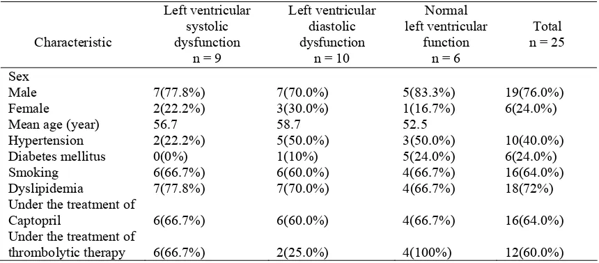
Table 1. Characteristic of patients suffering from acute coronary syndromes and profile of left ventricular function
Characteristic
Left ventricular systolic dysfunction
n = 9
Left ventricular diastolic dysfunction
n = 10
Normal left ventricular
function n = 6
Total n = 25
Sex Male Female
7(77.8%) 2(22.2%)
7(70.0%) 3(30.0%)
5(83.3%) 1(16.7%)
19(76.0%) 6(24.0%)
Mean age (year) 56.7 58.7 52.5
Hypertension 2(22.2%) 5(50.0%) 3(50.0%) 10(40.0%)
Diabetes mellitus 0(0%) 1(10%) 5(24.0%) 6(24.0%)
Smoking 6(66.7%) 6(60.0%) 4(66.7%) 16(64.0%)
Dyslipidemia 7(77.8%) 7(70.0%) 4(66.7%) 18(72%)
Under the treatment of
Captopril 6(66.7%) 6(60.0%) 4(66.7%) 16(64.0%)
Under the treatment of
thrombolytic therapy 6(66.7%) 2(25.0%) 4(100%) 12(60.0%)
The number of male patients suffering from left ventricular systolic dysfunction was 7 persons (77.8%) and that of female patients was 2 persons (22.2%). The number of male patients suffering from left ventricular diastolic dysfunction was 7 persons (70.0%) and that of female patients was 3 persons (30.0%). While, another 5 male patients (83.3%) and 1 female patient (16.7%) who
Brain Natriuretic Peptide
Folia Medica Indonesiana Vol. 41 No. 2 April – June 2005 The risk factors of patients suffering from acute
coronary syndromes with left ventricular systolic dysfunction were dyslipidemia (77.8%, found in 7 persons), smoking (66.7%, found in 6 persons), and hypertension (22.2%, found in 2 persons). The risk factors of patients suffering from left ventricular diastolic dysfunction were dyslipidemia (70.0%, found in 7 persons), smoking (60.0%, found in 6 persons), hypertension (50%, found in 5 persons), diabetes mellitus (10.0%, found in 1 person). The risk factors of patients suffering from acute coronary syndromes with normal left ventricular function were diabetes mellitus (83.3%, found in 5 persons), dyslipidemia (66.7%, found in 4 persons) smoking (64%, found in 4 persons), and hypertension (50.0%, found in 3 persons).
The number of patients suffering from acute myocardial infarction was 20 persons and 12 persons (60.0%) among them were under the treatment of thrombolytic therapy. There were 6 patients (66.7%) suffering from acute myocardial infarction with left ventricular systolic dysfunction who were under the treatment of thrombolytic therapy, 2 patients (25%) suffering from acute myocardial infarction with left ventricular diastolic dysfunction, and 4 patients (100%) with normal left ventricular function who were under the treatment of thrombolytic therapy. Meanwhile, the treatment of Captopril was given to 16 persons (64%), 6 persons (66.7%) of them suffered from left ventricular systolic dysfunction, another 6 persons (60.0%) of them suffered from left ventricular diastolic dysfunction and the last 4 persons (100%) had a normal left ventricular function.
Table 2. The Distribution of BNP level according to left ventricular function.
Left ventricular function
Number n
Mean pg/ml
Standard deviation
Minimum pg/ml
Maximum pg/ml
ANOVA test
P Systolic
dysfunction
9 301.11 189.62 88.00 712.00
Diastolic dysfunction
10 273.70 146.27 97.00 614.00
Normal 6 42.67 22.44 14.00 64.00
0.007
The mean of BNP plasma level of patients suffering from acute coronary syndromes with left ventricular systolic dysfunction was 301.11 pg/ml. The mean of BNP plasma level of patients suffering from acute coronary syndromes with left ventricular diastolic dysfunction was 273.70 pg/ml. Whereas, the mean of
BNP plasma level of patients suffering from acute coronary syndromes with normal left ventricular function was 42.67 pg/ml. P = 0.007 was obtained by conducting the ANOVA test on the BNP plasma level among the three groups (Table 2).
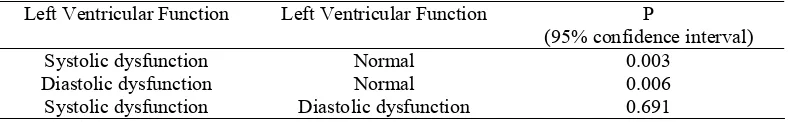
Table 3. Comparison BNP plasma level in left ventricular dysfunction
Left Ventricular Function Left Ventricular Function P
(95% confidence interval)
Systolic dysfunction Normal 0.003
Diastolic dysfunction Normal 0.006
Systolic dysfunction Diastolic dysfunction 0.691
According to comparison of BNP plasma level to the left ventricular function of patients suffering from acute coronary syndromes, there was a significant difference both between patients with systolic dysfunction and patients with normal ventricular function and between patients with diastolic dysfunction and patients with normal ventricular function (p < 0.05). On the other
hand, according to comparison of BNP plasma level to the left ventricular function of patients suffering from acute coronary syndromes, there was no significant difference between patients with systolic dysfunction and patients with diastolic dysfunction (p = 0.691) (Table 3).
Table 4. Ejection fraction to left ventricular function
Left ventricular
function
Number
n
Mean EF %
Standard deviation
Minimum %
Maximum %
ANOVA test
P Systolic
dysfunction
9 44.1 5.2 35 49.1
Diastolic dysfunction
10 64.5 9.2 54.3 79.2 0.0001
Normal 6 61.6 8.8 53.3 76.2
The mean of ejection fraction of patients suffering from acute coronary syndromes with systolic dysfunction was 44.1% and that of patients suffering from acute coronary syndromes with diastolic dysfunction was
64.5%. Meanwhile, the mean of ejection fraction of patients with normal left ventricular function was 61.6% (Table 4).
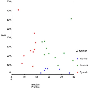
Figure 1. Relationship of BNP plasma level, ejection fraction and ventricular function
It was obtained that there was 1 patient (11.1%) suffering from left ventricular systolic dysfunction with a BNP plasma level less than 100 pg/ml. In the mean time, there was 1 patient (10.0%) suffering from left
ventricular diastolic dysfunction with a BNP plasma level less than 100 pg/ml. All patients with normal ventricular function had BNP plasma level less than 100 pg/ml as well (Table 5).
Ejection Fraction
80 70
60 50
40 3
0 BNP
800
700
600
500
400
300
200
100
0
LV function
Normal
Diastolic
Brain Natriuretic Peptide
Folia Medica Indonesiana Vol. 41 No. 2 April – June 2005 Table 5. Left ventricular dysfunction with cut of point 100 pg/ml
BNP Level
The number of patients suffering from acute coronary syndromes who experienced cardiovascular events within 30 days was 10 patients (40%), 3 of them were deceased and the remaining 7 patients suffered from heart failure. Among the 10 patients who experienced cardiovascular events, 3 persons were diagnosed acute large anterior wall myocardial infarction, 2 persons were diagnosed acute anteroseptal myocardial infarction, 1 person was diagnosed acute anterior wall myocardial infarction, 2 persons were diagnosed acute inferior wall myocardial infarction, and the last 2 persons were diagnosed acute inferior and right ventricular myocardial infarction.
The mean of BNP plasma level of patients suffering from acute anterior wall and acute large anterior myocardial infarction is 403 pg/ml, and the mean of BNP plasma level of other types of infarction is 177.4 pg/ml. Among 3 patients who were deceased, 2 patients were due to cardiogenic shock and the remaining 1 experienced ventricular fibrillation. The 3 cases of mortality occurred to patients suffering from acute large anterior wall and acute anterior wall myocardial infarction. The mean of BNP plasma level of the deceased patients was 582.33 pg/ml.
Table 6. BNP Plasma Level In Cardiovascular Events
Cardiovascular
The mean of BNP plasma level of patients who experienced cardiovascular events was 392.30 pg/ml, and that of patients who did not experience cardiovascular events was 118.67 pg/ml. The minimum limit of BNP plasma level of patients who experienced cardiovascular events was 248 pg/ml, and the maximum limit of BNP plasma level of patients who did not
experience cardiovascular events was 234 pg/ml. The difference of BNP plasma level between patients who experienced cardiovascular events and patients who did not experience cardiovascular events was fairly significant (p = 0.0001) (Table 6).
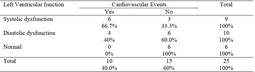
Table 7.Left Ventricular Dysfunction To Cardiovascular Events
Cardiovascular Events Total
Left Ventricular function
Yes No
Systolic dysfunction 6
66.7%
3 33.3%
9 100%
Diastolic dysfunction 4
The cardiovascular events within 30 days were occurred in 10 patients (40%) suffering from acute coronary syndromes, 6 of them (66.7%) happened to patients with systolic dysfunction and the remaining 4 (40.0%) happened to patients with diastolic dysfunction. And there were no cardiovascular events among patients with normal left ventricular function (Table 7).
DISCUSSIONS
This study was carried on patients suffering from acute coronary syndromes in all clinical spectrum including acute ST elevation myocardial infarction, acute non ST elevation myocardial infarction and unstable angina, and there was no history of heart failure. Clinical evaluation on patients suffering from acute coronary syndromes is extremely important to determine complication and prognosis. Early finding of left ventricular dysfunction due to acute coronary syndromes is useful to give the optimum therapy and to prevent heart failure that ultimately worsens the coronary perfusion.
In the current study, it was obtained that 76% of patients suffering from acute coronary syndromes experienced left ventricular dysfunction. The systolic dysfunction happened in 40% of them, and the diastolic dysfunction happened to the remaining 36%. The mean of BNP plasma level of patients with either systolic or diastolic dysfunction was significantly higher than in patients with normal left ventricular function. The mean of BNP plasma level of patients with systolic dysfunction was 301.11 pg/ml and that of patients with diastolic dysfunction was 273.70 pg/ml, which was not significantly difference (p = 0.691). In contrast, it was obtained a significant difference of the increase of BNP plasma level that happened to patients with systolic dysfunction (p = 0.003), and the increase of BNP plasma level of patients with diastolic dysfunction differed significantly in comparison with that of patients suffering from acute coronary syndromes with normal left ventricular function (p = 0.006).
On a guinea pig suffering from transmural myocardial infarction, the gene expression level of BNP on left ventricle increased within 4 hours after ligating coronary vessels and the level of BNP on non-infarcted tissue equals to that on infarcted tissue. Being gained from the evaluation of BNP plasma level of the guinea pigs, the increase of BNP plasma level was 5 times on the first day and 7 times on the third day. On the other studies, it was proved that BNP plasma level of patients suffering from unstable angina was higher than patients suffering from stable angina or normal people (Maisel A, 2001). Myocardial ischemia results in down
regulation contraction as a conservative adapting response of the myocyte and consequently it will reduce the energy necessity in order to maintain viability. Myocardial infarction can result in either ventricular systolic dysfunction or ventricular diastolic dysfunction (Nobuyoshi M et al, 1991).
The mean of BNP plasma level of patients suffering from acute myocardial infarction (419.1 pg/ml) in this study was significantly higher than that of patients suffering from unstable angina (124.5 pg/ml). The increase of BNP level was in line with regional disturbances of the ventricle. The experimental study proved that there was an immediate increase to the expression of BNP mRNA on myocyte located at the border of infarcted area (it was likely influenced by the change of growth factor and the expression of basic fibroblast growth factor). The stimulation of BNP increase was also affected by the increase of wall stress that directly influenced by the process of infarction. On a further process, as a result of the existence of ventricular load, it would enhance the stimulation of BNP increase. The increase of BNP produced systemic effect, natriuresis and vasodilatation that formed protection mechanism to the expansion of infarction.
The mean of BNP level of the deceased patients was 1582.33 pg/ml and all were with acute anterior wall and acute large anterior wall myocardial infarction. The causes of the mortality were cardiogenic shock and ventricular fibrillation in this study. At least there was 40% of myocardial damage of the left ventricular muscle mass from the autopsy conducted on patients with cardiogenic shock (Daly-Nee C et al, 1999) The increase of BNP plasma level during the period of post myocardial infarction had a relationship with the infarction area, the decrease of ejection fraction, and the increase of heart failure risk and death (Nobuyoshi M al, 1991). Ventricular dilatation that occurred after acute myocardial infarction was repetitively related to ventricular remodeling. On the basis of Laplace law, the thinning and dilatation of infarction area can increase ventricular wall tension during the systolic period and diastolic period (Daly-Nee C et al, 1999).
In the study on 72 patients suffering from symptomatic ventricular dysfunction with ejection fraction less than 50%, BNP can be used to predict the increase of left ventricular diastolic end pressure. The other study has proved the increase of left ventricular diastolic end pressure 18 mmHg higher with sensitivity 81% and specificity 85% (Braunwald E, 2001).
Brain Natriuretic Peptide
Folia Medica Indonesiana Vol. 41 No. 2 April – June 2005 disturbance in comparison with other groups of
natriuretic peptide. BNP in blood circulation regulates blood pressure and fluid balance. BNP acts physiologically through the receptor that makes a bond with guanylate cyclase on natriuretic peptide receptor A (NPR-A). NPR-A is found in various tissues that catalyze the conversion of guanosine triphosphate to cyclic guanosine monophosphate (cGMP). cGMP produces a potent vasodilator effect and acts as a second messenger BNP. BNP possesses natriuresis and diuresis effects. BNP excretes sodium and water by increasing glomerulal filtration rate and by inhibiting sodium reabsorption in kidneys. BNP prevents the effect of renin angiotensin system by reducing secretion of aldosteron and renin that causes the decrease of blood pressure and extracellular fluid volume. BNP is retaken from plasma by means of 2 different mechanisms, that is to say endocytosis and enzyme degradation by endopeptidase (Hama N, 1995: Levin ER et al, 1998: Lainchbury JG et al, 2002)
In this study, 24% of patients suffering from acute coronary syndromes have normal left ventricular function. All patients suffering from acute myocardial infarction that possessed normal left ventricular function were treated with thrombolytic agent in order to gain a reperfusion. If ischemia occurred short lasting by reperfusion, the major molecule is not damage and myocard disturbances can be reversible (Nobuyoshi M et al, 1991).
The diastolic dysfunction would be found in patients suffering from acute coronary syndromes whose BNP plasma level more than 97 pg/ml and echocardiography result without systolic dysfunction. On the other hand, the normal diastolic function would be found in patients suffering from acute coronary syndromes without systolic dysfunction and BNP plasma level less than 64 pg/ml.
Approximately 40%-50% of patients that were diagnosed heart failure had normal systolic function that implied the existence of diastolic dysfunction. Diastolic heart failure could not be differentiated by anamnesis, physical diagnostic, chest x-ray and electrocardiogram. It required the measurement of ventricular pressure and ventricular volume to optimize the evaluation of diastolic dysfunction measurement. This approach needed an invasive technique or echocardiography evaluation that needed time and measurement technique to be used to value diastolic property characteristic (Crilley JG & Farrer M, 1999)
Appleton et al measured diastolic function by using the transmitral doppler velocity. The assessment using doppler on diastolic dysfunction and transmitral velocity
pattern would be inconsistent due to the influence of heart rate, preload, afterload, contractility, valvular regurgitation, and the position of sample volume. On normal systolic condition, the simple blood examination in clinic to evaluate diastolic dysfunction will be very useful ((Crilley JG & Farrer M, 1999: Lubien E et al, 2002).
In this study, it was proven that the increase of BNP plasma of patients with normal systolic function had diastolic dysfunction. However, BNP plasma level cannot be used to differentiate between systolic dysfunction and diastolic dysfunction. In the mean time, by using echocardiography the existence of systolic dysfunction can be established. The low BNP plasma level of patients that previously checked with echocardiography without systolic dysfunction can be used to exclude diastolic dysfunction.
The mean of BNP plasma level of patients suffering from acute coronary syndromes with cardiovascular events was significantly higher than that of patients without cardiovascular events within 30 days. Patients suffering from acute coronary syndromes with the increase of BNP plasma level more than 248 pg/ml would have a risk to experience heart failure or death within 30 days. In contrast, patients suffering from acute coronary syndromes with left ventricular dysfunction whose BNP plasma level less than 234 pg/ml would not have a risk to experience cardiovascular events.
BNP that is used to predict the risk of patients suffering from acute coronary syndromes is not like traditional biomarker, that is to say BNP plasma has a role as contraregulator response against ischemia. The increase of BNP plasma level will have a relationship with the severity of ischemia and the degree of ventricular dysfunction (McClure S et al, 1998: Peacock WF, 2002).
The activation of neurohormonal system is a death risk after acute coronary syndromes. The relationship between BNP plasma level and long-term risk of death is an independent factor. The increase of BNP plasma level more than 80 pg/ml is indicating the activation of neurohormonal system on patients with heart failure. This limit is no different in acute coronary syndromes (Braunwald E, 2001). On this study the use of the limit as of BNP 80 pg/ml or higher indicated the presence of left ventricular dysfunction and it was considered as normal ventricular function if BNP level was less than 80 pg/ml.
The measurement of BNP of patients with heart failure that was conducted on one study showed the increase of BNP plasma level resulted in 52% of patients would die
or need to be rehospitalized within 30 days. On the other hand, the decrease of BNP level of 84% of patients showed a favorable result. 290 patients suffering from NYHA I and II heart failure with the mean of ejection fraction of 37% showed the increase of BNP level more than 56 pg/ml can be used independently to predict the progress of heart failure and death. On other study, the increase of BNP level to predict the death was much better than that of age, ANP, ejection fraction of pulmonal artery pressure, sex, etiology of heart failure or NYHA class on 12-month evaluation (Braunwald E, 2001: Poulsen SH et al, 2001: Peacock WF, 2002)
The study that compared normal people, patients suffering from coronary heart disease, and patients with heart failure showed that there was no increase of BNP level of patients suffering from coronary heart disease without ventricular dysfunction. On Veteran Administration Study, BNP level could be used to predict criticalness level of the patients, with the purpose of deciding whether the patients were supposed to be hospitalized or not. Patients that were hospitalized possessed the mean of BNP level 700 pg/ml, and patients that were not hospitalized had the mean of BNP level 254 pg/ml (Braunwald E, 2001: Peacock WF, 2002)
In other study on patients suffering from acute myocardial infarction, it was obtained that BNP can be used to predict left ventricular function, heart failure and long term survival rate. Patients suffering from myocardial infarction with the increase of BNP level were predicted having unfavorable prognosis that possibly reflected ventricular function disturbance. The study on 30 patients suffering from myocardial infarction showed BNP level 1 week post myocardial infarction had a relationship with heart remodeling. The BNP level had a relationship with the increase of left ventricular volume and the decrease of ejection fraction (Peacock WF, 2002: Struthers A, 2002)
CONCLUSIONS
Plasma BNP level can be used to detect the presence of diastolic as well as systolic LV dysfunction on echocardiography in early events of acute coronary syndromes. Moreover, plasma BNP level can also predict patients with cardiovascular events in 30-days after acute coronary syndromes.
REFERENCES
Braunwald E, 2001. Heart Disease: a Text Book of Cardiovascular Medicine. 6th edition, USA.
Crilley JG, Farrer M, 1999. Left Ventricular Remodelling and Brain Natriuretic Peptide After First Myocardial Infarction. Heart 82:254-255.
Daly-Nee C, Brunt H, Jairath N, 1999. Risk and Coronary Heart Disease. In Jairath N (Ed). Coronary Heart Disease & Risk Factor Management, 9th ed. Philadelpia, WB Saunders Company, 3-19.
De Lemos JA, Morrow DA, Bentley JH et al, 2001. The Prognostic Value of B-Type Natriuretic Peptide in Patients with Acute Coronary Syndromes. N Engl J Med 345: 1014-1021.
Hama N, Itoh H, Shirakami G et al, 1995. Rapid Ventricular Induction of Brain Natriuretic Peptide Gene Expression in Experimental Acute Myocardial Infarction. Circulation 92: 1558-1564.
Lainchbury JG, Richards M, Nicholls MG et al, 2002. The Effects of Pathophysiological Increments in Brain Natriuretuc Peptide in Left Ventricular Systolic Dysfunction. The Journal of American College of Cardiology 39: 798-803.
Levin ER, Gardner DG, Sampson WK, 1998. Natriuretic Peptides. N Engl J Med 339: 321-328. Lubien E, DeMaria A, Krishnawasmy P et al, 2002.
Utility of B-Natriuretic Peptide in Detecting Diastolic Dysfunction. Circulation 105: 595-601.
Maisel A, 2001. B-Type Natriuretic Peptide In the Diagnosis and Managementof Congestive Heart Failure. Cardiology Clinics.
McClure S, Caruana L, Davie AP, Goldthrop S et al, 1998. Cohort Study of Plasma Natriuretic Peptides for Idetifying Left Ventricular Systolic Dysfunction in Primary Care. BMJ 317: 516-519.
Nagaya N, Nishikimi T, Goto Y et al, 1998. Plasma Brain Natriuretic Peptide is a Biochemical Marker for the Prediction of Progressive Ventricular Remodeling After Acute Myocardial Infarction. American Heart Journal 135: 21-28.
Nobuyoshi M, Tanaka M, Nosaka H, 1991. Progression of Coronary Atherosclerosis: is Coronary Spasm Related to Progression? J Am Coll Cardiol 18: 904-910.
Peacock WF, 2002. The B-Type Natriuretic Peptide Assay: A Rapid Test for Heart Failure. Cleveland Clinic Journal of Medicine 69:243-250.
Poulsen SH, Jensen SE, Moller JE et al, 2001. Prognostic Value of Left Ventricular Diastolic Function and Association with Heart rate Variablity After a First Acute Myocardial Infarction. Heart 86: 376-380.