www.elsevier.comrlocateranireprosci
The effect of porcine parvovirus and porcine
reproductive and respiratory syndrome virus on
porcine reproductive performance
qW.L. Mengeling
), K.M. Lager, A.C. Vorwald
Virology Swine Research Unit, National Animal Disease Center, USDA, Agricultural Research SerÕice,
2300 Dayton AÕenue, P.O. Box 70, Ames, IA 50010, USA
Abstract
Ž .
From a worldwide perspective, porcine parvovirus PPV and porcine reproductive and
Ž .
respiratory syndrome virus PRRSV are the most common viral causes of porcine reproductive failure. A typical epidemic of PPV-induced reproductive failure is presented as an increased number of mummified fetuses and sometimes, entire litters are mummified. If infection with PPV is very early in gestation, the number of liveborn pigs may be further reduced as a result of embryonic death and resorption. During the acute stage of infection gilts and sows have few, if any, clinical signs, and it is unlikely that PPV is ever the direct cause of abortion. In contrast, a typical epidemic of PRRSV-induced reproductive failure is presented as a broader spectrum of clinical features including abortions, late-term dead fetuses, stillborn pigs, and weakborn pigs. In the later stages of an epidemic, there may also be an increase in the number of mummified fetuses, but their prevalence is likely to be far less than during an epidemic of PPV-induced reproductive failure. During the acute stage of infection with PRRSV, gilts and sows may have few, if any, clinical signs, or they may be severely affected and even die. This difference largely reflects the relative virulence of the strain of PRRSV causing the epidemic. A timely and reliable laboratory diagnosis of either disease can be made when appropriate tests are performed with appropriate
q
No endorsements are herein applied. Brand names are necessary to report factually on available data; however, the USDA neither guarantees nor warrants the standards of the products, and the use of the names by the USDA implies no approval of the products to the exclusion of others that may also be suitable.
)Corresponding author. Tel.:q1-515-663-7254; fax:q1-515-663-7458.
Ž .
E-mail address: [email protected] W.L. Mengeling .
0378-4320r00r$ - see front matter Published by Elsevier Science B.V.
Ž .
samples. Vaccines are available for prevention of both diseases. Published by Elsevier Science B.V.
Keywords: Porcine parvovirus; Porcine reproductive and respiratory syndrome virus; Reproductive failure;
Abortion; Mummification
1. Introduction
Successful swine production depends in part on preventing infectious diseases that affect reproductive performance. Among the most important are those caused by viruses and recognized mainly by their effect on pregnant gilts and sows. Some viruses cause severe maternal illness and abortion. Others cause few, if any, immediate clinical signs and are evident only at or near the expected time of farrowing when all or part of a litter is delivered dead, weak, or malformed. Still others can cause either immediate or delayed signs depending on a variety of circumstances. A few also affect the reproduc-tive performance of boars, e.g. reduced libido or fertility.
A large number of viruses have been reported to affect porcine reproductive performance, and it is likely that any virus capable of causing clinical illness in adult swine, or crossing the placental barrier to infect the porcine conceptus, is a potential reproductive pathogen. Two viruses that have been especially problematic worldwide are
Ž . Ž .
porcine parvovirus PPV Cartwright and Huck, 1967; Mengeling, 1978 and porcine
Ž . Ž
reproductive and respiratory syndrome virus PRRSV Keffaber, 1989; Wensvoort et
.
al., 1991 . In the remainder of this report we focus on these 2 viruses and the diseases they cause. Brief discussions of selected physical and biologic properties of each virus are included to help explain various aspects of the epidemiology, pathogenesis, diagno-sis, and prevention of the associated disease.
2. PPV-induced reproductive failure
2.1. Virus properties
PPV is classified in the family Parvovirdae, subfamily Parvovirina, genus Parvovirus. Other members of the same genus include parvoviruses of cattle, cats, dogs, geese, mice, rats, rabbits, mink, chickens, geese and raccoons. Although PPV is distinguishable from
Ž
parvoviruses of all other species, it is antigenically related to some Mengeling et al.,
.
However, differences have been reported in regard to the relative virulence of a few
Ž .
isolates Choi et al., 1987 .
A wide range of porcine cells support replication of PPV in vitro. Early-passage cells are generally thought to be more susceptible than established cell lines. Fetal porcine kidney cell cultures are commonly used for virus detection and propagation. Replication is dependent on mitotic activity in that enzymes produced by the cell for its own DNA replication are ‘‘borrowed’’ by PPV for its replication. Viral replication is cytocidal and is characterized by ‘‘rounding up’’, pyknosis, and lysis of cells. Nascent viral antigen can be detected by immunostaining techniques in the nucleus of an infected cell starting at about 8 h after initial infection. It accumulates there until about 16 h when the cell starts to show cytopathic changes. Eventually infected cells disintegrate and large clumps of antigen and virus are released and subsequently internalized by non-infected
Ž .
cells if any in the same culture. In the absence of conditions that promote virus replication, which would then result in the death of the infected cell, antigen may persist in the cytoplasm for at least several days.
2.2. Epidemiology
PPV is ubiquitous in swine throughout the world. In most herds infection is endemic. The typical cycle of infection is thought to be the following. At the time of farrowing most gilts and sows are immune and impart a high level of PPV antibody to their offspring via colostrum. This passively acquired antibody persists at progressively lower levels for 4 to 6 months, during which time pigs are relatively refractory to infection
ŽPaul et al., 1980, 1982 . As passively acquired antibody wanes pigs become progres-.
sively more likely to be infected and acquire active immunity. Active immunity apparently persists for life, perhaps as a result of repeated exposure to virus. If all gilts developed active immunity by natural exposure prior to conception PPV would rarely, if ever, be a problem. However, some gilts, for a variety of reasons, escape infection until sometime during their first gestation. If they are first exposed to PPV anytime during about the first one-half of gestation, transplacental infection and reproductive failure are
Ž .
likely to ensue Mengeling and Cutlip, 1976; Joo et al., 1976a . If they are infected anytime during about the second one-half of gestation there is still likely to be transplacental infection. In fact, the mean number of transplacentally infected fetuses per litter may be higher when infection is later in gestation because of the increase area of contact between the maternal and fetal placentas and the increased blood flow to the uterus. However, at least most fetuses infected after about 70 days of gestation produce antibody and survive infection. They may, however, shed PPV during at least the early postnatal period. Events during the development of the porcine conceptus that affect the
Ž .
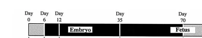
outcome of transplacental infection with PPV and other viruses are presented Fig. 1 . Additional details on the temporal aspects of transplacental infection are presented in the section on Pathogenesis.
Fig. 1. Benchmarks during gestation that affect the consequences of transplacental infection of the porcine conceptus with viruses. During the very early part of gestation, i.e. prior to hatching, the conceptus is protected by the zona pellucida and is therefore insusceptible to infection. Thereafter, during the stage of the embryo,
Ž
infection with PPV and perhaps PRRSV it has not yet been shown that PRRSV infects and kills the porcine
.
embryo results in embryonic death and resorption. The stage of the fetus begins at or about gestation day 35 when organogenesis is essentially complete and ossification begins. Infection of the conceptus during the stage of the fetus can result in death but resorption is precluded by the developing fetal skeleton. At or about day 70 of gestation, the fetus is able to produce sufficient antibody to protect itself against the otherwise fatal effects of PPV. Thereafter, fetal infection with PPV seems to be subclinical. The degree of mitotic activity of vital fetal tissues before and after about gestation day 70 may also play a role in relative susceptibility of fetuses to PPV. On the other hand, PRRSV can kill the fetus even very late in gestation. This is reflected in the large number of late-term dead fetuses that are part of some epidemics of PRRS.
environment. By adding naive pigs to a room previously inhabited by acutely infected pigs it was shown that PPV can remain infectious outside its host for at least 4 months. Conversely, it was shed for only a few weeks when the same acutely infected pigs were moved to a separate isolation room and put in contact with additional naive pigs
ŽMengeling and Paul, 1986 ..
PPV has little, if any, clinical effect on mature boars. However, it has been isolated from scrotal lymph nodes for as long as 7 weeks after acute infection, and from semen. Boars may also serve as non-infected carriers of PPV as they move among infected and non-infected females.
2.3. Clinical signs
The only well established clinical response to infection with PPV is reproductive failure. Acute infection usually goes unnoticed and the first evidence of a problem is at
Ž
the time of farrowing 2 or more months later i.e. keep in mind that infection later in
.
gestation is unlikely to result in reproductive failure . PPV-induced reproductive failure is typically signaled by an unusually large number of mummified fetuses delivered at or near term. When infection is very early in gestation litters may be smaller as a result of
Ž .
embryonic death and resorption Mengeling et al., 1980 , but such litters are still likely to contain dead fetuses because of intrauterine spread of the virus with littermates becoming infected at progressively later times in gestation. There may also be an increased number of stillborn pigs when, as a result of fetal death, farrowing times are delayed or farrowing intervals are prolonged.
2.4. Pathogenesis
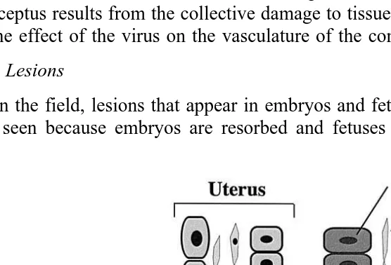
viruses move from one part of the body to another in one of three ways; as a passenger in body fluids such as blood or lymph, or in or on cells such as macrophages, or as a result of progressive replication through contiguous cells. Because fetal and maternal
Ž
circulations are clearly separated in pigs not even antibody passes from mother to
.
offspring in utero , and because experiments in which tissues were collected at various times after maternal infection have indicated that several of the layers of tissues
Ž .
comprising the so-called placental barrier Fig. 2 are insusceptible to PPV, we can probably eliminate the first and third possibilities. If we are correct, that leaves the transfer of virus in or on cells that traverse the placental barrier as the most likely possibility. Our hypothesis is that PPV reaches the conceptus as a passenger in or on maternal macrophages. The likelihood that this is a relatively rare event would explain why often only part of a litter is transplacentally infected.
Ž .
Once the virus reaches the conceptus embryo or fetus , it finds an environment particularly conducive to replication because of the high mitotic index of most tissues
Žsee the previous discussion on the relationship between PPV replication and mitotic .
index . Examination of tissues of embryos and fetuses collected at various times after infection has revealed extensive virus replication. It is probable that death of the conceptus results from the collective damage to tissues and organs. Especially important is the effect of the virus on the vasculature of the conceptus and placenta.
2.5. Lesions
In the field, lesions that appear in embryos and fetuses that succumb to infection are not seen because embryos are resorbed and fetuses are presented on at or near term
when they are in an advanced stage of mummification. However, a cascade of lesions
Ž
seen under experimental conditions when fetuses are collected at various times after
.
infection are the following: a variable degree of stunting and sometimes a loss of condition; occasionally, an increased prominence of blood vessels due to congestion and leakage of blood into surrounding tissues; congestion, edema, and hemorrhage with accumulation of serosanguineous fluids in body cavities; hemorrhagic discoloration of
Ž .
skin and other tissues becoming progressively darker after death ; and dehydration
Žmummification. ŽMengeling and Cutlip, 1976 . Fetuses that produce antibody and.
Ž .
survive infection i.e. those infected at or about gestation day 70 or later have tissue changes consistent with an immune response. Congenitally infected stillborn pigs may
Ž .
have meningoencephalitis Narita et al., 1975 .
2.6. Diagnosis
A highly reliable means to diagnose PPV-induced reproductive failure is to examine
Ž .
fetal tissues by immunofluorescence microscopy Mengeling and Cutlip, 1976 . Cryo-stat-microtome sections of fetal lung are reacted with fluorescein-labeled PPV antibody. Lung is recommended because it is relatively easy to identify in a mummified fetus and it has a minimum of autofluorescence. In most instances where fetuses have died as a result of PPV infection, viral antigen will be present throughout the lung tissue. Detection of viral antigen in a mummified fetus provides an almost incontrovertible connection between infection and death. To minimize the chance that the fetuses selected for testing produced some antibody that might interfere with detection of antigen, those selected should be F 70 days of gestational age, i.e. F 16 cm in length
Žcrown-rump measurement . Fetuses older than 70 days of gestational age can produce.
titers of antibody at or near those of postnatal pigs.
Some additional diagnostic procedures include examining tissues for viral
hemag-Ž .
glutinin Joo et al., 1976b , testing fetal sera for antibody, and testing fetal fluids and
Ž .
tissue extracts by the polymerase chain reaction PCR . The latter is very sensitive and
Ž .
can be used to identify PPV i.e. a segment of the viral genome under conditions that preclude virus isolation, e.g. in the presence of neutralizing antibody.
2.7. PreÕention
PPV-induced reproductive failure can be prevented by making sure that all females have developed an active immunity before they conceive for the first time. Because infection is endemic in most herds, immunity is often the result of natural exposure. However, to ensure immunity, it is a common practice to vaccinate gilts once or twice before conception and at least once annually thereafter. Inactivated vaccines are both
Ž .
safe and effective Mengeling et al., 1979 .
3. PRRSV-induced reproductive failure
3.1. Virus properties
Ž .
fever virus, and equine viral arteritis virus Meulenberg et al., 1993 . All are antigeni-cally distinguishable, but are classified in the same genus on the basis of common structural and replicative features. An infectious virion of PRRSV is 50 to 65 nm in diameter and comprises a lipid-containing outer envelope and 6 structural proteins: four
Ž . Ž .
glycoproteins GP identified as GP2, GP3, GP4, and GP5; a membrane M protein;
Ž . Ž .
and a nucleocapsid N protein Meulenberg et al., 1995 . The major structural proteins are N, M, and GP5. GP5 is believed to include the primary determinants for virus neutralization. PRRSV is relatively unstable once released from an infected cell; however, it can remain infectious for at least several years when maintained under selected conditions, e.g. stored ay708C in cell culture medium. Its genome is a single strand of positive-sense RNA comprising about 15 kb and eight open reading frames
ŽORFs designated ORF 1a, ORF 1b which code for the viral polymerases , and ORFs. Ž .
Ž .
2 through 7 which code for structural proteins . Two major serotypes, prototype
Ž .
Lelystad and prototype VR-2332, have been identified Wensvoort et al., 1992 . Most if not all field strains isolated in Europe are more closely related to the strain Lelystad than to strain VR-2332, whereas most if not all field strains isolated in North America and elsewhere in the world are more closely related to strain VR-2332 than to strain Lelystad. Within each serotype there are additional genomic differences that can be
Ž . Ž .
identified by restriction fragment length polymorphism RFLP Wesley et al., 1996 as
Ž .
well as by base sequencing Kapur et al., 1996 .
Only a few cell types are known to support replication of PRRSV in vitro. Those used most often for diagnostic and research purposes are African green monkey kidney
Ž
cells viz. an established cell line MA-104 and MA-104-derived cell lines CL-2621 and
. Ž . Ž
MARC-145 Kim et al., 1993 , and porcine alveolar macrophages Wensvoort et al.,
. Ž .
1991 . Virus-induced cytopathic effects CPE are usually evident within 1 to 4 days after infection. The exact time depends largely on the strain of PRRSV and the extent of its previous adaption to replication in cell culture. A few strains of PRRSV have been reported to replicate in only one of these cell types. However, most strains that have been tested have been shown to replicate in both.
3.2. Epidemiology
The known history of PRRS and PRRSV is relatively short. The first clinical case was reported in North Carolina in 1987. However, the virus was apparently present in the swine population in North America sometime earlier in that a retrospective serologic
Ž .
study revealed antibody in a few pigs in Canada as early as 1979 Carman et al., 1995 . It is possible that a PRRSV-like virus of another species became adapted to swine sometime in the late 1970s. But as yet there is no firm evidence to support this idea. The rapid spread of PRRSV throughout North America since 1987 and its emergence and spread throughout much of the rest of the world since 1987 attests to its transmissibility. PRRSV is spread both horizontally and vertically. Horizontal transmission is
proba-Ž .
bly most often by direct contact with virus present in secretions including semen and excretions. Circumstantial evidence for airborne transmission is provided by the
observa-Ž
tion that in some epidemics what appears to be the same strain of PRRSV as
.
in the same geographic area despite the lack of any known direct or indirect contact among them. On the other hand, in some trials under controlled experimental conditions PRRSV was not transmitted from groups of experimentally infected pigs to groups of
Ž .
naive pigs even when the infected and naive groups kept on separate raised decks were
Ž .
in the same room for 31 days and separated by as little as 41 cm Wills et al., 1997 . Vertical transmission is an equally important means by which PRRSV is disseminated and maintained in the swine population. Congenitally infected pigs have the potential to be persistently infected and shed PRRSV for at least the first several months of their life
ŽBenfield et al., 1997 ..
3.3. Clinical signs
Until relatively recently, the cardinal features of most PRRSV-induced epidemics of reproductive failure were late-term abortions and the delivery of late-term dead fetuses and weak, unthrifty pigs. Often, there were few, if any, clinical signs during the acute stage of infection of gilts and sows several weeks earlier. In the fall of 1996, the clinical picture in North America changed dramatically, at least in some epidemics, with the emergence, presumably through mutations, of more virulent strains of PRRSV. In these more severe epidemics, now often referred to as ‘‘acute’’ or ‘‘atypical’’ PRRS, there were abortions at all stages of gestation. Moreover, soon after infection, gilts and sows typically had markedly elevated body temperatures and became inappetent. Many also
Ž .
became recumbent and some died Zimmerman et al., 1997 . All of the clinical features of atypical PRRS were subsequently reproduced experimentally with strains isolated from relevant field cases. During the last year or so, the prevalence of atypical PRRS seems to have lessened. The reason or reasons are unknown.
3.4. Pathogenesis
Before the emergence of atypical PRRS, it was generally assumed that the reproduc-tive consequences of infection with PRRSV, including abortion, were due to the direct effect of the virus on the conceptus following transplacental infection. While this is certainly true for many cases of PRRSV-induced reproductive failure, it apparently does not explain all cases because many litters aborted during epidemics of atypical PRRS were found free of infection. The latter observation indicates that abortion can also be the result of a systemic reaction. The additional observation that abortions were common in herds that had previously been vaccinated raised the question of whether an acute
Žanamnestic immune response contributed to the clinical picture. If so, there is a.
Abortions account for only some of the reproductive losses following exposure to PRRSV during gestation. In many cases fetuses are infected transplacentally and yet gestation goes to term or longer. In these cases, fetuses often die in utero. As yet the means by which PRRSV kills the fetus is unknown. It is suspected that fetal circulation is affected. The means by which PRRSV crosses the placenta is also unknown, but like PPV it may cross in association with maternal macrophages.
3.5. Lesions
There may be segmental hemorrhages in the umbilical cord that are obvious at birth
ŽLager and Halbur, 1996 and cellular infiltration in the maternal placenta. Moreover,.
infected fetuses that survive in utero and produce antibody are likely to have cellular changes reflecting an immune response.
3.6. Diagnosis
The two most commonly used methods to diagnose PRRS are virus isolation and PCR. Each has its advantages and disadvantages, but both are highly reliable if appropriate samples are obtained. For virus, isolation it is critical that samples be
Ž .
obtained from pigs free of virus neutralizing VN antibody. In general, this is an issue only with samples from suckling pigs. The reasons are as follows. Within 7 to 10 days
Ž .
after initial infection with PRRSV field exposure or vaccination , pigs produce antibody that can be detected by tests such as indirect immunofluorescence and enzyme-linked immunosorbent assay. Maximum titers of such antibodies are usually reached by 14 to 21 days after exposure. However, VN antibodies are slow to develop and it may be 6 to 8 weeks before even low titers are evident. As a result, there is generally little difficulty in isolating virus from acutely infected pigs except for suckling pigs of dams vaccinated weeks or months before, e.g. before conception, and then exposed to virulent field virus during gestation. In such cases the colostrum is likely to contain VN antibody that will interfere with virus isolation. To circumvent this problem it is recommended that, in suspected cases of PRRSV-induced reproductive failure, samples be obtained from
Ž .
weakborn pigs which may reflect infection with PRRSV in utero before they suckle. Unlike PPV, PRRSV is a relatively unstable virus so that attempts to isolate it from dead fetuses, even if death is relatively recent, are seldom successful. Also keep in mind, as previously discussed in the section on Pathogenesis, that aborted fetuses, especially those aborted in early and middle gestation, are likely to be free of infection. For at least most strains of PRRSV, non-nested-set PCR is less sensitive than virus isolation and nested PCR is about as sensitive as virus isolation for detecting low concentrations of PRRSV — if the amounts of sample tested by each technique are taken into account. That is, nested-set is more sensitive than virus isolation but under practical conditions
Ž .
much less sample is tested Umthun and Mengeling, 1999 . Under certain conditions,
Ž . Ž
such as when the sample contains VN antibody or is cytotoxic e.g. semen
Chris-.
In epidemiologic investigations, it is also helpful to provide a presumptive identifica-tion of strain or strains of PRRSV involved. Techniques most commonly used for this purpose are RFLP analysis and base sequencing. Strains have also been grouped according to their reaction with panels of monoclonal antibodies.
3.7. PreÕention
Ž
The first PRRS vaccine was introduced in North America in 1994 Gorcyca et al.,
.
1995 . It was an attenuated strain of PRRSV derived by repeated cell culture passage of
Ž
what, by today’s standards, can be defined as a moderately virulent field strain i.e.
.
strain VR-2332 . It was licensed only for administration to pigs 3 to 18 weeks of age for preventing the respiratory facet of PRRS. However, from the beginning it was also used extensively ‘‘off label’’ for preventing the reproductive facet of PRRS. Today it is licensed for that use as well. The same vaccine is now also used in several countries in Europe. Other PRRS vaccines, both attenuated and inactivated, have been introduced more recently.
Attenuated-virus vaccines are generally believed to be more efficacious than inacti-vated vaccines. However, both field and experimental evidence has indicated that even attenuated-virus vaccines may fail to provide optimal protection when vaccinated gilts and sows are exposed during gestation to highly virulent field strains of PRRSV. It is assumed that this reflects antigenic differences among strains. Because attenuated
Žvaccine virus can persist for weeks and perhaps longer in vaccinated pigs, and because.
PRRSV mutates easily, it is advisable to keep vaccinated pigs apart from naive pigs for
Ž .
at least several weeks after vaccination Mengeling et al., 1999 . Studies are continuing on how to improve current vaccines and vaccination strategies.
4. Conclusions
A presumptive differential diagnosis of the two most common viral causes of porcine reproductive failure, namely PPV and PRRSV, can usually be made on the basis of clinical features. A number of laboratory tests for detecting virus or viral antigen are available for diagnostic confirmation. In North America and in several other parts of the world, vaccines are used extensively as a means to prevent both diseases. Most, if not all, of the vaccine used for preventing PPV-induced reproductive failure is inactivated. It is generally believed to be safe and efficacious. Most of the vaccine used for preventing PRRSV-induced reproductive failure is attenuated. It must be used judiciously because of the ability of vaccine virus to persist in vaccinated pigs for at least several weeks and the ability of PRRSV to mutate. The efficacy of PRRSV vaccine depends in part on the nature of the virulent field strain to which vaccinated pigs are subsequently exposed.
References
Benfield, D.A., Christopher-Hennings, J., Nelson, E.A., Rowland, R.R.R., Nelson, J.K., Chase, C.C.L., Rossow, K.D., Collins, J.E., 1997. Persistent fetal infection of porcine reproductive and respiratory
Ž .
Carman, S., Sanford, S.E., Dea, S., 1995. Assessment of seropositivity to porcine reproductive and respiratory
Ž .
syndrome PRRS virus in swine herds in Ontario: 1978 to 1982. Can. Vet. J. 36, 776–777.
Cartwright, S.F., Huck, R.A., 1967. Viruses isolated in association with herd infertility, abortions and stillbirths in pigs. Vet. Rec. 81, 196–197.
Choi, C.S., Molitor, T.W., Joo, H.S., Gunther, R., 1987. Pathogenicity of a skin isolate of porcine parvovirus in swine fetuses. Vet. Microbiol. 15, 19–29.
Christopher-Hennings, J., Nelson, E.A., Nelson, J.A., Hines, R.J., Swenson, S.L., Hill, H.T., Zimmerman, J.J., Katz, J.B., Yaeger, M.J., Chase, C.C.L., Benfield, D.A., 1995. Detection of porcine reproductive and respiratory syndrome virus in boar semen by PCR. J. Clin. Microbiol. 33, 1730–1734.
Gorcyca, D., Schlesinger, K., Chladek, D., Behan, W., Polson, D., Roof, M., Doitchenoff, D., 1995. RespPRRS: a new tool for the prevention and control of PRRS in pigs. Proc. 26th Annu. Meet. Am. Assoc. Swine Pract., 1–22.
Joo, H.S., Donaldson-Wood, C.R., Johnson, R.H., 1976a. Observations on the pathogenesis of porcine parvovirus infection. Arch. Virol. 51, 123–129.
Joo, H.S., Donaldson-Wood, C.R., Johnson, R.H., 1976b. Rapid diagnostic techniques for detection of porcine parvovirus infection in mummified foetuses. Aust. Vet. J. 52, 51.
Kapur, V., Elam, M.R., Pawlovich, T.M., Murtaugh, M.P., 1996. Genetic variation in porcine reproductive and respiratory syndrome virus isolates in midwestern United States. J. Gen. Virol. 77, 1271–1276.
Keffaber, K.K., 1989. Reproductive failure of unknown etiology. Am. Assoc. Swine Pract. Newsl. 1, 1–10. Kim, H.S., Kwang, J., Yoon, I.J., Joo, H.S., Frey, M.L., 1993. Enhanced replication of porcine reproductive
Ž .
and respiratory syndrome PRRS virus in a homogenous subpopulation of MA-104 cell line. Arch. Virol. 133, 477–483.
Lager, K.M., Halbur, P.G., 1996. Gross and microscopic lesions in porcine fetuses infected with porcine reproductive and respiratory syndrome virus. J. Vet. Diagn. Invest. 8, 275–282.
Mengeling, W.L., 1978. Prevalence of porcine parvovirus-induced reproductive failure: an abattoir study. J. Am. Vet. Med. Assoc. 172, 1291–1294.
Mengeling, W.L., Cutlip, R.C., 1976. Reproductive disease experimentally induced by exposing pregnant gilts to porcine parvovirus. Am. J. Vet. Res. 37, 1393–1400.
Mengeling, W.L., Paul, P.S., 1986. Interepizootic survival of porcine parvovirus. J. Am. Vet. Med. Assoc. 188, 1293–1295.
Mengeling, W.L., Brown, T.T. Jr., Paul, P.S., Gutekunst, D.E., 1979. Efficacy of an inactivated virus vaccine for prevention of porcine parvovirus-induced reprodctive failure. Am. J. Vet. Res. 40, 204–207. Mengeling, W.L., Paul, P.S., Brown, T.T. Jr., 1980. Transplacental infection and embryonic death following
maternal exposure to porcine parvovirus near the time of conception. Arch. Virol. 65, 55–62.
Mengeling, W.L., Ridpath, J.F., Vorwald, A.C., 1988. Size and antigenic comparisons among the structural proteins of selected autonomous parvoviruses. J. Gen. Virol. 69, 825–837.
Mengeling, W.L., Vorwald, A.C., Lager, K.M., Clouser, D.F., Wesley, R.D., 1999. Identification and clinical assessment of suspected vaccine-related field strains of porcine reproductive and respiratory syndrome virus. Am. J. Vet. Res. 60, 334–340.
Meulenberg, J.J.M., Hulst, M.M., de Meijer, E.J., Moonen, P.L.J.M., den Besten, A., de Kluyver, E.P., Wensvoort, G., Moorman, R.J.M., 1993. Lelystad virus, the causative agent of porcine epidemic abortion
Ž .
and respiratory syndrome PEARS , is related to LDV and EAV. Virology 192, 62–72.
Meulenberg, J.J.M., Petersen-den Besten, A., de Kluyver, E.P., Moorman, R.J.M., Schaaper, W.M.M., Wensvoort, G., 1995. Characterization of proteins encoded by ORFs 2 to 7 of Lelystad virus. Virology 206, 155–163.
Narita, M., Inui, S., Kawakami, Y., Kitamura, K., Maeda, A., 1975. Histopathological changes of the brain in
Ž .
swine fetuses naturally infected with porcine parvovirus. Natl. Inst. Anim. Health Q. Tokyo 15, 24–28. Paul, P.S., Mengeling, W.L., Brown, T.T. Jr., 1980. Effect of vaccinal and passive immunity on experimental
infection of pigs with porcine parvovirus. Am. J. Vet. Res. 41, 1368–1371.
Paul, P.S., Mengeling, W.L., Pirtle, E.C., 1982. Duration and biological half-life of passively-acquired colostral antibodies to porcine parvovirus. Am. J. Vet. Res. 43, 1376–1379.
Wensvoort, G., Terpstra, C., Pol, J.M.A., ter Laak, E.A., Bloemraad, M., de Kluyver, E.P., Kragten, C., van Buiten, L., den Besten, A., Wagenaar, F., Broekhuijsen, J.M., Moonen, P.L.J.M., Zestra, T., de Boer, E.A., Tibben, H.J., de Jong, M.F., van’t Veld, P., Groenland, G.J.R., van Gennep, J.A., Voets, M.Th., Verheijden, J.H.M., Braamskamp, J., 1991. Mystery swine disease in The Netherlands: the isolation of Lelystad virus. Vet. Q. 13, 121–130.
Wensvoort, G., de Kluyver, E.P., Luijtze, E.A., den Besten, A., Harris, L., Collins, J.E., Christianson, W.T., Chaldek, D., 1992. Antigenic comparison of Lelystad virus and swine infertility and respiratory syndrome
ŽSIRS virus. J. Vet. Diagn. Invest. 4, 134–138..
Ž
Wesley, R.D., Mengeling, W.L., Andreyev, V., Lager, K.M., 1996. Differentiation of vaccine Strain
.
RespPRRS and field strains of porcine reproductive and respiratory syndrome virus by restriction enzyme analysis. Proc. 27th Annu. Meet. Am. Assoc. Swine Pract., 141–143.
Wills, R.W., Zimmerman, J.J., Swenson, S.L., Yoon, K.-J., Hill, H.T., Bundy, D.S., McGinley, M.J., 1997. Transmission of porcine reproductive and respiratory syndrome virus by direct, close, or indirect contact. Swine Health Prod. 6, 213–218.