www.elsevier.com / locate / bres
Research report
Are autonomic signals influencing cortico-spinal motor excitability?
A study with transcranial magnetic stimulation
a,b b a a,c
Maria Maddalena Filippi
, Massimiliano Oliveri , Fabrizio Vernieri , Patrizio Pasqualetti
,
a,c ,
*
Paolo Maria Rossini
a
AFaR CRCCS Ospedale Fatebenefratelli, Isola Tiberina, 39-00100 Rome, Italy
b
IRCCS’S. Lucia’, Rome, Italy
c
IRCCS S. Giovanni di Dio, A.Fa.R Fatebenefratelli, Brescia, Italy
Accepted 8 August 2000
Abstract
In order to investigate the role of visceral afferent inputs flowing along autonomic fibers on corticospinal tract excitability, the variability of Motor Evoked Potentials (MEPs), elicited by Transcranial Magnetic Stimulation (TMS), was analysed during simultaneous monitoring of electrocardiogram (EKG) phases, breathing phases and sudomotor skin responses (SSRs) in a group of 10 healthy subjects. A cascade of at least 60 consecutive magnetic stimuli, with an interstimulus interval randomly varying between 20 and 40 s, was acquired. At the end of the recording session, the subject was asked to make at random five not consecutive self-paced forced inspirations. TMS was carried out at an intensity 10% above motor threshold excitability via a circular coil placed over the motor area of the right hemisphere. MEPs were recorded from the contralateral abductor digiti minimi muscle (ADM). Sudomotor Skin Responses (SSRs) were recorded on both hand palms. MEPs latency and amplitude did not show significant correlation with any of the EKG and respiratory phases. During forced inspiration, a significant latency shortening was found. TMS elicited SSRs, whose amplitudes were not correlated with MEP parameters. During forced inspiration a significant SSR amplitude increment, not correlated with MEP latency shortening, was also observed. These results assign a minor if any role to the considered autonomic parameters in modulating corticospinal motor excitability. 2000 Elsevier Science B.V. All rights reserved.
Theme: Motor system and sensorimotor integration
Topic: Cortex
Keywords: TMS; MEP; Cortical excitability; Autonomic system; Skin test
1. Introduction feed-back as causes of the variability of motor cortex
excitability [7,16,17,20,21].
Motor evoked potentials (MEPs) from resting muscles TMS is supposed to activate the cortico-spinal fibers produced in response to TMS, show a high degree of mainly through cortico-cortical connections after one or amplitude variability even with apparently stable stimulus more synaptic interruptions; therefore, threshold for MEPs and subject’s condition. This variability appears to be excitation is profoundly influenced by the level of cortical generated spontaneously [8] and has no known cause. It activation. In fact, mental activity with a low-voltage could result from physical changes in the parameters of (desynchronized EEG) is facilitating larger MEPs during stimulation or from neurophysiological changes in the TMS, while EEG rhythms in a range, during relaxed excitability of the corticospinal pathway [5]. wakefulness with closed eyes, are associated to MEPs
Previous reports have emphasized the role of mental amplitude reduction [17].
activity, background EEG oscillating rhythms and sensory The time-varying fluctuations of MEPs amplitude may also reflect modulation of cortical and spinal a
motoneurons excitability by somatic afferent inputs [9,20]. The possible influence of visceral inputs, travelling *Corresponding author. Tel.:139-6-683-7300; fax:139-6-683-7360.
E-mail address: [email protected] (P.M. Rossini). along autonomic fibers, on corticospinal descending
leys has not yet been investigated in detail. The simulta- Heart Rate (HR) was measured in the 5 s preceding and neous analysis of motor cortical outputs in the Central following TMS.
Nervous System (CNS), as studied with TMS, and of some Pneumogram recorded by means of a band tight round functional parameters under the control of Autonomic the thorax connected to a mechanical transducer sensitive Nervous System (ANS) could therefore shed light on the to thoracic excursions and through a mask connected to a potential interactions between the two systems, classically transducer sensitive to the air in and outflow during thought as anatomically and functionally independent. respiratory phases.
In the present study we aimed to investigate the interac- Sudomotor skin response (SSRs) recorded by means of tions between MEP latency and amplitude and phasic Ag /AgCl exploring electrodes placed on both hand palms, changes of some autonomic parameters, such as cardiac referred to the hand dorsum (0.01–10 Hz-3 dB / oct) with cycle, respiratory phases and sudomotor activity in a group an amplification level ranging from 200 to 500 mV/ div, of healthy subjects. This is in order to verify whether these individually determined for each subject at the beginning ANS parameters play some role on cortical excitability of the recording session.
fluctuations. All these parameters (EKG, Pneumogram, SSRs) were
recorded together with the TMS trigger on paper at a velocity of 15 mm / s. The magnetic stimulus produced an
2. Materials and methods artifact that allowed the evaluation of the chronological
correlation between MEPs and the EKG phase, the respira-The study was performed in 10 healthy subjects, includ- tory phase (inspiration / expiration), the SSR amplitude (see ing the authors, (4 males, 6 females; mean age 33.465.15 Fig. 1).
years). TMS was performed using a flat positioned regular
The examined subject was lying fully relaxed on a bed round coil lateralized on the right hemiscalp until overly-with the eyes open in a quiet room. Care was taken to ing the right motor strip (Cadwell MES-10 stimulator; I.D. avoid sleep or drowsiness. 5 cm). The site was carefully located where MEPs with the Poligraphy was carried out as follows: lowest intensity and with minimal latency could be elicited Electrocardiogram (EKG) recorded by means of two in the contralateral abductor digiti minimi (ADM). Intensi-Ag /Intensi-AgCl electrodes filled with conductive jelly, placed on ty of TMS was 10% above the threshold, defined — both wrists. The cardiac cycle was analyzed with respect to following international standards [19] — as the intensity the different phases: P wave, QRS complex, ST tract, T which elicits reproducible MEPs of above 50% mV in wave, TP tract (interval from the end of the preceding T to amplitude, in 50% of 10 consecutive stimuli.
the P wave of the next phase). A continuous sequence of at least 60 magnetic stimuli,
with an average interstimulus interval of 30 s, was z-scores were computed both for MEP parameters (latency acquired and stored on floppy disk for off-line analysis. and amplitude) and for SSR amplitude.
During the recording session the subject was asked to The relationship between MEP and cardiorespiratory make maximal inspirations, randomly intermingled with parameters was investigated by means of ANOVA and physiological respiratory phases, and at the moment of the graphically represented by error bars (95% confidence maximal thoracic excursion the magnetic pulse was de- intervals).
livered. Pearson’s r correlation index was used to assess the
MEPs were recorded throughout the experiment with a possible correlation between MEPs and SSR amplitudes 50 ms analysis time, 5–5000 Hz bandpass filters, 5000 Hz during maximal inspiration. The chronological behavior of sampling rate and a 200 mV/ div amplification. Trials the MEP and SSR amplitudes during the recording session contaminated by even minimal EMG activity were dis- was analyzed by regression techniques with time as carded from further analysis. MEPs selected on the polig- independent variable; the correlation index (Pearson’s r) raphic recordings were retrieved from floppy disk for was calculated both for each subject and for the whole detailed analysis of latency / amplitude characteristics. sample.
Throughout the statistical analysis, the significance level was set at 0.05.
2.1. Data analysis
Amplitude values of MEPs and SSRs were measured 3. Results between the two largest peaks of opposite polarity and
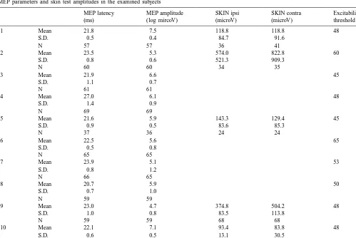
were analyzed after logarithmic transformation, in order to Table 1 summarizes latency, log amplitude values and achieve a better fit to normal distribution. MEP latencies excitability thresholds for each subject. The reported were measured at the onset of the initial reproducible statistics were computed without including data recorded
deflection. during maximal inspiratory effort. All values were
trans-Since a large inter-individual variability was observed, formed to obtain the z-scores.
Table 1
MEP parameters and skin test amplitudes in the examined subjects
MEP latency MEP amplitude SKIN ipsi SKIN contra Excitability
(ms) (log mircoV) (microV) (microV) threshold (%)
s1 Mean 21.8 7.5 118.8 118.8 48
S.D. 0.5 0.4 84.7 91.6
N 57 57 36 41
s2 Mean 23.5 5.3 574.0 822.8 60
S.D. 0.8 0.6 521.3 909.3
s5 Mean 21.6 5.9 143.3 129.4 45
S.D. 0.9 0.5 83.6 85.3
s9 Mean 23.0 4.7 374.8 504.2 48
S.D. 1.0 0.8 83.5 113.8
N 59 59 68 68
s10 Mean 22.1 7.1 93.4 83.8 48
S.D. 0.6 0.5 13.1 30.5
not significantly different between the group with a reliable number of SSRs and the other one (t-test, P50.595; see Table 1). Concerning the former group, neither the SSR presence–absence nor their amplitudes correlated with MEP parameters. In these subjects, no clear pattern of SSR changes was identifiable during the recording session, not allowing detecting an adaptation or a facilitation process as a function of the time. Therefore, the large standard deviation (shown in Table 1) could not be accounted for by any of the considered parameters. In addition, no signifi-cant SSR amplitude differences were observed between the side contralateral and ipsilateral to TMS. During forced maximal inspiration SSRs were significantly larger in amplitudes compared to those recorded in the physiologi-cal respiratory phases, but without any evident correlation with MEP latency.
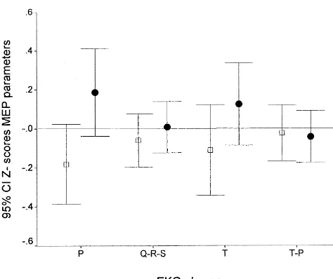
TMS did not induce any significant HR modification. In addition, the HR measured before and after each TMS Fig. 2. MEP latency (open squares) and amplitude (closed circles) as a
function of EKG phases (P, QRS, T, T–P interval). stimulus was constant throughout the experiment.
In addition, no significant changes could be observed in MEP parameters in the course of the recording session. MEP latency and amplitude did not show significant The lack of significance found in bivariate analyses was relationship with any of the EKG phases (F50.56; df5 also confirmed when simultaneous effects were taken into
5.524; P50.730 NS; see Fig. 2). account.
Similarly, physiological breathing phases (inspiration and expiration) did not influence MEP parameters. On the
other hand, during forced maximal inspiration a significant 4. Discussion MEP latency shortening was found (F514.3, df52.481
P#0.001; Fig. 2), without any related amplitude modi- Results of the present study show a lack of correlation fication (F51.3, df52.481 P50.256; Fig. 3). between MEP parameters and cardiac cycle, sudomotor SSRs were present in all the tested subjects. However in skin responses and respiratory phases. On the other hand, five of them SSR rapidly attenuated and disappeared. In during maximal inspiratory effort, a significant, but in-the remaining five subjects a reliable (.60%) number of dependent MEP latency decrement and SSR amplitude
SSRs to TMS was recorded. increment, were observed.
Intensities of TMS (and the related acoustic noise) were These data seem to suggest that visceral signals origina-ting from the mechanical cyclic impact of the heart action and chest expansion do not significantly influence motor excitability either at the cortical or at the spinal levels.
It is known that somatosensory mechanoreceptors in the chest pick up the cyclic impact of the heart muscle on the surrounding tissue and that mechanoreceptors, located near the large arteries, are stimulated by small translocations of tissues induced by the pulse wave [14]. All these signals, originating at different locations in the body, could flow synchronously with each heart action and have been associated to a heart action-related EEG potential in frontal and parietal regions [3]. It remains an open question whether the above mentioned visceral inputs can modulate the motor outputs.
Connections of the cerebral cortex with the heart, blood vessels and viscera are well known. Frontal lobe, insular cortex and limbic areas establish a rich mutual exchange of information with vegetative centers located in the brain stem and medulla [1,12,25]. Results of stimulation and Fig. 3. MEP latencies (open squares) and amplitudes (closed circles) as a
lesion studies suggest that insular cortex is involved in function of breathing phases (inspiratory, expiratory, maximal inspiratory
According to these results, repetitive TMS induced a independent of the cortical region and the brain hemi-clearly marked autonomic response with HR acceleration sphere underlying the stimulating coil.
and decrease in blood pressure in normal subjects [6]. In conclusion, our study suggests that the investigated At more fundamental anatomic levels, it has been shown autonomic parameters have a minor influence in modu-that noradrenergic fibers have a global extent in the animal lating motor cortical and / or spinal excitability. Although a cortex. They form through the neocortex a three dimen- large amount of data was collected for each subject and we sional matrix, where multiple contacts are possible, permit- never found a clear pattern of influence on motor ex-ting a synchronous modulation of afferent neuronal activity citability, a limit of the present study is the small number throughout this network [10]. of recruited subjects, which is a very critical point
espe-All the above observations suggest the potential for the cially for ‘negative’ results.
autonomic afferent fibers to modulate cortical and / or In addition, the approach adopted was limited to a spinal motor excitability. description of autonomic function in normal subjects, in a Our findings show a significant reduction of MEP state of equilibrium. For a more complete knowledge on latencies during forced inspirations. It has been previously this topic to occur, future research should include the study demonstrated that hyperventilation increases motor cortical of patients with selective autonomic disorders, in whom excitability [15,22], as measured with TMS, and that this the altered balance between somatic and visceral inputs effect is related to a reduction in pCO2. could reveal other contributions of autonomic signals to
On the other hand, in the present study, reduced levels cortical and spinal excitability. of pCO can not account for the finding of MEP latency2
decrement, since our subjects did not hyperventilate and it
is known that pCO becomes significantly lower only after2 References 5 min of hyperventilation [22].
Therefore, a possible explanation for the latency short- [1] A. Albanese, G. Macchi, Suprasegmental control of vegetative ening during forced inspirations could be referred to an nervous system, Funct. Neurol. 4 (1987) 407–416.
increase of afferent inputs from cardiopulmonary system to [2] E. Balzamo, G. Gayan-Ramirez, Y. Jammes, Pulmonary vagal sensory afferents and spontaneous EEG rhythms in the cat sen-the cerebral cortex and spinal motoneurons. In fact,
sorimotor cortex, J. Auton. Nerv. Syst. 30 (1990) 149–157. inflation of lungs is known to activate pulmonary stretch
[3] G. Dirlich, T. Dietl, L. Vogl, F. Strian, Topography and morphology receptors of tracheobronchial tree [24], that could target
of heart action-related EEG potentials, Electroencephalogr. Clin. the motor centers, influencing their excitability. In this Neurophysiol. 108 (1998) 299–305.
context, it has been demonstrated an interaction between [4] B. Elie, P. Guiheneuc, Sympathetic skin response: normal results in different experimental conditions, Electroencephalogr. Clin. Neuro-vagal afferent fibers and spontaneous
electroencephalog-physiol. 76 (1990) 258–267. raphic (EEG) activity of the sensorimotor cortex, evident
[5] P.H. Ellaway, N.J. Davey, D.W. Maskill, S.R. Rawlinson, H.S. during the mechanical activation of pulmonary afferents
Lewis, N.P. Anissimova, Variability in the amplitude of skeletal [2]. However, a spread of motor cortical excitability due to muscle responses to magnetic stimulation of the motor cortex in the voluntary act of inspiration, as well as the involvement man, Electroencephalogr. Clin. Neurophysiol. 109 (1998) 104–113. of unspecific factors, such as an arousal reaction, during [6] A. Foerster, J.M. Schmitz, S. Nouri, D. Claus, Safety of rapid-rate transcranial magnetic stimulation: heart rate and blood pressure self-paced maximal inspiration can not be excluded.
More-changes, Electroencephalogr. Clin. Neurophysiol. 104 (1997) 207– over, since maximum voluntary inspiration is a voluntary
212.
act, it might induce a transient spread of cortical excitabili- [7] S.I. Izumi, T.W. Findley, T. Ikai, J. Andrews, M. Daum, N. Chino, ty similar to the one induced by voluntary contraction of Facilitatory effect of thinking about movement on motor evoked muscles of the hand or foot opposite to the one where potentials to transcranial magnetic stimulation of the brain, Am. J.
Phys. Med. Rehab. 74 (1995) 207–213. MEP are recorded from [23].
[8] L. Kiers, D. Cross, K.H. Chiappa, J. Fang, Variability of motor In the present study, TMS was able to elicit a
sympa-potentials evoked by transcranial magnetic stimulation, Electroence-thetic skin reflex (SSR) in both contralateral and ipsilateral phalogr. Clin. Neurophysiol. 89 (1993) 415–423.
hands. This finding confirms previous reports showing an [9] R. Mariorenzi, F. Zarola, M.D. Caramia, C. Paradiso, P.M. Rossini, SSR induced by TMS of the motor area in normal subjects Non-invasive evaluation of central motor tract excitability changes following peripheral nerve stimulation in healthy humans, Elec-[4,18]. This response has been explained by the activation
troencephalogr. Clin. Neurophysiol. 81 (1991) 90–101. of cortical structures influencing palmar and generalized
[10] J.H. Morrison, M.E. Molliver, Noradrenergic innervation of cerebral sweating. Therefore, the significant SSR amplitude incre- cortex: widespread effects of local cortical lesions, Science 205 ment observed during phases of deep breathing probably (1979) 313–316.
reflects a combined summation of two stimuli (TMS and [11] L. Niehaus, B.U. Meyer, S. Roricht, Magnetic stimulation over different brain regions: no differential effects of the elicited sympa-deep breathing), each individually able to elicit this
thetic skin responses, Electroencephalogr. Clin. Neurophysiol. 109 response.
(1998) 94–99.
[13] S.M. Oppenheimer, G. Kedem, W.M. Martin, Left insular cortex Lucking, A.L. Martens de Noordhout, C.D. Marsden, N.M.F. lesions perturb cardiac autonomic tone in humans, Clin. Auton. Res. Murray, J.C. Rothwell, M. Swash, C. Tomberg, Non-invasive 6 (1996) 131–140. electrical and magnetic stimulation of the brain, spinal cord and [14] A.S. Paintal, Cardiovascular Receptors, in: E. Neil (Ed.), Handbook roots: basic principles and procedures for routine clinical
applica-of Sensory Physiology : Enteroceptors, Vol. 3 / 1, Springer Verlag, tion, Electroencephalogr. Clin. Neurophysiol. 91 (1994) 79–92. `
Berlin, 1972. [20] P.M. Rossini, F. Tecchio, A. Sabato, A. Finazzi-Agro, P. Pasqualetti, [15] A. Priori, A. Berardelli, B. Mercuri, M. Inghilleri, M. Manfredi, The S. Rossi, The role of cutaneous inputs during transcranial magnetic
effect of hyperventilation on motor cortical inhibition in humans: a stimulation, Muscle Nerve 19 (1996) 1302–1309.
study of electromyographic silent period evoked by transcranial [21] P.M. Rossini, S. Rossi, P. Pasqualetti, F. Tecchio, Corticospinal magnetic stimulation, Electroencephalogr. Clin. Neurophysiol. 97 excitability modulation to hand muscles during movement imagery,
(1995) 69–72. Cereb. Cortex 9 (1999) 161–167.
[16] S. Rossi, P. Pasqualetti, F. Tecchio, F. Pauri, P.M. Rossini, Cor- [22] M. Seyal, B. Mull, B. Gage, Increased excitability of the human ticospinal excitability modulation during mental simulation of wrist corticospinal system with hyperventilation, Electroencephalogr. movements in human subjects, NeuroSci. Lett. 243 (1998) 147–151. Clin. Neurophysiol. 109 (1998) 263–267.
[17] P.M. Rossini, M.T. Desiato, F. Lavaroni, M.D. Caramia, Brain [23] A. Stedman, N.J. Davey, P.H. Ellaway, Facilitation of human first excitability and electroencephalographic activation: non invasive dorsal interosseus muscle responses to transcranial magnetic stimu-evaluation in healthy humans via transcranial magnetic stimulation, lation during voluntary contraction of the contralateral homonymous Brain Res. 567 (1991) 111–119. muscle, Muscle Nerve 21 (8) (1998) 1033–1039.
[18] P.M. Rossini, R.J. Opsomer, P. Boccasena, Sudomotor skin re- [24] J.G. Widdicombe, Site of pulmonary stretch receptors in the cat, J. sponses following nerve and brain stimulation, Electroencephalogr. Physiol. 125 (1954) 336–351.
Clin. Neurophysiol. 89 (1993) 442–446. [25] Y. Yasui, C.D. Breder, C.B. Saper, D.F. Cecchetto, Autonomic [19] P.M. Rossini, A.T. Barker, A. Berardelli, M.D. Caramia, G. Caruso, responses and efferent pathways from the insular cortex in the rat, J.