Hippocampus of Individuals with Major Depression
John E. Piletz, He Zhu, Gregory Ordway, Craig Stockmeier, Ginny Dilly,
Donald Reis, and Angelos Halaris
Background: A downregulation of I2-imidazoline binding
sites has been reported in frontal cortices of depressed suicide victims, according to I2-radioligand binding and
confirmed by Western blotting. We now report Western blots of imidazoline receptor proteins in hippocampi of subjects with and without depression at the time of death. Methods: Postmortem diagnoses were obtained from 17 cases of Axis I major depressive disorder and 17 cases without Axis I psychopathology. No psychotropic com-pounds were found in body fluids. Hippocampi were removed, sectioned, and assessed histologically. Through-out the analysis, each major depressive disorder sample was paired with a sample from a psychiatrically healthy subject based on equivalent life spans and postmortem delays. The antiserum was identical to that used in previous studies that reported a downregulation of corti-cal 29/30-kd imidazoline receptor– binding proteins in depression.
Results: A triad of imidazoline receptor– binding protein bands (40 –50 kd) was detected in the human hippocam-pus. Subjects with major depressive disorder had signifi-cantly less intensity in each imidazoline receptor– binding proteins band compared with control subjects (p5.01 for overall bands).
Conclusions: The present results can be aligned with previous reports of downregulation of I2-radioligand
binding sites in both cortices and platelets of depressed patients. Biol Psychiatry 2000;48:910 –919 © 2000 So-ciety of Biological Psychiatry
Key Words: Imidazoline receptors, monoamine oxidases,
immunoreactivity, hippocampus, major depression, suicide
Introduction
I
midazoline receptors (more conservatively called I-sites) and a2-adrenoceptors (a2ARs) are distinct mem-brane proteins that share high affinity for imidazoline compounds such as clonidine and idazoxan (Ernsberger et al 1995). I-sites are not adrenergic in nature because they lack appreciable (nanomolar) affinities for all known monoamines. There are two subtypes of I-sites (I1and I2),and these are differentially distributed throughout the brain (DeVos et al 1994). Recently, an imidazoline recep-tor– binding protein (IRBP) was cloned from human hip-pocampus and shown to have I1-like properties (Ivanov et
al 1998b; Piletz et al 2000). The function of the hippocam-pal IRBP remains controversial (Guyenet 1997), but stud-ies of brainstem nuclei have linked an I1site to the central
control of blood pressure (Bousquet et al 1984; Molder-ings 1997).
Additionally, I2 sites are of interest to psychiatry
be-cause they are physically associated with monoamine oxidases A (MAO-A) and B (MAO-B; Tesson et al 1995). An I2 binding site has been localized within a 73 amino
acid sequence (K149 –M222) of MAO-B (Raddatz and Lanier 1997; Raddatz et al 1997). This I2site lies outside
the catalytic domain proposed for MAO-B enzymatic inhibition, but a point mutation (T1583A of MAO-B)
within the same region (K149 –M222) causes complete loss of MAO-B activity (Cesura et al 1996). On the other hand, not all I2 sites are accounted for by MAO-A or
MAO-B molecules. In fact, a relatively small subpopula-tion of MAO-A or MAO-B molecules is actually accessi-ble for I2radioligand binding, and this varies in a
tissue-dependent manner (Raddatz and Lanier 1997; Raddatz et al 1997). The source and significance of this subpopula-tion of MAO molecules remains unknown.
Both I1 and I2 sites have been found to be altered in
depressed patients (Piletz et al 1994; Sastre et al 1995; Sastre and Garcia-Sevilla 1997), as have a2AR agonist
binding sites for clonidine (Callado et al 1998). First, an increase in radioligand binding density (BMAX) of I1sites
on platelet plasma membranes of depressed patients was reported (Piletz et al 1994, 1996a, 1996b) relative to From the Departments of Psychiatry and Human Behavior (JEP, HZ, GO, CS, AH),
Pharmacology and Toxicology (JEP, GO), and Physiology and Biophysics (JEP), University of Mississippi Medical Center, Jackson; the Department of Psychiatry, Case Western Reserve University, College of Medicine, Cleveland, Ohio (GD); and Division of Neurobiology, Department of Neurology and Neurosciences, Cornell University Medical College, New York, New York (DR).
Address reprint requests to John E. Piletz, Ph.D., Department of Psychiatry, University of Mississippi Medical Center, Rm. G128, 2500 North State Street, Jackson MS 39216.
Received August 23, 1999; revised March 21, 2000; accepted March 23, 2000.
© 2000 Society of Biological Psychiatry 0006-3223/00/$20.00
healthy control subjects. Then, I2sites were shown to be
decreased in platelet internal membranes of depressed patients (Piletz et al 1994) and in frontal cortices of suicide victims (Sastre et al 1995; Sastre and Garcia-Sevilla 1997) compared with control subjects. Furthermore, studies with radiolabeled clonidine (or its analogs) have consistently revealed an up-regulation ofa2AAR agonist binding sites in platelets of depressed patients (Garcia-Sevilla et al 1981; Piletz et al 1994) and in some brain regions of suicide victims (Callado et al 1998; Meana et al 1992; Ordway et al 1994). Thus, numerous studies have now revealed alterations in I-sites,a2AAR agonist sites, or both
in depression.
The first purification of an IRBP was reported by Wang et al (1992) using bovine adrenal chromaffin cells. A human hippocampal homologue of this protein was sub-sequently cloned by Ivanov et al (1998b; Piletz et al 2000). Based on retentions of the bovine IRBP by two affinity chromatography resins and selective elutions from those columns by an imidazoline displacing agent, this protein was reported (Wang et al 1992) to possess a combination of I1and I2binding properties. This protein was further
shown to run as a single band on a SDS gel with a molecular weight (MW) of 70 kd (Wang et al 1992). The same protein was later used as an immunogen to produce polyclonal antiserum, designated IRBP antiserum (Wang et al 1993), which we have used in our study.
Several related polypeptides react with IRBP antiserum on Western blots. These include its progenitor 70-kd immunogen from bovine adrenal chromaffin cell mem-branes (Wang et al 1993), an 85-kd protein from freshly prepared rat brain membranes (Ivanov et al 1998a, 1998c), and several breakdown polypeptides (Ivanov et al 1998c). Correlations have been established between the intensities of doublet 29/30-kd IRBP bands versus Bmaxvalues for I2
sites across several rat and human tissues and conditions (Escriba et al 1994; Garcia-Sevilla et al 1995). Con-versely, 33- and 45-kd bands detected by IRBP antiserum have been correlated with I1 sites in human tissues
(Garcia-Sevilla et al 1999; Ivanov et al 1998a). IRBP antiserum fails to cross-react with MAO-A, MAO-B, or any a2AR subtypes (Escriba et al 1999; Ivanov et al 1998a). In summary, there appear to be multiple subtypes of I-sites:
1. One I2 sites represents a subpopulation of MAO
molecules encoded by the K149 –M222 sequence within MAO-B, but it cannot be detected by IRBP antiserum.
2. Another I2 site is related to a 29/30-kd doublet
protein in brain and to a 70-kd protein in bovine adrenal chromaffin cells according to cross-reactiv-ity with IRBP antiserum.
3. An I1-like site is related to a 45-kd protein in brain
according to cross-reactivity with IRBP antiserum. 4. Another I1-like site is related to a 33-kd protein in
platelets according to cross-reactivity with IRBP antiserum (Ivanov et al 1998a).
We have proposed the latter three subtypes may be breakdown products of a common 85-kd precursor protein (Ivanov et al 1998c). They may also all be derived from the same cloned hippocampal IRBP (Piletz et al 2000).
In our study, IRBP antiserum has been used to quantify Western blots of hippocampi from suicide subjects who were retrospectively diagnosed with MDD at the time of death, compared with paired control subjects who lacked a major (Axis I) psychiatric diagnosis. Based on the previ-ous studies of Garcia-Sevilla et al (1996), we hypothesized that hippocampi from depressed suicide victims might be low in 29/30-kd IRBP bands relative to control subjects, but a 45 kd band might be increased in MDD subjects compared with control subjects.
Methods and Materials
Brain Tissue
Human brains were obtained at the time of autopsy by the Medical Examiner’s Office of Cuyahoga County, Ohio, in accordance with an approved institutional review board protocol. Cadavers were immediately refrigerated when arriving at the medical examiner’s office. The coroner determined the cause of death. Brain sections were dissected, coded to protect the subject’s identity, and frozen (282°C) in tightly sealed contain-ers. Samples from both groups of subjects were stored for comparable lengths of time before thawing.
Information on lifetime events and the recent (within the last month of life) psychiatric status of all subjects was obtained from next-of-kin during structured clinical interviews by a trained interviewer. The interviews were according to the Schedule for Affective Disorders and Schizophrenia: Lifetime Version (SADS-L) supplemented by questions from the Diagnostic In-terview Schedule (DIS-III-R) to make diagnoses compatible with the DSM-IV (Rush and Weissenburger 1994). The SADS has obtained adequate validity when comparing the patient report with that of an informant (Andreasen et al 1977). Evaluations of drug and alcohol abuse and dependency were assessed using the DIS-III-R (Kelly et al 1998). Axis I diagnoses were made by a psychiatrist and a clinical psychologist, based on the data gathered from the structured interview and, when available, hospital and medical records.
depen-dence, and two had histories of alcohol abuse (Table 1). Subjects in the control group consisted of 4 females and 13 males, and the causes of death in this group were: cardiovas-cular failure (n511), gunshot (n51), pulmonary embolism (n 5 1), aneurysm (n 5 1), pancreatitis (n 51), lightning strike (n51), and bike accident (n51). All control subjects were assessed retrospectively through structured interviews with family members and had no active major psychiatric diagnoses (Axis I; DSM-IV) at the time of death. Among the control subjects, one had experienced an episode of adjust-ment disorder with depressed mood 5 months before death, and one had a history of alcohol abuse 7 years before death. A toxicologic screen of blood, bile, and urine from all subjects was performed at the county coroner’s office. Qualitative and quantitative analyses were performed to detect the following compounds or classes of compounds: ethanol, barbiturates, benzodiazepines, sympathomimetic drugs, and antidepressant and antipsychotic drugs and their metabolites. The toxicology results from MDD subjects are shown in Table 1. Postmortem records revealed that within the month before death, three MDD subjects had received antidepressant drug prescriptions (one for fluoxetine, one for nortriptyline, and one for sertraline); however, these substances were not found in body fluids at the time of death (an exclusion criteria was evidence of antidepressants or antipsychotics in the toxicologic screens). The toxicologic screens of six control subjects revealed the following: two subjects had low but detectable levels of ethanol, one had ethanol plus a low level of cocaine, one had flurazepam, one had phenobarbital plus phenytoin, and one had diazepam plus co-deine in the blood. They were not excluded from the study.
Dissection
At the time of autopsy, the brain was dissected into small tissue blocks. Particular care was taken to maintain gross morphology during freezing. Tissue containing the right hippocampus was placed on hard cardboard and then dipped into isopentane (250°C) for 5 sec. Blocks of hippocampus were placed on dry ice for 10 min, and then stored at282°C. Frozen blocks were then mounted on a specimen chuck of a cryostat microtome (Leica, Cryocut 1800, Reichert-Jung, Deerfield, IL). All brain tissue surrounding Ammons horn of the hippocampus was dissected away with a razor blade. Tissue sections through Ammons horn (including the dentate gyrus) were cut at216°C, and sections (60mm thick) from each subject were place in an ice-cold microcentrifuge tube. The adjacent section (40 mm thick) was cut for morphology, dried at room temperature, and stained with cresyl violet. Histologic sections were used to confirm anatomic equivalence of the hippocampal sections for all MDD subjects and their paired control subjects.
Immunodetection of Imidazoline Receptor Proteins
Frozen hippocampal sections were sonicated for 5 sec in ice-cold 220 mL of 5 mmol/L Tris-HCl buffer, pH 7.4, containing 0.25 mol/L sucrose and 1 mmol/L MgCl, plus a previously described cocktail of eight protease inhibitors (Ivanov et al 1998c). Aliquots were assayed for total protein according to the Lowry-Biuret reagent kit from Sigma (St. Louis). Remaining homoge-nates were flash frozen. Samples were thawed and diluted to 0.8
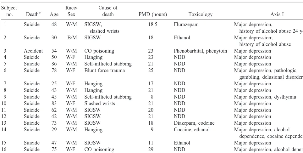
mg protein/mL with ice-cold 40 mmol/L Tris-HCl buffer, pH 6.8, plus 4% SDS. Samples (12mg) were denatured and electropho-Table 1. Demographic Information for Subjects with Major Depression
Subject
no. Deatha Age
Race/ Sex
Cause of
death PMD (hours) Toxicology Axis I
1 Suicide 48 W/M SIGSW,
slashed wrists
18.5 Flurazepam Major depression,
history of alcohol abuse 24 years ago
2 Suicide 30 B/M SIGSW 18 Ethanol Major depression;
history of alcohol abuse
3 Accident 54 W/M CO poisoning 23 Phenobarbital, phenytoin Major depression
4 Suicide 50 W/F Hanging 23 NDD Major depression
5 Suicide 86 W/M Self-inflicted stabbing 21 NDD Major depression
6 Suicide 78 W/F Blunt force trauma 25 NDD Major depression, pathologic
gambling, delusional disorder
7 Suicide 25 W/F Hanging 17 NDD Major depression
8 Suicide 43 W/M Hanging 21 NDD Major depression
9 Suicide 45 W/M Self-inflicted stabbing 8 NDD Major depression, dysthymia
10 Suicide 83 W/F Slashed wrists 21 NDD Major depression
11 Suicide 62 W/M SIGSW 20 NDD Major depression
12 Suicide 42 W/M SIGSW 21 NDD Major depression
13 Suicide 73 W/M SIGSW 18 Diazepam, codeine Major depression
14 Suicide 29 W/M Hanging 9 Cocaine, ethanol Major depression, alcohol
dependence, cocaine dependence
15 Suicide 47 W/M SIGSW 11 Ethanol Major depression
16 Suicide 75 W/F CO poisoning 29 NDD Major depression, alcohol dependence
17 Suicide 68 W/M CO poisoning 4 NDD Major depression
resed through two identical 13.7% polyacrylamide gels contain-ing SDS (SDS-PAGE) accordcontain-ing to our previous studies (Ivanov et al 1998c). Platelet total membrane proteins (1.0, 2.5, and 5.0
mg) were prepared from a common bag of platelet-rich plasma (Mississippi Blood Services, Jackson, MS) and run as standards on each gel. The proteins were electrotransferred onto nitrocel-lulose membranes (Hybond ECL, Amersham, Arlington Heights, IL). After electrotransfer, the blots were blocked with milk and incubated with IRBP antiserum (1:3000 dilution in TBST/10% milk) at room temperature (Ivanov et al 1998c). Using an Amersham ECL detection system, IRBP-immunoreactive bands were detected by exposure to film for 2 to 6 min (Amersham ECL Hyperfilm). To detectb-actin as a standard, the same blots were stripped (Ivanov et al 1998c) and incubated with a 1:9000 dilution of b-actin antiserum (Chemicon International, Te-mecula, CA). Detection by ECL was performed identically with
b-actin antiserum except using 1:3000 dilution of anti-mouse IgG antibody (Amersham), and the films were exposed for 30 sec to develop the bands. Samples from subjects with MDD were loaded beside paired control subjects, and each sample was run into two SDS-PAGE gels (duplicate Western blots).
A Microcomputer Controlled Imaging Device (MCID model M2; Imaging Research, St. Catharines, Canada) was used for densitometry. Optical densities (ODs) of sample bands were compared with those of standard platelet membranes on each blot. A standard curve from each blot assured that sample OD values were within the linear response range for each film and therefore useful for comparison to previous publications (Ivanov et al 1998c). We standardized OD values using an optical density step-wedge (Imaging Research). Coomassie blue staining of duplicate gels was also quantified using a Molecular Dynamics Personal Densitometer (Molecular Dynamics, Sunnyvale, CA). Coomassie blue intensity provided verification that comparable amounts of protein were loaded onto each lane and that there was sample integrity (i.e., no generalized proteolysis). Finally, OD values were obtained from the Western blots forb-actin and used to normalize IRBP OD values (subject by subject). As with IRBP, the OD values of b-actin bands were derived from two blots, and the values were averaged.
Note on IRBP Antiserum
The IRBP antiserum used herein is identical to that described repeatedly by Garcia-Sevilla’s group (e.g., Garcia-Sevilla et al 1996). Despite that group’s emphasis on a 45-kd band in human brain, both our laboratory and their laboratory have obtained comparable results when analyzing fresh rat cerebral cortex. With rat cortex, their method yielded 29/30-kd (60 – 63%), 45-kd (31–33%) and 84/85-kd (4 –9%) bands, compared to our meth-od’s 29/30-kd (47–56%), 45-kd (39 – 48%), and 84/85-kd (5– 6%) bands (Garcia-Sevilla, personal communication, 1996). Thus, our study and theirs (Garcia-Sevilla et al 1996) are likely to differ only in tissue sampling, rather than methodologically (also see Discussion).
Statistics
Statistical analyses were performed using GraphPAD InStat (GraphPAD Software, San Diego). All results are expressed as
mean 6 SEM. Student’s paired t tests (two-tailed) were per-formed on the raw data and on their logarithms. The test of significance was p # .05. A paired t test was used because samples from MDD subjects were paired for age and postmortem interval and because the pairs were processed together and run side by side on the same gels.
Results
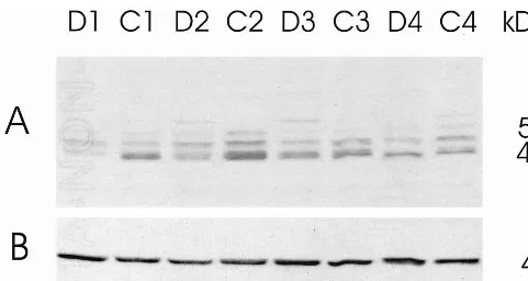
Western blots revealed at least three IRBP-immunoreac-tive bands in the human hippocampus. These bands ranged in size between 40 and 50 kd (Figure 1A). In some subjects, the smaller of this triad (40 kd) appeared as a closely aligned doublet, but it could not be fully resolved (Figure 1A, section C2). In a few subjects, there was an additional band of '55 kd (Figure 1A, sections D2, D3, C4). By extending the ECL reaction time to 15 hours and redeveloping the films, it was also possible to visualize a weak 30-kd band and an even weaker 33-kd band similar to that found in platelets (data not shown). The OD values for the 30- to 33-kd bands were close to background levels and therefore deemed unreliable. After stripping the blots of IRBP antiserum, a sharp band was also detected with
b-actin antiserum at MW543 kd (Figure 1B). Densitom-etry was performed on all prominent bands (individually). Although the 40-kd IRBP-immunoreactive band appeared as a doublet in some cases, it was traced as a singlet to maintain uniformity across samples.
The IRBP-immunoreactive bands were normalized per
b-actin protein in each sample as a means of internally correcting for slight variances in protein loading. No significant differences inb-actin were observed between MDD subjects compared with matched control subjects (p5.390, paired t test, two-tailed). Further verification of the uniformity of protein loading was seen on gels stained with Coomassie blue. Each lane was almost identical in Coomassie staining (data not shown).
Western blots revealed a significant decrease in the ratio of IRBP/b-actin in IRBP bands between 40 and 50 kd in MDD subjects compared with paired control subjects (Figure 2). Greater statistical significance was achieved using log-transformed values, indicating that relative dif-ferences were more consistent than absolute difdif-ferences. From higher MW to lower MW, the significance levels for log-transformed IRBP data were as follows (by paired t tests, two-tailed): 50-kd band, p5.007; 45-kd band, p5
together, an overall decrease was verified in depressed patients compared with matched control subjects (p5.01 for log-transformed data; Figure 2D). Thus, 14 of the 17 pairs of subjects had total IRBP immunodensities lower in those with MDD than in the paired control subjects (Figure 2D).
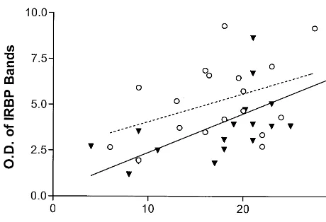
Finally, postmortem delay was found to be a significant covariable (Table 1). All IRBP bands, regardless of diagnos-tic group, were found positively correlated in intensity with postmortem delay (Figure 3). From higher MW to lower MW, the p values and r2 values for correlating IRBP immunodensities with postmortem delays were as follows: 50-kd band, p5.0430, r25.115; 45-kd band, p5.0030, r25.229; and 40-kd band, p,.0001, r25.407. The result of pooling all three bands together is shown in Figure 3 (p5
.0010, r25.264). The effects of postmortem delay were not significantly different between diagnostic groups (i.e., nearly identical slopes).
Discussion
In our study, the ODs of IRBP-immunoreactive bands were found decreased by '25% in the hippocampi of subjects with MDD compared with paired control subjects
lacking a psychiatric diagnosis (Figures 1 and 2). This finding was significant using untransformed data (Figure 2), but for 45-kd and 50-kd bands, it was even more statistically significant when comparing log-transformed values. There was also an effect of postmortem delay to increase IRBP bands (Figure 3), but because both patients and control subjects had identical postmortem delays, this did not change the conclusion. Thus, imidazoline receptors appear to be less abundant in the hippocampi of depressed suicide victims compared with control subjects matched for age and postmortem delay.
Previous studies have shown that IRBP-immunoreac-tive bands are susceptible to proteolysis even in fresh brain tissue (Ivanov et al 1998c) and cultured cells (Ivanov et al 1998a). Therefore, a critical aspect of our experimental design was pairing of subjects with MDD and control subjects for identical postmortem delays. Our samples were homogenized in a cocktail of eight protease inhibi-tors to avoid proteolysis (Ivanov et al 1998a). Nonetheless, much intersubject variability in the intensity of IRBP bands could be due to postmortem delay (Table 1 and Figure 3).
(Piletz et al 1990, 1996a, 1996b) have been consistent in revealing an elevation in the density of platelet I1binding
sites in depressed patients compared with healthy control subjects. Normalization (downregulation) of platelet I1
binding sites also has been documented 6 weeks after antidepressant treatment with either desipramine or fluox-etine (Piletz et al 1996a, 1991, 1996b). Rats injected with imipramine for 3 weeks also have been shown to down-regulate brainstem I1 binding sites (Zhu et al 1997).
Reports also exist that IRBP-immunodensities of platelet 33-kd bands (Zhu et al 1999) and 45-kd bands (Garcia-Sevilla et al 1996) are elevated in depressed patients. Various antidepressants can also downregulate the IRBP-immnoreactive bands in platelet membranes of depressed patients after antidepressant treatments (Garcia-Sevilla et al 1996; Zhu et al 1999). In one study, Garcia-Sevilla’s group reported (1996) a 45-kd band in frontal cortices
from suicide victims was increased compared with sud-den-death control subjects lacking a history of psychopa-thology. Thus, an abundance of evidence has accumulated that I1 sites, in conjunction with 33- and 45-kd
IRBP-immunoreactive bands, are elevated in depressed patients. Nonetheless, previous evidence also has suggested that I2sites are changed in a reciprocal fashion with I1sites in
depression. Research findings support a lower density of I2
binding sites in depressed patients, with a normalization (up-regulation) following treatment with fluoxetine (Piletz et al 1994). Reciprocity with I1sites was also observed in
a 25-day treatment study of rats with imipramine, which led to up-regulation of I2 binding sites in the midbrain
(Zhu et al 1996). Furthermore, 29/30-kd doublet bands associated with I2sites were reported (Escriba et al 1994)
to be downregulated in frontal cortices of suicide victims, whereas a 45-kd band related to I1sites was increased in
suicide victims relative to control subjects (Garcia-Sevilla et al 1996). None of these findings were directly related to MAO-B because MAO-B sites labeled by [3 H]-lazabe-mide were not altered in depressed suicide victims (Sastre and Garcia-Sevilla 1997). Thus, a number of previous studies have demonstrated reciprocal changes in densities of I1 sites (higher) and of I2 sites (lower) in depressed
patients.
In our opinion, the results under discussion align closest with two previous studies of I2sites by Garcia-Sevilla et al
(1996; Sastre and Garcia-Sevilla 1997). They reported 19% lowered IRBP-immunoreactivity for a 29/30-kd dou-blet in frontal cortices (Broadmann’s area 9) of suicide victims compared with sudden-death controls. [3 H]-ida-zoxan binding to I2 sites in frontal cortices of suicide
victims was also reported (Sastre and Garcia-Sevilla 1997) to be lowered (240%) in suicide victims compared with sudden death control subjects. In human cortex, the 29/30-kd doublet is associated with I2 sites labeled by
[3H]-idazoxan as a function of human aging (parallel changes; Garcia-Sevilla et al 1995). Furthermore, our results compare favorably with our earlier study (Piletz et al 1994) wherein lowered (243%) I2radioligand binding
sites on internal membranes of platelets were found in depressed patients; the platelet I2site also normalized (i.e.,
upregulated) following 6 weeks of treatment of depressed patients with fluoxetine (Piletz et al 1994). Thus, our present results seem in best agreement with previous studies of I2sites in depression (Garcia-Sevilla et al 1996;
Piletz et al 1994; Sastre and Garcia-Sevilla 1997). The 45-kd IRBP band may be of particular interest
because Greney and coworkers (Greney et al 1997; Vonthron et al 1998) identified a 45-kd protein as a candidate for the I1receptor protein. Moreover,
Garcia-Sevilla et al (1996) reported an elevation in the immuno-reactivity (151%) of a 45-kd IRBP band in frontal cortices of depressed suicide victims. Furthermore, plate-let 33-kd (Zhu et al 1999) and 45-kd IRBP bands (Garcia-Sevilla et al 1996) have been reported to be higher (140%) in depressed patients relative to healthy control subjects. In this light, the middle band in our hippocampal triad of IRBP bands (Figure 1) is identically sized as the 45-kd candidate I1protein found in frontal cortex
(Garcia-Sevilla et al 1996). Thus, we cannot exclude that I1sites,
rather than or in addition to I2sites, might be the subtype
that is decreased in the hippocampi of depressed patients. A variety of speculations might be considered to fully reconcile our findings with those of Garcia-Sevilla and colleagues ( 1996). Modern theories about psychiatric illnesses have invoked “neuronal circuitry” mechanisms, rather than unidirectional alterations in neurotransmitter concentrations or in receptor regulations in all brain regions. One relevant example comes from postmortem studies of schizophrenic brains, where completely oppo-site changes in dopaminergic neuronal density have been observed when comparing prefrontal cortices to hip-pocampi from the same schizophrenic subjects (Selemon and Goldman-Rakic 1999). Similar to the reciprocal change noted for neurons in different brain regions in schizophrenia, IRBP regulation might also be expressed reciprocally in prefrontal cortex versus hippocampus in depression. If this hypothesis proves true, then levels of the same IRBP (i.e., the 45-kd IRBP) could be increased in the prefrontal cortex (Garcia-Sevilla et al 1996) and decreased in the hippocampus of depressed patients (the study under discussion).
Our criterion for subject selection was more rigorous than that of Garcia-Sevilla and coworkers ( 1996). The psychiatric status of our subjects before death was evalu-ated via postmortem interviews with next of kin to establish firm retrospective diagnoses (Table 1). All de-pressed subjects in our study had experienced an active phase of MDD at the time of their deaths. By contrast, only 4 of 13 suicide victims in the study of Garcia-Sevilla et al (1996) had any historic evidence of MDD. Secondly, the average postmortem delay in our study was 17 6 1 hour (identical for both groups of subjects); however, the postmortem delays in the study of Garcia-Sevilla et al (1996) were 2663 hours for suicide victims and 3064 hours for the control subjects. Blood, urine, and bile were also analyzed in our study at the time of autopsy to confirm the absence of antidepressants or antipsychotic compounds (Table 1). No biochemical screening of psy-choactive substances was reported by Garcia-Sevilla et al
Figure 3. Correlation between the sum of imidazoline receptor bands in the hippocampus (Figure 2, panel D) and postmortem delays. Seventeen pairs of subjects were studied with identical postmortem delays: with major depressive disorder at the time of death (, MDD, solid regression line) compared with matched
control subjects (V, CTRL, dashed regression line). Overall p5 .001 and r2
(1996), except for ethanol. Ethanol was found in 8 of 13 suicide victims and 9 of 11 control subjects in the study of Garcia-Sevilla et al (1996). This high incidence of ethanol could have caused changes in IRBP in the study by Garcia-Sevilla et al. It should also be noted that neither of our studies were able distinguish whether changes in IRBP bands were due to the depressive illness itself or to committing suicide.
Our laboratory has reported (Ivanov et al 1998a) that two endogenous brain substances, agmatine and norepi-nephrine, are capable of up-regulating IRBP bands when these agents are applied to cultured cells. We also recently reported (Halaris et al 1999) an elevation in plasma agmatine concentration in depressed patients. Agmatine is a metabolite of arginine, formed in the brain by arginine decarboxylase (Reis and Regunathan 1998). Agmatine competes for radioligand binding to I-sites (Li et al 1994; Piletz et al 1995). In rats’ hippocampi the concentration of agmatine was reported (Feng et al 1997) to average 2857 pmol/g wet weight, which is well in excess of its affinity constant (KiHrange of two studies533–127 nmol/L) at
the high-affinity state of I1binding sites (Piletz et al 1995).
Furthermore, following a 6-hour treatment with 10
mmol/L agmatine, IRBP immunodensity in a human cell line (MEG-01) was reported (Ivanov et al 1998a) to be upregulated on cell membranes (i.e., ligand-induced up-regulation). Conceivably, if hippocampal concentrations of agmatine were low in patients with MDD, this might provide a mechanism to downregulate IRBP immunoreac-tivity (Halaris et al 1999).
Endogenous norepinephrine (NE) has also been pro-posed as a regulator of I-sites in vivo (Piletz et al 1998). In healthy women of reproductive age, a positive correlation was found between steady-state plasma NE concentrations and platelet I1 binding site densities (Piletz et al 1998).
Moreover, in a study of tissue culture effects (Ivanov et al 1998a), IRBP-immunoreactivity was upregulated in re-sponse to treating cultured cells with 10mmol/L NE for 6 hours. Thus, changes in brain NE, as well as agmatine, could underlie the observed downregulation of IRBP in postmortem hippocampi of depressed subjects.
Finally, the possible physiologic significance of a downregulation of imidazoline receptors in the hippocam-pus of depressed patients should be discussed. Beyond some role in the pharmacologic modulation of blood pressure via brainstem nuclei (Molderings 1997), nothing is certain about the function of imidazoline receptors in other brain regions. Although a subpopulation of MAO-B proteins possess I2binding sites (Tesson et al 1995), it is
not known if other I-sites also have a relationship with enzymes that metabolize monoamines. Neuronal inhibi-tion of NE release in pulmonary arteries is also ascribed to a peripheral subtype of imidazoline receptors, but this site
may possess neither I1 nor I2 pharmacologic properties
(Fuder and Schwarz 1993; Molderings and Gothert 1995). It is also believed that I1 sites reside on neurons
(Erns-berger et al 1995). A deficiency in, or dysregulation of, monoaminergic neurons in the central nervous system remains the prevailing hypothesis for the etiology of depression. Thus, tentative associations of I-sites with monoaminergic neurons might signify some role for imi-dazoline receptors in the pathophysiology or treatment of depression.
This project was supported in part by NIH Grant Nos. MH57601 (AH), MH46922 (GO), MH45488 (CS), and MH65183 (JP). We thank Dr. Herbert Meltzer and Dr. James Overholser for their expert assistance during the postmortem diagnoses at Case Western Reserve University, Cleveland, Ohio. We also appreciate Mr. Josh Farley and Dr. Violetta Klimek (University of Mississippi Medical Center) for help in sectioning tissues and with densitometric measurements.
References
Andreasen NC, Endicott J, Spitzer RL, Winokur G (1977): The family history method using diagnostic criteria: Reliability and validity. Arch Gen Psychiatry 34:1229 –1235.
Bousquet P, Feldman J, Schwartz J (1984): Central cardiovas-cular effects of a adrenergic drugs: Differences between catecholamines and imidazolines. J Pharmacol Exp Ther 230:232–236.
Callado LF, Meana JJ, Grijalba B, Pazos A, Sastre M, Garcia-Sevilla JA (1998): Selective increase of a2A-adrenoceptor agonist binding sites in brains of depressed suicide victims.
J Neurochem 70:1114 –1123.
Cesura AM, Gottowik J, Lahm H-W, Lang G, Imhof R, Mal-herbe P, et al (1996): Investigation on the structure of the active site of monoamine oxidase B by affinity labelling with the selective inhibitor lazabemide and by site-directed mu-tagenesis. Eur J Biochem 236:996 –1002.
DeVos H, Bricca G, DeKeyser J, DeBacker J, Bousquet P, Vauquelin G (1994): Imidazoline receptors, non-adrenergic idazoxan binding sites, and a2 adrenoceptors in the human central nervous system. Neuroscience 59:589 –598.
Ernsberger P, Graves ME, Graff LM, Zakieh N, Nguyen P, Collins LA, et al (1995): I1-Imidazoline receptors: Definition,
characterization, distribution and transmembrane signalling.
Ann N Y Acad Sci 763:22– 42.
Escriba PV, Ozaita A, Garcia-Sevilla JA (1999): Pharmacologic characterization of imidazoline receptor proteins identified by immunologic techniques and other methods. Ann N Y Acad
Sci 881:8 –25.
Escriba PV, Sastre M, Wang H, Regunathan S, Reis DJ, Garcia-Sevilla JA (1994): Immunodetection of putative imi-dazoline receptor proteins in the human and rat brain and other tissues. Neurosci Lett 178:81– 84.
Feng Y, Halaris AE, Piletz JE (1997): Determination of agmatine in brain and plasma using high-performance liquid chroma-tography with fluorescence detection. J Chromatogr B
Fuder H, Schwarz P (1993): Desensitization of inhibitory prejunctionala2-adrenoceptors and putative imidazoline
re-ceptors on rabbit heart sympathetic nerves. Naunyn Schmiedebergs Arch Pharmacol 348:127–133.
Garcia-Sevilla J, Escriba P, Guimon J (1999): Imidazoline receptors and human brain disorders. Ann N Y Acad Sci 881:392– 409.
Garcia-Sevilla JA, Escriba PV, Sastre M, Walzer C, Busquets X, Jaquet G, et al (1996): Immunodetection and quantitation of imidazoline receptor proteins in platelets of patients with major depression and in brains of suicide victims. Arch Gen
Psychiatry 53:803– 810.
Garcia-Sevilla JA, Sastre M, Escriba PV (1995): Age-dependent increases of immunoreactive imidazoline receptors in the human brain: Possible association of a 29/30 kD protein with the I2-imidazoline receptor identified by [
3
H]idazoxan.
Neu-rosci Lett 184:133–136.
Garcia-Sevilla JA, Zis AP, Hollingsworth PJ, Greden JF, Smith CB (1981): Plateleta2adrenergic receptors in major
depres-sive disorder. Arch Gen Psychiatry 38:1327–1333.
Greney H, Dontenwill M, Vonthron C, Bousquet P (1997): Further biochemical characterization of imidazoline binding sites from the human brainstem. Fundam Clin Pharmacol 11:63– 67.
Guyenet PG (1997): Is the hypotensive effect of clonidine and related drugs due to imidazoline binding sites? Am J Physiol 273:R1580 –R1584.
Halaris A, Zhu H, Feng Y, Piletz JE (1999): Plasma agmatine and platelet imidazoline receptors in depression. Ann N Y
Acad Sci 881:445– 451.
Ivanov T, Feng Y, Wang H, et al (1998a): Imidazoline receptor proteins are regulated in platelet-precursor MEG-01 cells by agonists and antagonists. J Psychiatr Res 32:65–79. Ivanov TR, Jones JC, Dontenwill M, Bousquet P, Piletz JE
(1998b): Characterization of a partial cDNA clone detected by imidazoline receptor-selective antisera. J Auton Nerv Syst 72:98 –110.
Ivanov TR, Zhu H, Regunathan S, Reis DJ, Dontenwill M, Vonthron C, et al (1998c): Co-detection by two imidazoline receptor protein antisera of a novel 85 kilodalton protein.
Biochem Pharmacol 55:649 – 655.
Kelly B, Raphael B, Judd F, Perdices M, Kernutt G, Burrows GD, et al (1998): Psychiatric disorder in HIV infection. Aust
N Z J Psychiatry 32:441– 453.
Li G, Regunathan S, Barrow CJ, Eshraghi J, Cooper R, Reis DJ (1994): Agmatine: An endogenous clonidine-displacing sub-stance in the brain. Science 263:966 –969.
Meana JJ, Barturen F, Garcia-Sevilla JA (1992): Alpha2
-adreno-ceptors in the brain of suicide victims: Increased receptor density associated with major depression. Biol Psychiatry 31:471– 490.
Molderings GH, Gothert M (1995): Inhibitory presynaptic imi-dazoline receptors on sympathetic nerves in the rabbit aorta differ from I1 and I2-imidazoline binding sites. Naunyn Schmiedebergs Arch Pharmacol 351:507–516.
Molderings GJ (1997): Imidazoline receptors: Basic knowledge, recent advances and future prospects for therapy. Drug
Future 22:757–772.
Ordway GA, Widdowson PS, Streator-Smith K, Halaris A
(1994): Agonist binding toa2adrenoceptors is elevated in the
locus coeruleus from victims of suicide. J Neurochem 63: 617– 624.
Piletz JE, Andrew M, Zhu H, Feng YZ, Rains J, Halaris A (1998): Alpha2-adrenoceptors and I1-imidazoline binding
sites: Relationship with catecholamines in women of repro-ductive age. J Psychiatr Res 32:55– 64.
Piletz JE, Chikkala DN, Ernsberger P (1995): Comparison of the properties of agmatine and endogenous clonidine-displacing substance at imidazoline anda2adrenergic receptors. J Phar-macol Exp Ther 272:581–587.
Piletz JE, Halaris A, Ernsberger PR (1994): Psychopharmacol-ogy of imidazoline anda2-adrenergic receptors: Implications for depression. Crit Rev Neurobiol 9:29 – 66.
Piletz JE, Halaris A, Nelson J, Qu Y, Bari M (1996a): Platelet I1-imidazoline binding sites are elevated in depression but not
generalized anxiety disorder. J Psychiatr Res 30:147–168. Piletz JE, Halaris A, Saran A, Marler MR (1991): Desipramine
lowers 3
H-para-aminoclonidine binding in platelets of de-pressed patients. Arch Gen Psychiatry 48:813– 820. Piletz JE, Halaris A, Saran A, Marler M (1990): Elevated
3
H-para-aminoclonidine binding to platelet purified plasma membranes from depressed patients.
Neuropsychopharma-cology 3:201–210.
Piletz JE, Halaris AE, Chikkala D, Qu YS (1996b): Platelet I1
imidazoline binding sites are decreased by two dissimilar antidepressant agents in depressed patients. J Psychiatr Res 30:169 –184.
Piletz JE, Ivanov TR, Sharp JD, Ernsberger P, Chang C-H, Pickard RT, et al (2000): Imidazoline receptor antisera-selected (IRAS) cDNA: Cloning and characterization. DNA
Cell Biol 19:319 –330.
Raddatz R, Lanier SM (1997): Relationship between imidazo-line/guanidinium receptive sites and monoamine oxidase A and B. Neurochem Int 30:109 –117.
Raddatz R, Parini A, Lanier SM (1997): Localization of the imidazoline binding domain on monoamine oxidase B. Mol
Pharmacol 52:549 –553.
Reis DJ, Regunathan S (1998): Agmatine: An endogenous ligand at imidazoline receptors may be a novel neurotransmitter in brain. J Auton Nerv Syst 72:80 – 85.
Rush AJ, Weissenburger JE (1994): Melancholic symptom features and DSM-IV. Am J Psychiatry 151:489 – 498. Sastre M, Escriba PV, Reis DJ, Garcia-Sevilla JA (1995):
Decreased number and immunoreactivity of I2 imidazoline
receptors in the frontal cortex of suicide victims. Ann N Y
Acad Sci 763:520 –522.
Sastre M, Garcia-Sevilla JA (1997): Densities of I2-imidazoline
receptors, a2 adrenoceptors and monoamine oxidase B in
brains of suicide victims. Neurochem Int 30:63–72. Selemon L, Goldman-Rakic P (1999): The reduced neuropil
hypothesis: A circuit based model of schizophrenia. Biol
Psychiatry 45:17–25.
Tesson F, Limon-Boulez I, Urban P, Puype M, Vandekerckhove J, Coupry I, et al (1995): Localization of I2-imidazoline
binding sites on monoamine oxidases. J Biol Chem 270: 9856 –9861.
imdazoline receptor. Naunyn Schmiedebergs Arch Pharmacol 358:R69.
Wang H, Regunathan S, Meeley MP, Reis DJ (1992): Isolation and characterization of imidazoline receptor protein from bovine adrenal chromaffin cells. Mol Pharmacol 42:792– 801. Wang H, Regunathan S, Ruggiero DA, Reis DJ (1993):
Production and characterization of antibodies specific for the imidazoline receptor protein. Mol Pharmacol 43:509 – 515.
Zhu H, Halaris A, Madakasira S, Pazzaglia P, Goldman N,
DeVane CL, et al (1999): Effect of bupropion on immuno-density of putative imidazoline receptors on platelets of depressed patients. J Psychiatr Res 33:323–333.
Zhu H, Halaris A, Piletz JE (1997): Chronic imipramine treat-ment downregulates I1imidazoline receptors in rat brainstem. Life Sci 61:1973–1983.
Zhu H, Paul IA, McNamara M, Redmond A, Nowak G, Piletz JE (1996): Chronic imipramine treatment upregulates IR2