CHAPTER II
LITERATURE REVIEW
2.1 COLORECTAL CANCER
Colorectal cancer could grow in all sections of the large intestine. Even though its position to some extent influences the prognosis and obviously, the approach to surgical treatment, from a biological point of view, it could be considered less than one heading, i.e. colon cancer. Captivatingly, cancers of the small intestine are very rare and certainly signify a distinct disease. There is a good morphological and molecular genetical evidence that colon carcinoma develops through several precursor stages. The earliest recognizable preneoplastic changes result in hyperplastic or dysplastic crypts. It is generally agreed that adenomatous polyps are a precursor stage for many carcinomas. They are usually found as single benign tumors protruding into the lumen of the bowel and consist of a thickened, more or less disorganized epithelium. Multiple polyps are found in
after adjustment for age. The causes for these differences in incidence are not really understood. The best evidence points to dietary factors as being responsible.
A large body of evidence supports the idea that accumulated genetic changes underlie the development of neoplasia. This multistep process is well illustrated by colorectal cancers, which typically develop over decades and appear to require at least seven genetic events for completion [13]. Even so, inheritance of a single altered gene can result in a marked predisposition to colorectal cancer in two distinct syndromes, Familial Adenomatous Polyposis (FAP) and Hereditary Nonpolyposis Colorectal Cancer (HNPCC). Recent evidence suggests that the genetic defect in FAP affects the rate of tumor initiation by targeting the gatekeeper function of the APC gene [14]. In contrast, the defect in HNPCC largely affects tumor progression by targeting the genome guardian of DNA mismatch repair [14]. Studies of these syndromes have provided unique insights into both inherited and sporadic forms of human tumors.
2.2 GENETIC OF FAMILIAL CRC
Between 2% to 5% of all colorectal cancers incidence, arise in the setting of inherited syndromes. These syndromes include Lynch syndrome, familial adenomatous polyposis, MUTYH-associated polyposis and certain hamartomatous polyposis conditions [18]. Each of the syndromes is associated with a high risk of CRC. In addition to the syndromes, up to 20% of CRC exhibit increased familial risk, likely related to inheritance. A number of less penetrant, but possibly more frequent susceptible genes might have involved in inheritance process of the disease.
microsatellite alterations in human colorectal cancer are: type A alterations, which hich are more [23]. Both types of MSI were associated with MSH2 or MLH1 mismatch repair gene alterations.
Familial adenomatous polyposis (FAP) is the best known familial CRC syndrome. Affected individuals will inevitably develop CRC by the age of 20-30. FAP is a classical, rare, mainly inherited disease that affects one in 13,000 births [24]. FAP was one of the first relatively common Mendelian diseases whose gene responsible for the disease, Adenomatous polyposis coli [25], was identified by positional cloning and linkage analysis. APC is located on chromosome 5q21 which acts as a tumor suppressor [21]. It inhibits the Wnt signalling pathway in -catenin [26]. APC acts as a negative regulator of Wnt -catenin for degradation. Mutation in APC impaired the interaction between APC -catenin, therefore
- -catenin then activated the
germline mutations are located throughout the entire APC gene, and more than 90% of mutations introduce a premature stop codon that results in a truncated protein product [27]. However, APC mutations are not detected in 10-50% of FAP patients. Recently Thean et al [28] has searched for a new cancer gene by performing genome-wide genotyping on members of an APC mutation-negative FAP variant family and ethnicity-matched healthy controls. Their results showed no common copy number change was found in all affected members using the unaffected members and healthy controls as baseline. Though a 111 kb copy number variable (CNV) region at 3q26.1 was shown to have copy number loss in all eight polyps compared to match lymphocytes of two affected members [28]. A common region of loss in all polyps, which are precursors to CRC, is likely to harbor disease-causing gene in accordance to Knudsen's "two-hit" hypothesis. Through these result CNVs showed to have an important role in familial CRC.
2.3 CNVs IN FAMILIAL CRC
Human cancer is caused in part by irreversible structural mutations. These can produce changes in DNA copy number at distinct locations in the genome [30]. Deletions, insertions, duplications, and complex multi-site variants of DNA segments, collectively termed copy number variants (CNVs) or copy number polymorphisms (CNPs), are found in all humans [31].Recent genome-wide studies have shed light on CNVs, an unexpectedly frequent, dynamic and complex form of genetic diversity, and have quickly overturned the idea of a single diploid
location of these regions in healthy genomes is far from complete, many groups are actively trying to determine the clinical impact of CNVs in patient populations.
additional and more distal CNVs that have long range effect on cancer gene transcription [8].
An ongoing studies performed by a colleague, Venkatachalam et al. (2010) [12] has shown that in cases of unknown causes of familial CRC (MSS) CNVs occurs in regions that affecting microRNA (miR/miRNA) genes. These discoveries lead to the possibility that not only CNVs of large size occur but also smaller CNVs, that might has been missed by many techniques of screening, contribute to the predisposition of familial CRC.
Recent discovery of thousands of members of the class of noncoding RNAs (ncRNAs, which are genes without a significant open reading frame [ORF]), has added the genomic complexity of the cancer cells than previously anticipated. At present, cancer is considered a complex genetic disease involving structural and expressive abnormalities of both coding and noncoding oncogenes (OG) and tumor suppressor genes (TSG). Data accumulated in the last couple of years show that alterations in microRNA (miRNA) genes play a critical role in cancer initiation and progression. The genetic identification of hot spots for chromosomal abnormalities showed that miRNAs, a small ncRNA class of genes, frequently reside in such genomic regions.
2.4 MicroRNA
(~30% of the human gene set) are implicated as miRNA targets, therefore putting miRNA one of the most abundant classes of regulatory genes in humans [32].
2.4.1 MicroRNA DISCOVERY
The first miRNA was first discovered by Ambros et al. in 1993. On the genetic screening of the round worm Caenorhabditis elegans (C. elegans), one gene, lin-4, did not encode a protein but, instead, a 22-nucleotide small RNA [33, 34]. In 2000, another 22-nucleotide miRNA, let-7, was discovered in C. elegans. This miRNA was found to be involved in coordinating developmental timing and conserved across species, including humans, thus suggesting that it has an important biological function. Subsequently, many small regulatory RNAs similar to lin-4 and let-7 were identified in almost all multicellular organisms and were named miRNAs.
2.4.2 BIOGENESIS OF microRNA
pri-miRNA is cleaved by the RNaseDrosha on the non-loop end and forming 60 70 bp length precursor microRNA (pre-miRNA). Pre-miRNA then exported from the nucleus into the cytoplasm of a cell via a transporter on the nuclear membrane. The nature of this transporter was previously suspected but remained unknown until 2003, when two independent teams published their finding that the transporter is RanGTP dependent Exportin 5 [38-40]. In the cytoplasm, pre-miRNA is cleaved by Dicer forming a pre-miRNA-microRNA duplex that is unwound by a helicase. It releases two mature microRNAs of which one or both may be active [41].
Mature microRNAs inhibit protein expression in two different ways. Firstly, mature microRNA act through the RNA-induced silencing complex (RISC) to target and cleave mRNA [42]. RISC, well known in its association with small interference RNA (siRNA), joins with Argonaute 2 proteins Gemin 2 and Gemin 3 when it is charged by microRNA. Both microRNA and siRNA are small RNAs associated with RISC, but they differ in that siRNA matches exactly to its target mRNA and leads to cleavage of the mRNA [36]. Without perfect complimentary of miRs towards their target mRNA they can cleave their target, though the introduction of a synthesized miR that has a perfect complimentary has an action identical to that of siRNA [36, 43].
ribosomal complexes, along with lin-14miRNA [45]. Their study also demonstrated that the lin-4 mechanism of action was not mediated by cleavage or destabilisation of the mRNA, because the level of lin-14 remained steady and the poly-A tails were not shortened [45].
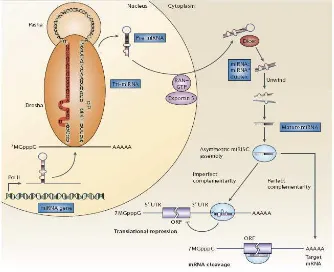
Figure 1.The biogenesis of microRNAs.MicroRNA (miRNA) genes are generally transcribed by RNA Polymerase II (Pol II) in the nucleus to form large pri-miRNA transcripts, which are capped (7MGpppG) and polyadenylated (AAAAA). These pri-miRNA transcripts are processed by the RNase III enzyme Drosha and its co-factor, Pasha, to release the ~70-nucleotide pre-miRNA precursor product. (Note that the human let-7a-1 miRNA is shown here as an example of a pre-miRNA hairpin sequence. The mature miRNA sequence is shown in red.) RAN GTP and exportin 5 transport the pre-miRNA into the cytoplasm. Subsequently, another RNase III enzyme, Dicer, processes the pre-miRNA to generate a transient ~22- nucleotide miRNA:miRNA* duplex. This duplex is then loaded into the miRNA associated multiprotein RNA-induced silencing complex (miRISC) (light blue), which includes the Argonaute proteins, and the mature single-stranded miRNA (red) is preferentially retained in this complex. The mature miRNA then binds to complementary sites in the mRNA target to negatively regulate gene expression in one of two ways that depend on the degree of complementation between the miRNA and its target. miRNAs that bind to mRNA targets with imperfect complementary block target gene expression at the level of protein translation (lower left). However, recent evidence indicates that miRNAs might also affect mRNA stability (not shown). Complementary sites for miRNAs using this mechanism are
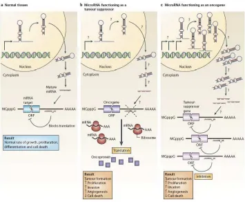
Figure 2.MicroRNAs can function as tumor suppressors and oncogenes. A. In normal tissues, proper microRNAs (miRNA) transcription, processing and binding to complementary sequences on the target mRNA results in the repression of target-gene expression through a block in protein translation or altered mRNA stability (not shown). The overall result is normal rates of cellular growth, proliferation, differentiation and cell death. B. The reduction or deletion of a miRNA that functions as a tumor suppressor leads to tumor formation. A reduction in or elimination of mature miRNA levels can occur because of defects at any stage of miRNA biogenesis (indicated by question marks) and ultimately leads to the inappropriate expression of the miRNA-target oncoprotein (purple squares). The overall outcome might involve increased proliferation, invasiveness or angiogenesis, decreased levels of apoptosis, or undifferentiated or de-differentiated tissue, ultimately leading to tumor formation. C. The amplification or over expression of a miRNA that has an oncogenic role would also result in tumor formation. In this situation, increased amounts of a miRNA, which might be produced at inappropriate times or in the wrong tissues, would eliminate the expression of a miRNA-target tumor-suppressor gene (pink) and lead to cancer progression. Increased levels of mature miRNA might occur because of amplification of the miRNA gene, a constitutively active promoter, increased efficiency in miRNA processing or increased stability of the miRNA (indicated by question marks). ORF, open reading frame. (Taken from [46])
2.5 MicroRNA IN CANCER
the BIC gene, is highly expressed in many types of B cell lymphomas, including diffused large B-cell lymphoma and pediatric Burkitt lymphoma [48]. Takamizawa et al. [49] reported decreased expression of let-7miRNA in non-small cell lung cancers, ranging from adenocarcinomas, to squamous carcinomas, to adenosquamous carcinomas, to large cell carcinomas of the lung. These authors also found that depressed let-7 expression is correlated with poorer patient prognosis.
Michael et al. in 2003 published the first study of miRNA in colon cancer, identified miR-143 and miR 145 as potential factors in colon tumorigenesis [50]. Total RNA from matched normal and colon adenocarcinoma were collected by these researchers and they cloned the 18-22 nucleotide RNA fragments, which sequences had been compared to microRNA databases. The result was that miR-143 and miR-145, being located close together on chromosome 5, is expressed at reduced levels in colon cancer epithelial cells [50]. The targets of miR-143 and miR-145 remain speculative but there are several candidates such as ERK5 and IRS1.
The let-7 family consists of 14 isomers, which majorities are dysregulated in colorectal cancer. Akao et al. has reported down regulation of let-7a-1 in colon cancer tumors and colon cancer cell lines. Reduced expression of let-7a-1 in colon cancer cell line shows increased RAS and c-myc protein expression level lead to increase cell growth [51].
analysis, they demonstrate the elevated levels of miR-31, miR-96, miR-31, miR-135b, and miR-183 in colorectal tumor cells. The authors also find the association of cells showing up regulation of miRNA which also harbored oncogenic mutation of either KRAS or BRAF [52].
2.6 DNA COPY NUMBERABNORMALITIES AFFECTING microRNA
GENES
[11]. Recently, this finding was experimentally confirmed by an array-based comparative genomic hybridization (aCGH) study in 227 human tumors [59]. Common fragile sites are specific chromosomal areas that are susceptible to form gaps and breaks when cells are exposed to stresses that affect DNA synthesis [64]. The latest compendium comprises 103 Common fragile sites [65] in colon cancer includes: chromosome 16q23 [66], 8q24 (128.14-128.62 Mb) [67], 8p21 [68], 15q15 [69], 2q31 33 [65], 3p14.2 [70], 5q21 [65], Xp23.3 [64], 17q21 [71] and 6q26 [72]. The rearrangements are usually one or more large deletions of tens to hundreds of kilobase pairs within the fragile sites. To date, all fragile sites that have been cloned are practically AT-rich and they do not have expanded di- or trinucleotide repeats [64]. A more recent study has suggested that genomic copy number loss may account for the downregulation of approximately 15% of miRNAs in advanced ovarian tumors. These findings support the notion that DNA copy number alterations of miRNAs are highly prevalent in cancer and may account in part for the frequency of miRNA gene dysregulation.
2.7 OLIGONUCLEOTIDE ARRAY COMPARATIVE GENOMIC
HYBRIDIZATION (Oa-CGH)