The Antiplatelet Aggregation Effect of Extract and Ethyl Acetate Fraction
of Velvet Bean Seed (
Mucuna pruriens
L.) in Dyslipidemic Rat
Wahyu Widowati
1, Hana Ratnawati
1, Udju
Djunaedi Rusdi
2,
Wahyu Winarno
3, Felix Kasim
11Medical Research Center, Faculty of Medicine, Maranatha Christian University, Jl. Prof. Drg. Suria Sumantri 65
Bandung,40164; 2 Department of Microbiology, Faculty of Medicine, University of Jenderal Achmad Yani,
Jl. Terusan Jend Sudirman, PO Box 148, Cimahi 40533; 3Department of Agricultural Production, StatePolytechnic of Jember,
Jl Mastrip PO Box 164, Jember 68101
Email: [email protected]
ABSTRACT
Cardiovascular disease (CVD) is the irst cause of death in the world, CVD has complex and multifactorial process
including atherogenic lipoprotein, oxidized low density lipoprotein (LDL), endothelial dysfunction, plaque stability,
vascular inlammation, thrombotic and ibrinolytic disorder, exercises and genetic factor. Inhibiting the platelet ag -gregation is one of the CVD prevention. Velvet bean seed (Mucuna pruriens L.) can be found abundantly in Indonesia,
but has not been used as herbal medicine. Ethanol extract and ethyl acetate of velvet bean seed contain high lavonoids
and antioxidants properties which is expected could inhibit platelet aggregation. The objectives of the research were to determine the activity of ethanol extract and ethyl acetate fraction of velvet bean seed towards clotting and bleeding time in dyslipidemic rats. This research used completely randomized design in dyslipidemic rats which were given by ethanol extract of velvet bean seed at the concentrations of 50, 100 and 200 mg/kg BW/day and ethyl acetate fraction
of velvet bean seed at the concentrations of 15, 30 and 60 mg/kg BW/day and 42.2 mg/kg BW/day aspirin for ten days.
Clotting and bleeding time were measured at days 0, 10, and 20. Data were analyzed using One way analysis of
vari-ance and continued with Duncan’s post Hoc test with 95 % level of signiicancy. The results showed that administration of 60 mg/kg BW/day ethyl acetate fraction of velvet bean seed and at the concentrations of 100 and 200 mg/kg BW/
day ethanol extract of velvet bean seed, prolong the clotting time at day 10, ethyl acetate fraction at the concentration
of 60 mg/kg BW/day, 200 mg/kg BW/day ethanol extract of velvet bean prolong bleeding time at day 10.
Keywords : antiaplatelet, lavonoid, platelet aggregation, Mucuna pruriens L., cardiovascular disease, dyslipidemic
INTRODUCTION
Cardiovascular disease (CVD) is the irst cause of death in the world; around 19.8 % from total death in 1993 and be
-come 24.4 % in 1998. An estimated 17.1 million people died from CVD in 2004, representing 29% of all global deaths. Of these deaths, an estimated 7.2 million were due to CVD and 5.7 million were due to stroke (AHRQ, 2003; WHO, 2009).
Incidence of CVD is increasing in developed countries, the incidences are estimated to be three to four times higher in developing countries. Globally, CVD is estimated 31 % of
the worldwide mortality. Each year 17.2 million people die
of CVD, 80 % of them in the developing world and
emerg-ing economies. By 2030, almost 23.6 million people will die
from CVD, mainly from heart disease and stroke. These are
projected to remain the single leading causes of death. The largest percentage increase will occur in the Eastern Medite-ranian Region. The highest increase in number of death will
occur in the South-East Asia Region (WHO, 2009). Based on
the data of Medical Department and Yayasan Penyakit Jan-tung (Cardiac Disease Foundation) showed that CVD is the leading cause of death in Indonesia. Based on World Health Organization (WHO) data, around 12 million people died of heart attack in the world. According to the family health
sur-vey (survei kesehatan rumah tangga) in 1992, CVD causes around 16 % of death and become 26.4 % in 2001 (Irawan, 2007).
oxidized low-density lipoprotein (LDL), endothelial
dysfunc-tion, plaque instability, vascular inlammadysfunc-tion, thrombotic and ibrinolytic disorder, exercises and genetic factor (Wijaya, 1998; Halliwel and Gutteridge, 1999). Atherosclerosis is an inlammatory disease and high plasma cholesterol concentra -tions, in particular those of LDL cholesterol, is one of the prin-cipal risk factor of atherosclerosis. If LDL particles become trapped in an artery, they can undergo progressive oxidation and be internalized by macrophages by means of the
scaven-ger receptors on the surfaces of these cells (Ross, 1999). The
internalization leads to the formation of lipid peroxides and fa-cilitates the accumulation of cholesterol esters. Oxidized LDL may also be involved in atherogenesis by inducing smooth muscle cell proliferation and smooth muscle foam cell gen-eration (Holvoet et al., 1998). Subendothelial accumulation of foam cells plays a key role in the initiation of atherosclerosis. These foam cells may be generated by the uptake of oxidized
LDL and/or malondialdehyde (MDA)-modiied LDL (Holvo
-et et al., 1998; Ross, 1999). Numerous in vitro studies using a
variety of oxidation methods and measurements have shown that polyphenolics from red wine, green tea and chocolate can inhibit LDL oxidation (Anderson et al., 2001).
Platelet aggregation is an important factor in thrombosis with uncontrolable clotting blood (Wu et al., 2007). Formation of platelet aggregates in an atherosclerotic vessel can cause
total blockage of blood low, leading to myocardial infarc -tion and thromboembolic diseases. Atherosclerosis promotes increased adhesiveness in the endothelial wall for platelets, and a lost of anticoagulant properties, which contribute to atherosclerotic plaque formation. Platelets adhesion is initi-ated when platelets contact with exposed connective tissue of injured tissues. Adhesion stimulates platelets to secrete a
vari-ety of factors, including ibrinogen and von Willebrand factor,
which facilitates aggregation and platelet plaque on the ves-sel wall. Activated platelets also secrete thromboxane A2 that further stimulates platelet aggregation, and the activated im-mune cells also secrete platelet activating factor (PAF) which
stimulate platelet aggregation (Boik, 1996; Geraldo, 2010).
The activated platelets change shape, put out pseudopodia, discharge their granules, and stick to other platelets thus ini-tiating the process of platelets aggregation (Resh and Ernst,
1995; Klepser and Klepser, 1999; El-Sabban, 2009). The
rupture of plaques is the most important mechanism for the progression of vascular disease because this rupture exposes thrombogenic subendothelial matrix of protein and collagen, triggering a cascade of platelet activity (Geraldo, 2010).
Flavonoids are component of a wide variety of ed-ible plants, fruits, and vegetables and of beverage appears to have antioxidative properties toward LDL, lipid peroxi-dation (Fuhrman et al, 1995; Sesso et al., 2003). Flavonoid from Garcinia cambogia inhibit atherosclerosis, endothelial
damaged, leucocyte activation, platelet adhesion and platelet aggregation (Koshy et al., 2001). According to the previous
research by Widowati and Retnaningsih (2007) antioxidant
activity in ethanol extract and ethyl acetate fraction of velvet bean seed (Mucuna pruriens L.) compared with α-tocopherol, ascorbic acid and butylated hydroxytoluen (BHT), ethanol extract and ethyl acetate fraction of velvet bean seed contain
higher lavonoid compared to the others fraction (hexane
fraction, butanol and water fraction) (Widowati et al., 2007; Widowati et al., 2010), thus this compound can be possibly used as antiaggregation platelet agent.
Many drugs as antiplatelet agents for treating CVD, however some have several collateral effects and resistance in long term therapy, such as the known clinical aspirin resistance (Geraldo, 2010). Thus, the purpose of this research is to de-velop new antiplatelets agents with low collateral effects, from ethanol extract and ethyl acetate fraction of velvet bean seed.
MATERIALS AND METHODS
Extraction and Fractionation
The velvet bean seed (Mucuna pruriens L.) was collect-ed from Sukoharjo Dstrict, Central Java, Indonesia, during
April, 2007. Five kilograms of velvet bean seeds were milled and soaked in distilled ethanol (EtOH) and extracted by
mac-eration technique; evaporated iltrate and yielded 516.845 g
(10.335 %) ethanol extract of velvet been seed. The ethanol
extract (133.889 g) was partitioned with solvent mixture of 75% n-hexane and 25 % water, produced 30.466 g (22.71 %)
hexane fraction and the water iltrate residue was partitioned with solvent mixture of 95 % ethyl acetate and 5 % water, produced 0.498 g (0.37 %) ethyl acetate fraction and the wa
-ter iltrate residue was partitioned with solvent mixture of 90 % n-buthanol and 10 % water, produced 3.578 g (2.67 %) ethyl acetate fraction. Finally, the water iltrate residue was evaporated and produced 4.358 g (3.26 %) water fraction. In
this study, we used ethanol extract and ethyl acetate fraction of velvet bean seed, since the previous in vitro study showed that ethanol extract and ethyl acetate fraction of velvet bean seed had the highest antioxidant activity.
Animals and Diets
Thirty adult male Wistar rats obtained from School of Life Science, Bandung Institute of Technology, were housed in standard cages provided with food and water ad libitum.
Rats were adapted for 7 days until the body weight were 175-200 g. The rats were feed by high fat diet (3000 g stan -dard diet mixed with 250 g duck egg yolk, 500 g palm oil,
1250 g wheat lour, 500 g lamb fat and hot water). The rats
of crude fat was 20.78 % compared to the standard diet rats which was only 7.37 % .
In spite of high fat diet, each rat was also given with 1
mL/day fructose liquid 60 % (120 g/200 mL aquadest). The
high fat diet and fructose liquid were given for 2 weeks until the body weight achieved 200 – 250 g, but the high fat diet was still given continually for 20 days. The ethanol extract and ethyl acetate fraction of velvet bean seed were given only for 10 days.
Rats were devided into 9 groups (3 rats) with differ
-ent treatm-ents. The irst group, as negative control, was given
standard diet. The second group, as positive control, was
given high fat diet. The third, fourth, and ifth groups were
treated with high fat diet plus ethyl acetate fraction 15 mg/
kg BW, 30 mg/kg BW and 60 mg/kg BW daily. The sixth,
seventh and eighth groups were treated with high fat diet plus ethanol extract 50 mg/kg BW, 100 mg/kg BW and 200 mg/ kg BW daily. Group ninth was treated with high fat diet and aspirin 42,2 mg/kg BW daily.
Sample Preparation for The Lipid Test and MDA
After the treatments, 1.5 mL blood from orbital vein was collected in the tube contained heparin. It was centrifuged at 3,000 rpm for 10 minutes and the plasma was used for mea-suring the total cholesterol, LDL-cholesterol, high-density li-poprotein (HDL-cholesterol), triglyceride, and MDA level.
Total plasma cholesterol and triglyceride were measured according to the instruction manuals by the diagnostic kits from
Abbott Clinical Chemistry (Abbott Clinical Chemistry, 2006),
HDL-cholesterol, LDL-cholesterol were measured according to the instruction manuals of the diagnostic kits from Daiichi Pure Chemicals Co., Ltd (Daiichi Pure Chemical, 2008).
After feeding high fat diet and fructose for 2 weeks,
the lipid proile of negative control group (standard diet) was
as follow : total cholesterol 58.33 mg/dL, LDL 8.00 mg/dL,
HDL 33.00 mg/dL, triglyceride 63.67 mg/dL and the dyslipi
-demic rats had total cholesterol 70.67 mg/dL, LDL 14.33 mg/
dL, HDL 24.33 mg/dL, and triglyceride 112.33 mg/dL. Thus, feeding with high fat nutrient induced dylipidemic rats.
Clotting Time Assay
The rat’s tail was injured, blood collected with capillary tube for 30 seconds. Capillary tube was broken every 15
sec-onds until the ibrin ilament appeared at the broken capillary
tube as clotting time. Clotting time was measured at days 0, 10 and 20.
Bleeding Time Assay
The rat’s tail was injured, blood absorbed with ilter pa
-per for 15 seconds. The interval time between the irst dropping blood until the lowing blood stopped was calculated as bleed -ing time. Bleed-ing time was measured at days 0, 10 and 20.
Statistical Analysis
To verify the statistical signiicance of all parameters,
the data were calculated the values of means and standard
deviation (M ± SD) and 95 % conidence interval (CI) of
means. This research used completely randomized design. To compare several groups, analysis of variance (ANOVA) was used. P-values of less than 0.05 were considered as
sta-tistically signiicant. Furthermore to know the best treatmet, Duncan’s post-Hoc test at 95 % conidence interval was used. Statistical analysis was done using SPSS 16.0 version.
RESULTS AND DISCCUSSION
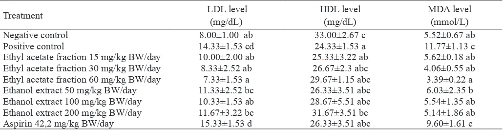
Based on the statistical analysis, at day 10 showed that
HDL, LDL and MDA signiicantly different among treatmet, high fat diet signiicantly increased the LDL-cholesterol, de -creased the HDL-cholesterol and de-creased MDA compared to standard diet (Table 1). Extract and ethyl acetate fraction of
Table 1. HDL, LDL and MDA level in dyslipidemic rats treated with ethanol extract and ethyl acetate fraction of velvet bean seed for 10 days
Treatment LDL level
(mg/dL)
HDL level (mg/dL)
MDA level (mmol/L)
Negative control 8.00±1.00 ab 33.00±2.67 c 5.52±0.67 ab
Positive control 14.33±1.53 cd 24.33±1.53 a 11.77±1.13 c
Ethyl acetate fraction 15 mg/kg BW/day 10.00±2.00 ab 25.33±3.22 ab 5.62±0.18 ab Ethyl acetate fraction 30 mg/kg BW/day 8.33±2.52 ab 26.67±2.3 abc 4.06±0.55 ab
Ethyl acetate fraction 60 mg/kg BW/day 7.33±1.53 a 29.67±1.15 abc 3.39±0.22 a
Ethanol extract 50 mg/kg BW/day 11.33±2.52 bc 26.33±3.51 abc 6.03±2.35 b
Ethanol extract 100 mg/kg BW/day 10.33±1.53 ab 28.67±5.51 abc 5.54±1.35 ab
Ethanol extract 200 mg/kg BW/day 11.67±3.22 bc 31.67±3.51 bc 5.14±1.86 ab
Aspirin 42,2 mg/kg BW/day 15.33±1.53 d 26.33±3.51 abc 9.60±1.61 c
velvet bean seed decreased the LDL level and ethanol extract of velvet bean seed 200 mg/kg BW/day increased HDL level in dyslipidemic rats. Extract and ethyl acetate fraction of vel-vet bean decreased the MDA level in dyslipidemic rats.
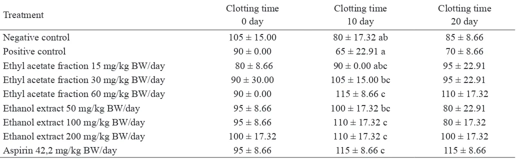
After treatment with various doses of ethanol extract, ethyl acetate fraction of velvet bean seed, and aspirin 42.2 mg/ kg BW/day for 10 days, the effect on clotting time was shown in Table 2. Based on the statistical analysis, at day 0, clotting
time showed no signiicant difference among groups because
at day 0 rats had not been given with ethanol extract, ethyl acetate and aspirin yet. At day 10, clotting time showed
sig-niicant difference among groups because rats had been given
ethanol extract and etyhl acetate fraction. At day 20, clotting
time showed no signiicant difference among groups because
the ethanol extract, ethyl acetate and aspirin had been stopped at day 10, indicated that ethanol extract and ethyl acetate frac-tion had no effect on clotting time after the treatment had been stopped. To determine the difference of the clotting time (days 0, 10 and 20) among treatment groups, we analyzed using Duncan’s post Hoc test (Table 2), which showed the effect of treatments (ethanol extract and ethyl acetate fraction of velvet bean seed) towards clotting time. At day 10, the clotting time
of the dyslipidemic rats treated with ethyl acetate fraction 60
mg/kg BW/day and ethanol extract 100 and 200 mg/kg BW/
day were signiicant difference compared to positive control
and had similar effect with aspirin as anticoagulant.
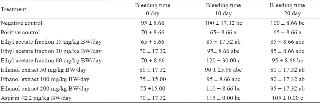
Based on statistical analysis, at day 0, the bleeding time
showed no signiicant difference among treatmet groups. While, at day 10, bleeding time showed signiicant difference
among treatment groups, since rats had been given ethanol extract and etyhl acetate fraction. At day 20, bleeding time
showed signiicant difference among treatment groups
al-though the ethanol extract, ethyl acetate and aspirin had been
stopped at day 10, indicated that ethanol extract and ethyl
acetate still inluenced the bleeding time. To determine the
difference of the bleeding time (0, 10 and 20days) among treatment groups, we analysed using Duncan’s post Hoc test
(Table 3), which showed that on day 0, no signiicant dif
fe-rence in bleeding time among treatment groups. At day 10,
the bleeding time showed that ethyl acetate group (60 mg/ kg BW/day) was signiicant difference compared to positive
control, even showed a better result compared to group ed with aspirin as anticoagulant. At day 20, the shortest bleed -ing time was only in positive control (dyslipidemic rats),
negative control, rats given ethyl acetate fraction 60 mg/kg BW/day and rats given with aspirin were signiicant
diffe-rence compared to positive control. Although aspirin and ethyl acetate fracti on had been stopped at day 10, it means that aspirin and ethyl acetate fraction could prolong the bleeding time until day 20.
Based on the clotting time data (Table 2) at day 10 indi-cated that dyslipidemic rats were given with extract and ethyl acetate prolonged clotting time compared to positive control. The bleeding time (Table 3) at days 10 and 20 in dyslipidemic rats were shorter than negative control. At day 10 showed that the dyslipidemic rats which were given ethanol extract and ethyl acetate fraction prolonged the bleeding time. This
re-search was veriied by previous study that hyperlipidemic in rats exhibit signiicant increase in ADP or collagen-induced
platelet aggregation and cholesterol/phospholipid molar ratio in platelets. The increase in cholesterol/phospholipid ratio was responsible for hyperaggregation of platelet in animals
(No-bukata, 1999). This data was veriied with previous research
that high cholesterol consumption can induce dyslipidemic and endothelial damaged, also effect thrombocyte adhesion
in collagen furthermore activate thombocyte (Aprami, 1993;
Tabel 2. The clotting time of dyslipidemic rats treated with ethanol extract and ethyl acetate of velvet bean seed
Treatment Clotting time
0 day
Clotting time 10 day
Clotting time 20 day
Negative control 105 ± 15.00 80 ± 17.32 ab 85 ± 8.66
Positive control 90 ± 0.00 65 ± 22.91 a 70 ± 8.66
Ethyl acetate fraction 15 mg/kg BW/day 80 ± 8.66 90 ± 0.00 abc 95 ± 22.91
Ethyl acetate fraction 30 mg/kg BW/day 90 ± 30.00 105 ± 15.00 bc 95 ± 22.91
Ethyl acetate fraction 60 mg/kg BW/day 90 ± 0.00 115 ± 8.66 c 110 ± 17.32
Ethanol extract 50 mg/kg BW/day 95 ± 8.66 100 ± 17.32 bc 80 ± 22.91
Ethanol extract 100 mg/kg BW/day 95 ± 8.66 110 ± 17.32 c 80 ± 17.32
Ethanol extract 200 mg/kg BW/day 100 ± 17.32 110 ± 17.32 c 100 ± 17.32
Aspirin 42,2 mg/kg BW/day 95 ± 8.66 115 ± 8.66 c 115 ± 8.66
Wijaya, 1998). Active thrombocyte release ADP and throm -boxane A2 and initiate aggregation of thrombocyte and vaso-constriction. Thrombocyte aggregation is important factor in thrombose formation in uncontrol blood clotting (Wu et al.,
2007). The high LDL and low HDL level in dyslipidemic rats
(Table 3) were shorter clotting and bleeding time compared
to negative control, this research was veriied that elevating
levels of LDL and low levels of HDL are widely used for pre-dicting risk to atherosclerosis (Sevanian et al., 1998).
Velvet bean seed ethanol extract and ethyl acetate
frac-tion contain high antioxidant and lavonoids (Widowati and Retnaningsih, 2007; Widowati et al., 2007, Widowati et al., 2010), were capable to prolong the clotting and bleeding time at day 10 in dyslipidemic rats (Table 2,3). This research was
veriied with previous study that lavonoids could reduce
LDL oxidation, an important step in atherogenesis (Setorki
et al., 2009) through free radicals scavenger mechanism.
Based on the data (Table 1) displayed that velvet bean seed extract and ethyl acetate fration had high antioxidant activity
(Widowati and Retnaningsih, 2007), and were capable to re -duce lipid peroxidation and decreased MDA, LDL level com-pared to dyslipidemic rats which were not given extract and
ethyl acetate fraction as antioxidant and lavonoid sources.
Antioxidant activity of polyphenols were shown to possess many biological properties including the inhibition of platelet aggregation, vasorelaxing activity, modulation of lipid me-tabolism, and inhibition of LDL oxidation (Agli et al., 2004). In Table 2, the LDL in dyslipidemic rats 14.33 mg/dL was higher than negative control (8.00 mg/dL) and will highly
in-luenced the oxidative damage (MDA 11.77 mmol/L), due to
the high LDL level accelerating atheroginicity
(Parthasara-thy et al., 1999). The oxidation of LDL was led by free rad
-ical-mediated chain reaction, yielding phosphatidylcholine
hydroperoxide (PCOOH) cholesteryl ester hydroperoxide (CEOOH) as the primary oxidations product (Noguchi et al.,
1998). It means that high LDL level increases oxidized LDL
(ox-LDL) and MDA level (Table 2) and play role in athero-sclerosis. LDL level increases amounts of thiobarbituric acid reactive materials (often attributed to increase amounts of MDA, 4-hydroxynonenal, and related carbonyl compounds), lipid peroxides, cholesterol oxides, decrease amounts of
spe-ciic phospholipids, showed increase lysophospholipids and
diminish antioxidants level (Sevanian et al., 1998). The evi
-dence indicate that oxidative modiication of LDL may play
a causative role in atherosclerosis (Sevanian et al., 1998). Oxidized LDL is cytotoxic to endothelial cells, and nLDL is not toxic (Sevanian et al., 1998; Halliwel and Gutteridge,
1999). The previous research indicated that atherogenicity was due to oxidative damage to LDL cholesterol. Modiied
or oxidized LDL (ox-LDL) has been shown to accelerate several steps in atherosclerosis including endothelial dam-age, monocyte/macrophage recruitment, increased uptake
of LDL by foam cells (Hannekens, 1999). The oxidation of
LDL is important in atherosclerosis, it is likely to occur in the subendothelial intima rather than the plasma (Parthasarathy,
1999). In Table 1, showed that dyslipidemic rats, high LDL
concentration increased ox-LDL. The MDA level in
dyslipi-demic rat was higher 11.77 mmol/L compared to normal rat
was 5.52 mmol/L, furthermore the bleeding time at 10 day of dyslipidemic rats were shorter than those in normal rats. This research result supports previous research, that oxLDL could accelerate atherosclerosis stages including endothelial dam-age, monocyte/macrophage recruitment, increased uptake of LDL by foam cells, alteration in vascular tone (Hannekens,
1999; Parthasarathy et al., 1999). The oxidative modiied
LDL is taken up by macrophage, which results in its unregu-Tabel 3. The bleeding time of dyslipidemia rats given ethanol extract and ethyl acetate of velvet bean seed
Treatment Bleeding time
0 day
Bleeding time 10 day
Bleeding time 20 day
Negative control 95 ± 8.66 100 ± 17.32 bc 100 ± 8.66 bc
Positive control 70 ± 8.66 65± 8.66 a 65 ± 8.66 a
Ethyl acetate fraction 15 mg/kg BW/day 65 ± 8.66 85 ± 17.32 ab 85 ± 8.66 abc Ethyl acetate fraction 30 mg/kg BW/day 70 ± 17.32 95± 8.66 abc 85 ± 8.66 abc
Ethyl acetate fraction 60 mg/kg BW/day 70 ± 8.66 120 ± 30.00 c 95 ± 8.66 bc
Ethanol extract 50 mg/kg BW/day 80 ± 17.32 90 ± 25.98 abc 80 ± 17.32 ab
Ethanol extract 100 mg/kg BW/day 75 ± 15.00 95 ± 8.66 abc 80 ± 17.32 ab
Ethanol extract 200 mg/kg BW/day 75 ±15.00 110 ± 8.66 bc 95 ± 17.32 ab
Aspirin 42,2 mg/kg BW/day 70 ± 17.32 115 ± 0.00 bc 105 ± 0.00 c
lated uptake and eventual formation of foam cells as the early stages in cardiovasular disease (Noguchi et al., 1998). Ath
-erosclerosis is a systemic inlammatory disease characterized
by the accumulation of monocytes/macrophages and lympho-cytes in the intima of large arteries. Rupture or erosion of the advanced lesion initiates platelet activation and aggregation on the surface of the disrupted atherosclerotic plaque (Mass-berg et al., 2002). The term of endothelial dysfunction has been used to refers several pathological conditions,
includ-ing altered anticoagulant and anti-inlammatory properties of
the endothelium, impaired modulation of vascular growth, and dysregulation of vascular remodeling (Cai and Harrison, 2000), damaging the platelet activity, leukocyte adhesion, and thrombosis and is intimately involved in the development of atherosclerosis (Heitzer et al., 2001). Increased production of oxygen-derived free radicals such as the superoxide anion has been linked to impaired endothelial vasomotor function (Heitzer et al., 2001). Ox-LDL is the key event in endothelial injury and dysfunction. LDL in the subendothelial space, un-dergoes progressive oxidation and activates the expression of macrophage chemotatic protein 1 (MCP-1) and macrophage colony stimulating fator (M-SF) in the endothelium. MCP-1 and M-CSF promote the entry and maturation of monocytes
to macrophages, which further ox-LDL. Ox-LDL is specii -cally recognized by the scavenger receptor of macrophages and once internalized, formation of foam cells. Ox-LDL and
modiied LDL induce endothelial, associated with changes of
the adhesivenes to leukocytes or platelet and to wall perme-ability (Virgilli and Scaccini, 2001).
Extract and ethyl acetate fractionof velvet bean could
prolong the clotting and bleeding time, and was veriied with the previous research that using apple juice as lavonoids source (total lavonoids 1.36 ± 0.03 g/100 mL equivalent cat -echin and total anthocyanin 3.05 ± 0.85 mg/100 g) in
cho-lesterolemic rabbit, signiicantly decreased ibrinogen and
factor VII levels compared to high-cholesterol group (Setorki
et al., 2009). Based on the data (Table 2,3) displayed velvet
bean seed extract and ethyl acetate fraction could prolong the
clotting and bleeding time due to contained high lavonoid
(Widowati et al., 2007). The previous research showed that ethyl acetate fraction of velvet bean seed 500 µg/mL exhibi-ted antiaggregation platelet against adhenosin diphosphate (ADP) inducer by in vitro test (Widowati and Ratnawati,
2009). Flavonoids are subgroup of polyphenol, capable of :
1). inhibiting atherosclerosis, endothelial disruption, leuco-cyte activity, adhesion, aggregation platelet, decrease LDL level in normal and hypercholesterolemic rats, also inhibit in vitro LDL oxidation; 2). decreasing atherogenesis, inhibit hyperlipidemia, inhibit triglyceride accumulation in blood and liver, decrease phospholipid and fatty acids in rat’s tis-sue (Koshy et al., 2001; Hidgon, 2005). Giving 10 mL and 5
mL apple juice in cholesterolemic rabbits, the atherosclerotic thickness of right and left coronary arteries was 0.8 – 1.4 and
0.92 -1.92, respectively and the plaque degree for both was
grade 1. High cholesterol groups, right and left coronary ar-teries, the atherosclerotic thickness of right and left coronary
arteries was 3.47 ± 0.37 and 3.28 ± 0.26, respectively and
the plaque degree in both was grade 3. Giving apple juices
as lavonoid sources decreased the plaque degrees (Setorki et
al., 2009). Atherosclerotic changes were absent in normal diet
group, whereas in the intimal surface of the coronary arteries from high-cholesterol diet group were seen many fat-laden
macrophages. The cytoplasm of the macrophages illed with
lipid droplets (foam cell) as the result of lipid digestion by the
macrophage. Apple juice as lavonoids source may be useful
in preventing hypercholesterolemic atherosclerosis and low-ering the related risk of coronary artery disease (Setorki et
al., 2009).
Flavonoids play role as an antiinlammation, antioxi -dant, antiallergic, hepatoprotective, antitrombotic, neuropro-tective and anticarcinogenic (Ganapaty et al., 2007). Antiag
-gregation platelet activity of lavonoid is through inhibition
of thromboxane A2 mechanism. The inhibited thromboxane A2 stimulatesadenyl cyclase further increase cAMP and the increasing cAMP will reduce ion Ca (calcium) concentration in thrombocyte, furthermore inhibit aggregation and adhesion
(Hoffbrand and Pettit, 1987), prolong clotting and bleeding
time.
Flavonoids protect against atherosclerosis, through re-ducing the susceptibility of LDL to oxidation (Benito et al., 2002), and vasodilator properties observed in vitro (Benito et
al., 2002)
The research results showed that giving velvet bean
seed extract 200 mg/kg BW/day increased HDL level (31.67
mg/dL) compared to positive control (24.33 mg/dL) (Table
2). This results were veriied with previous study that HDL
concentration inhibited atherosclerosis progression and ad-vance cardiovascular disease (Sevanian et al., 1998; Santoso and Setiawan, 2005).
Based on Table 2 showed that aspirin prolonged clot-ting time in dyslipidemic rats and prolonged bleeding time in dyslipdemic rats (Table 3), aspirin is antiplatelet agent, medication that blocks the formation of blood clots by pre-venting the clumping of platelets. Aspirin as anti-thrombotic compounds is through the inhibition of platelet
cyclooxyge-nase-1 (COX-1) by irreversible acetylation of a speciic ser -ine moiety, thereby blocking the formation of thromboxane A2 (TXA2) for the life time of the platelets (McKee et al.,
2002; Ohmori et al., 2006). Table 1 showed that aspirin has
CONCLUSIONS
Ethanol extract and ethyl acetate fraction of velvet bean seed exhibited antiplatelet aggregation in dyslipidemic rats, treated with ethanol extract 100 mg/kg BW/day, 200 mg/kg
BW/day and ethyl acetate fraction 60 mg/kg BW/day pro -longed the clotting time at day 10, ethanol extract 200 mg/
kg BW/day and ethyl acetate fraction 60 mg/kg BW/day pro -longed the bleeding time at day 10.
ACKNOWLEDGEMENT
We are grateful to Directorate General for Higher Edu-cation, Ministry of National Education of Republic
Indone-sia, for Research Grant of Hibah Bersaing (2007, 2008) for inancial support.
REFERENCES
Abbott Clinical Chemistry. (2006). Triglyceride.Abbott Park, IL 60064 USA.
Abbott Clinical Chemistry. (2006). Cholesterol.Abbott Park, IL 60064 USA.
Agli, M.D., Busciala, A. and Bosisio, A. (2004). Vascular ef-fects of wine polyphenols. Cardiovascular Research
63: 593 – 602.
AHRQ (2003). Effect of supplemental antioxidants vitamin C, vitamin E, and coenzyme Q10 for the prevention and
treatment of cardiovascular disease. PubMed
Naviga-tion. The Agency for Healthcare Research and Quality.
http://www.ahrq.gov/clinic/epcsums/ antioxsum.htm. [15th April 2009].
Anderson, K.J., Teuber, S. S., Gobeille, A., Cremin, P., Wa-terhouse, A. L. and Steinberg, F. (2001). Walnut poly-phenolics inhibit in vitro human plasma and LDL oxida-tion. J. Nutr. 131: 2837–2842.
Aprami, T.M. (1993). Peranan antioksidan dalam pencegahan
aterosklerosis. Bagian/UPF Ilmu Penyakit Dalam Fak. Kedokteran UNPAD. Bandung.
Benito,S., Lopez,D., Sáiz, M.P., Buxaderas, S., Sánchez,J.,
Puig-Parellada, P. and Mitjavila., M. T. (2002). A la -vonoid-rich diet increases nitric oxide production in rat aorta. Br J Pharmacol135: 910–916.
Boik J. (1996). Cancer and natural medicine. Oregon Medical
Press. Princeton, Minnesota.
Cai, H. and Harrison, D.G. (2000). Endothelial dysfunction in cardiovascular diseases: the role of oxidant stress. Circ Res 87:840–844.
Daiichi Pure Chemicals Co. Ltd. (2008). High density lipo-protein cholesterol test. Ibaraki.
Daiichi Pure Chemicals Co. Ltd. (2008). Low density lipo-protein cholesterol test. Ibaraki.
El-Sabban, F. (2009). Garlic as an antithrombotic and anti -platelet aggregation agent. J. Chin. Clin. Med. 4
:288-294.
Fuhrman, B., Lavv, A. and Aviram, M. (1995). Consumption
of red wine with meals reduces the susceptibility of hu-man plasma and low-density lipoprotein to lipid peroxi-dation. Am.J. Clin. Nutr.61:549-54.
Ganapaty S., Chandrashekhar V.M., Chitme H.R. and Narsu
M.L. (2007). Free radical scavenging activity of Gos -sypin and Nevadensin: An in-vitro evaluation. Indian J.
Pharmacol. 6: 281-283.
Geraldo, R.B., Belo, M.L., Dias, L.R.S., Vera, M.A.F., Na-gashima, T., Abreu, P.A., Santos, M.B., Albuquerque, M.G., Cabral, L.M., Freitas, A.C.C., Kalil, M.V., Rodri-gues, C.R., Castro, H.C. (2010). Antiplatelet activity and structure-activity relationship study of Pyrazolopiridine Derivatives as poetntial series for treating thrombotic diseases. J Atheroscler Tromb17: 2-11.
Halliwel, B., and J.M.C. Gutteridge. (1999). Free Radicals in
Biology and Medicine. Oxford University Press, New
York.
Hannekens, C.H. (1999). Antioxidant vitamins and cardio -vascular disease. In : Papas, A.M. (ed.). Antioxidant
Status, Diet, Nutrition and Health, p 463-478. CRC
Press, Washington, D.C.
Heitzer, T., Schlinzig, T., Krohn, K., Meinertz, T. and Münzel, T. (2001). Endothelial dysfunction, oxidative stress, and risk of cardiovascular events in patients with coronary artery disease. Am. Heart Assoc.104:2673-2678.
Hoofbrand, A. V. and Pettit, J.E. (1987). Kapita selekta hae -matologi (Essential Haematology). Edisi 2. Aru W. S, S. Bambang, A. Idrus. (eds.). Pusat Penerbitan Departe-men Ilmu Penyakit Dalam FK UI, Jakarta.
Holvoet, P., Vanhaecke, J., Janssens, S., de Werf, F.V. and
Collen, D. (1998). Oxidized LDL and malondialde
-hyde-modiied LDL in patients with acute coronary
syndromes and stable coronary artery disease. J. Am.
Heart Assoc. 98:1487-1494.
Irawan. B.2007. Penyakit Jantung Koroner. http://www. nursyifa.net /index.html. [5th November 2008].
Klepser, T.B. and Klepser, M.E. (1999). Unsafe and poten -tially safe herbal therapies. Am J Health Syst Pharm
56:125-138.
Koshy, A.S., Anila, L., and Vijayalaksmi, N.R. (2001). Fla-vonoids from Garcinia combagia lower lipid levels in hypercholesterlemic rats. Food Chemistry 72:289-294.
of platelet adhesion in the initiation of atherosclerotic lesion formation. J. Exp. Med. 196:887-96.
McKee, S.A., Sane, D.C. and Deliargyris, E.N. (2002). As-pirin resistance in cardiovascular disease: a review of
prevalence, mechanisms, and clinical signiicance.
Thromb Haemost88: 711–715.
Nobukata, H., Ishikawa, T., Obata, M. and Shibutani, Y.
(1999). Age-related changes in coagulation, ibrinoly -sis, and platelet aggregation in male WBN/Kob rats.
Thromb. Res.98:507-516
Noguchi, N., Okimoto, Y., Cynshi, O., Kodama, T. and Niki,
E. (1998). Inhibition of oxidative modiication of Low Density Lipoprotein by novel antioxidant BO-653 pre -pared by theoretical design. In : Packer, L and Ong, A.S.H. (eds.). Biological Oxidants and Antioxidants,
Molecular Mechanism and Health Effects, p:139-152.
OACS Press, Illionis.
Parthasarathy, S, Santanam, N. and Auge, N. (1999). Antioxi -dants and Low Density Lipoprotein Oxidation. In : Pa-pas, A.M. (ed.). Antioxidant Status, Diet, Nutrition and
Health, p:347-370. CRC Press. Washington, D.C.
Ohmori, T, Yatomi, Y., Nonaka, T., Kobayashi, Y., Madoiwa,
S., Mimuro, J., Ozakis, Y. and Sakata, Y. (2006). As -pirin resistance detected with aggregometry cannot be explained by cyclooxygenase activity: involvement of other signaling pathway(s) in cardiovascular events of aspirin-treated patients. J. Thromb. Haemost. 4: 1271–
1278.
Resh, K.I. and Ernst, E. (1995). Garlic (Allium sativum) a po-tent medicinal plant. Fortschr Med 113:311-315.
Ross, R. (1999). Atherosclerosis an inlammatory disease.
The New England J. Med.340:115-126.
Santoso, M., dan Setiawan, T. (2005). Penyakit Jantung Ko-roner. Artikel Cermin Dunia Kedokteran No.147. Sesso, H.D., Gaziano, J.M., Liu, S. and Buring, J.E. (2003).
Flavonoid intake and the risk of cardiovascular disease in women. Am.J. Clin. Nut, 7:1400-1408.
Setorki, M., Asgary, S., Eidi, A., Rohani, A.H. and Esmaei, N.
(2009). Effects of apple juice on risk factors of lipid pro
-ile, inlammation and coagulation, endothelial markers
and atherosclerotic lesions in high cholesterolemic rab-bits. Lipids in Health and Dis.8:1-9.
Sevanian, A., Duncan, R., Hwang, J. and Hodis, H.N. (1998).
Human LDL oxidation, atherosclerosis and cardiovascu-lar disease. In : Packer, L and Ong, A.S.H. (eds.).
Biolog-ical Oxidants and Antioxidants, Molecular Mechanism
and Health Effects, p:127-138. OACS Press, Illionis.
Virgilli, E. and Scaccini, C. (2001). Cardiovascular disease and nutritional phenolics. In: Pokorny, J., Yanishlieva, N. and Gordon, M. (eds.). Antioxidants in Food,
p:87-96. CRC Press, Washington D.C.
World Helath Organization (2009). Cardiovascular diseases (CVDs). Fact sheet No 317. WHO Media centre. Widowati, W. and Retnaningsih, Ch. (2007). Antioxidant
activity of ethanol extract and fraction of Velvet bean
(Mucuna pruriens L.). Poster presentation at the Sixth
Princess Chulabhorn International Science Congres (PC-VI). The Interface of Chemistry and Biology in the “OMICS” Era: Environment & Health and Drug
Dis-covery” November 25-29, 2007, Bangkok Thailand. Widowati, W., Lili, M.P. dan Ratnasari, A.I. (2007). Potensi
FraksiAktif Antioksidan, Antikolesterol Kacang Koro
(Mucuna pruriens L.) Dalam Pencegahan
Ateroskle-rosis. Laporan Hasil Penelitian Hibah Bersaing Tahun
Anggaran 2007/2008. Direktorat Jenderal Pendidikan
Tinggi, Departemen Pendidikan Nasional.
Widowati, W., Ratnawati, H., Rusdi, U.D., Winarno, W, Im-manuel, V. (2010). Phytochemical Assay and Antiplate-let Activity of Fractions of Velvet Bean Seeds (Mucuna pruriens L.). Hayati Journal of Biosciences.17:85-90
Wijaya, A. (1998). Faktor Resiko Penyakit Kardiovaskuler
Perspektif Baru. Forum Diagnosticum. Laboratorium Klinik Prodia. Bandung.
Wu, C.M., Wu, S.C., Chung, W.J., Lin, H.C., Chen, K.T., Chen, Y.C., Hsu, M.F., Yang, J.P., Wang, J.M. and Lin,
C.N. (2007). Antiplatelet effect and selective binding to
cyclooxygenase (COX) by molecular docking analysis