SHONE’S SYNDROME: TWO CASE REPORTS Meisa Puspitasari, Sri Endah Rahayuningsih
Division of Cardiology, Departement of Child Health, Faculty of Medicine Padjadjaran University,
Bandung, Indonesia
Abstract
Background:The Shone’s syndrome, defined by four cardiovascular defects such as a supravalvular mitral membrane, valvular mitral stenosis by a parachute mitral valve, subaortic stenosis, and aortic
coarctation, is a rare entity, which occurs most frequently in its incomplete form.
Objective:To report a case series of two girl with Shone’s syndrome.
Case Report:The first case is a 9 month old girl came to a cardiology polyclinic with chief complaint recurrent cough. The girl also had poor weight gain, no episode of dyspneu or cyanosis.
Echocardiography revealed aortic and pulmonal stenosis, mitral regurgitation-mitral stenosis with
shortening and thickening of mitral valve, small VSD and left ventricle hipoplasia. The second case
is a 5 years old girl presented at the emergency departement for progressively worsening dyspnea,
fever, and productive cough, due to bronchopneumonia, cyanosis. Echocardiography revealed the
co-existence of left ventricle and aortic hipoplastic, severe mitral stenosis due to parachute mitral valve,
VSD and a large PDA.
Conclusion: Echocardiography is particularly important in patients with congenital heart disease where lesions often co-exist and can be missed in the absence of a comprehensive examination.
Keyword: shone’s syndrome, echocardiography
Abstrak
Latar belakang: Sindrom Shone merupakan suatu kelainan yang sangat jarang, terdiri dari empat kelainan kardiovaskuler, yaitu membran mitral supravalvular, stenosis katup mitral yang disebabkan
karena katup mitral berbentuk seperti parasut, stenosis subaorta, dan koarktasio aorta. Sindrom ini
paling sering terjadi dalam bentuk inkomplit.
Tujuan: Untuk melaporkan dua kasus anak perempuan dengan Sindrom Shone.
Laporan Kasus: Kasus pertama, seorang anak perempuan usia 9 bulan datang ke poliklinik kardiologi anak dengan keluhan utama batuk berulang, disertai dengan riwayat berat badan sulit naik.
Keluhan tidak disertai dengan sesak nafas ataupun sianosis. Ekokardiografi menunjukkan adanya
stenosis aorta dan pulmonal, stenosis mitral-regurgitasi mitral dengan pemendekan dan penebalan
katup mitral, VSD kecil dan hipoplasia ventrikel kiri. Kasus kedua, seorang anak perempuan usia 5
tahun datang ke instalasi gawat darurat dengan keluhan sesak nafas yang semakin bertambah berat,
disertai panas badan, batuk, dan sianosis akibat bronkopneumonia. Ekokardiografi menunjukkan
hipoplasia ventrikel kiri dan aorta, stenosis mitral berat akibat katup mitral parasut, VSD dan PDA
Kesimpulan: Ekokardiografi sangat penting dilakukan pada pasien dengan penyakit jantung bawaan dimana kelainan yang ada dapat saling tumpang tindih atau tidak terdeteksi saat dilakukan
pemeriksaan yang komprehensif.
Kata kunci: sindrom shone, ekokardiografi.
Introduction
Shone's syndrome is a rare case, consists of four obstructive or potentially obstructive
left-sided lesions: supravalvular mitral ring, parachute deformity of the mitral valve, subaortic stenosis
and coarctation of the aorta. When all four lesions are present it is the complete form of Shone's
syndrome. When less than four of the lesions are present it is considered the incomplete form.1-5We present two cases of Shone’s syndrome from Hasan Sadikin Hospital, Bandung.
Case 1
A 9 month old girl came to a cardiology polyclinic with chief complaint recurrent cough. The
girl also had poor weight gain, no episode of dyspneu, cyanosis, fever or edema on palpebra or
extrimities. She was born from a P2A0, 22 years old mother, term, naturally by midwife, with
asphyxia, APGAR score was not known. Birth weight was 3000 grams, birth length was unknown.
There was a history of consuming herbal therapy in the first trimester of gestation from the mother.
There was also history of feeding intolerance, no history of cyanosis after crying, and no family
history of cardiac disease.
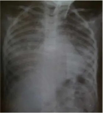
Figure 1. Thorax x-ray from a shone’s syndrome revealed no cardiomegaly.
At physical examination, her level of consciousness was clear, blood pressure were normal in
four extrimities, other vital signs were in normal limit. She was failure to thrive. Inspection showed no
chest retraction. Pulmonary auscultation within normal limit. Cardiac auscultation revealed sistolik
murmurs grade 3/6 at the third intercostal space left sternal border and holosystolic murmur grade 4/6
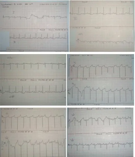
Figure 2. The ECG revealed sinus rhytm, RAH, and biventricular hypertrophy.
From the laboratory examination, complete blood count was in normal limit. Thorax x-ray
showed no cardiomegaly. Electrocardiography revealed sinus ritme, right atrium hypertrophy (RAH),
biventricular hypertrophy. Echocardiography revealed aortic and pulmonal stenosis, mitral
regurgitation-mitral stenosis with shortening and thickening of mitral valve, small VSD and left
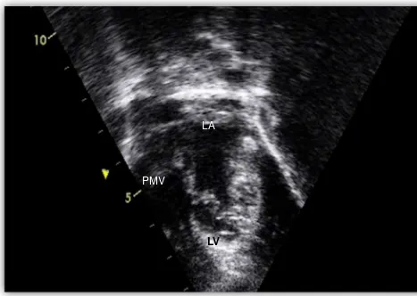
Figure 3. The apical view from transthoracal echocardiography showed PMV with shortening and thickening of the cordae, attached single posteriorly to free wall, and LV hipoplasia.
(PMV: Parachute Mitral Valve, LV: Left ventricle, LA: Left Atrium)
Figure 4. Transthoracal Echocardiography (TTE), parasternal long axis view showed small VSD, from apical view, systolic frame showed MR-MS.
(VSD: Ventricular Septal Defect, MR: Mitral regurgitation, MS: Mitral Stenosis)
Case 2
LV
LA
PMV
A 5 years old girl, was admitted to pediatrics emergency departement due to progressively
worsening dyspnea since 10 days before admission, without wheezing and stridor, with fever,
productive cough, and mild cyanosis on lips and most noticable cyanosis on the toes. There was
history of reccurent respiratory track infections.
At physical examination, her level of consciousness was clear, she was tachypnoeic (70
breaths/min), pulse 110 beats/min, temperature 38°C, blood pressure 90/60 mmHg in four extrimities,
Oxygen saturation was 88%. There were no sign failure to thrive. Inspection showed intercostal and
epigastric retraction. Pulmonary auscultation revealed crackles over both lung fields without
wheezing. Cardiac auscultation revealed sistolik murmurs grade 3/6 at the third intercostal space left
sternal border, continues murmur grade 3/6 at second intercostals space left sternal border and
diastolic murmur at the apex. There were no hepatomegaly or peripheral oedema. The extrimities
showed clubbing fingers and acrocyanotic on lower extrimities.
From the laboratory examination, complete blood count was slight increase in hematocrit
count, others within normal limit. The chest x-ray showed a mild increased cardio-thoracic index
(CTR 58%), there were enlargement of the left atrium and right ventricle. The confluent alveolar
opacities suggesting, in clinical context, the diagnosis of bronchopneumonia.
Figure 4. Chest X ray, child with Shone’s anomaly showed cardiomegaly (CTR 58 %) and confluent alveolar opacities.
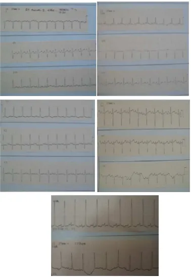
The electrocardiogram indicated sinus tachycardia (122 bpm), right axis deviation and
Figure 5. Electrocardiography indicated sinus tachycardi, RAD, RAH, RVH
Transthoracic echocardiography (TTE) revealed the presence of mitral stenosis with
parachute mitral valve. the chordae was thick and shorth with a narrow opening 3 mm, producing
severe mitral stenosis (mean transmitral pressure gradient 19 mmHg). Transthoracic
echocardiography also established the presence of hypoplastic aortic with the co-existence of large
PDA with right to left shunt, the aorta filled with PDA. Transthoracic echocardiography also revealed
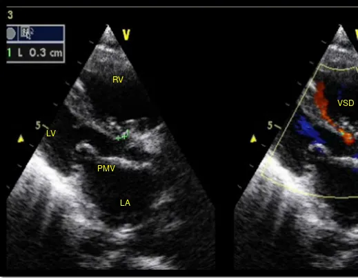
Figure 6. Transthoracic echocardiography, parasternal long axis view, showing hipoplastic LV, PMV and VSD 3 mm. (PMV: Parachute Mitral Valve, VSD: Ventricular Septal Defect, LV: Left Ventricle, RV: Right ventricle)
This patient were treated with oxygen and intravenous antibiotics for 8 days, and had slight clinical improvement, but she was discharge against medical advise because of economical issue.
DISCUSSION
The Shone’s complex defined by four cardiovascular defects such as a supravalvular mitral
membrane, valvular mitral stenosis by a parachute mitral valve, subaortic stenosis, and aortic
coarctation. In addition, these patients may have a component of endocardial fibroelastosis which
manifests as restrictive ventricular function, and some have a degree of LV hypoplasia which can
contribute to morbidity and mortality.1-5
Shone’s syndrome is a rare congenital heart disease (only 4 cases have been discovered
among 12.520 echo studies in a series published by Zucker et al), described by Shone et al initially in
1963, which occurs most frequently in its incomplete form.1,4 An incomplete form of Shone’s
PMV
LV
RV
LA
syndrome is defined by less than four criteria. The syndrome represents 0.6% of all cardiac
malformations with a male: female ratio of 1.5:1.6
It is mostly detected in childhood as the patient becomes symptomatic by the age of 2 years.
The usual symptoms are dyspnea, nocturnal cough, tachypnea, poor feeding, failure to thrive, fatigue,
and signs and symptoms of heart failure and reduced cardiac output. The child usually has recurrent
episodes of wheezing and respiratory tract infections due to pulmonary congestion and exudation of
fluid into the lungs. It is extremely unusual for a patient to remain largely asymptomatic throughout
childhood and get incidentally detected during adulthood while evaluating for some unrelated illness.2 Both cases in this report were diagnosed shone’s syndrome before age one year, preceeded by
recurrent respiratory tract infection, then confirmed by echocardiography.
Supravalvar mitral ring is a circumferential ridge or membrane, which arises from the left
atrial wall overlying the mitral valve and is frequently attached to the mitral valve. The ring may
range from a thin membrane to a thick discrete fibrous ridge. It may vary in its extent. Adhesion to the
valve may impair opening of the leaflets causing mitral-valve inflow obstruction in some patients. In
other patients, the ring may be large and protrude into the mitral-valve inflow thus causing
obstruction.1,4,7
Parachute mitral valve is a severe form of congenital mitral valve stenosis. In this anomaly,
all chordae tendineae are thickened and shortened and they attach to a single posteriorly located
papillary muscle, producing severe mitral stenosis. The anterior papillary muscle is usually absent.
The unifocal attachment of the chordae results in a restricted valve opening and subvalvar obstruction
and, rarely, valvar regurgitation. The diagnosis can be suspected by two-dimensional echo on the
parasternal views. In the parasternal short-axis view, only one papillary muscle is imaged.1,4,8
Mitral valve obstruction during early embryogenesis is considered the first pathological event
in Shone’s syndrome, causing underdevelopment of the LV cavity, thus leading to various degrees of
LV outflow tract obstruction and aortic coarctation. The embryologic origin of supramitral ring is
unclear, but is considered the result of incomplete division of endocardial cushion tissue.4,6
The first case, was found with milder symptoms, recurrent respiratory tract infection, poor
weight gain and late development. While the second case, was found in more severe condition, wich
experience severe dyspneu due to bronchopneumonia. After the further examination, the first case
revealed aortic and pulmonal stenosis, mitral regurgitation-mitral stenosis with shortening and
thickening of mitral valve, small VSD and left ventricle hipoplasia. And the second case, revealed the
co-existence of left ventricle and aortic hipoplastic, severe mitral stenosis due to parachute mitral
valve, VSD and a large PDA. These two cases were presented as incomplete form of shone’s
syndrome.
Patients with Shone’s syndrome have a poor long-term prognosis, with a perioperative
mortality rate of 24–27%, often requiring multiple interventions at an early age. In their first
critical lesion. Other studies confirmed that the severity of mitral valve obstruction correlates with
poor long-term outcome. Patients with the most severe forms of mitral obstruction presented with
severely elevated pulmonary artery pressure and had the poorest prognosis.4
In patients with aortic coarctation, the partial disappearance of elastic tissue at the level of
aortic media seems to be an important feature. Aortic coarctation occurs in 20–59% of cases with
mitral valve anomalies, whereas the mitral supravalvular ring is associated with other defects in
almost 90% of cases. Therefore, the finding of these defects should prompt for search of other cardiac
and vascular anomalies.4
A good outcome is possible in patients with Shone’s complex, provided the surgical
intervention is undertaken early before the onset of pulmonary hypertension. Mitral valve repair along
with resection of supramitral ring is preferable over valve replacement. Other surgical procedures
depend upon existence of associated cardiac anomalies, which ultimately define late surgical
outcome.2
This case study raises the awareness about this rare syndrome and highlights the importance
of a carefully performed echocardiogram particularly in patients with congenital heart disease where
lesions often co-exist and can be missed in the absence of a comprehensive examination.4
References:
1. Roche KJ, Genieser NB, Ambrosino MM, Henry GL. MR findings in Shone-s complex of left
heart obstructive lesions. Pediatr Radiol. 1998(28): 841-5.
2. Narvencar K, Costa AK, Patil VR. Shone’s complex. JAPI. 2009(57):415-7.
3. Shone JD, Sellers RD, Anderson RC, et al. The developmental complex of parachute mitral
valve, supravalvular ring of left atrium, subaortic stenosis, and coarctation of aorta. Am J
Cardiol.1963(11): 714-725.
4. Bogdan AP, Jurcut R, SerbanM, Parascan L, Ginghina C. Shone’s syndrome diagnosed with
echocardiography and confirmed at pathology. European Journal of Echocardiography.
2008(9): 865–867.
5. Joffe D, Gurvitz M, Oxorn D. An unusual presentation in a patient with shone’s anomaly.
Anesthesia-analgesia. 2008:1825-7.
6. Cinteza E, Ancha I, Gherghina I. Incomplete forms of Shone’s syndrome. Medika-a journal of
clinical medicine. 2008(3);255-61.
7. Serra W, Testa P, Ardissino D. Mitral supravalvular ring: a case report. Cardiovascular
ultrasound. 2005;1-4.
8.