The Effects of Aerobic Exercise and Detraining on Left
Ventricular Cardiomyocyte Apoptosis
Mustika A. Putri1, Dewi I.S. Santoso2, Ria Kodariah3 1Faculty of Medicine and Health Science, Syarif Hidayatullah State Islamic University
2Department of Physiology, Faculty of Medicine, Universitas Indonesia 3Department of Anatomic Pathology, Faculty of Medicine, Universitas Indonesia
KEYWORDS aerobic exercise, detraining, caspase-3, apoptosis, cardiomyocyte
ABSTRACT Apoptosis can occur in several pathological heart conditions. Physical exercise, particularly aerobic exercise may reduce apoptosis on cardiomyocytes. Detraining can restore adaptation after exercise. This study aimed to see the effect of aerobic exercise and detraining on left ventricular cardiomyocyte apoptosis using caspase-3 as the parameter. This was an in vivo experimental study on Wistar rats Rattus Novergicus. Rats divided to 8 groups: 4 sedentary control groups: 4-week (C4), 8-week (C4D), 12-week (C12), 16-week control (C12D), and 4 aerobic exercise treatment groups: 4-week (A4) and 12-week (A12), and 4 & 12-week post aerobic exercise treatment followed by 4 weeks detraining (A4D, A12D). Caspase-3 protein in rat left ventricular tissue was identified by immunohistochemistry staining. Data were analized with ANOVA test using SPSS proggramme version 20.
Data analysis showed an increase percentage of caspase-3 expression on post-aerobic exercise (A), be compared with conntrol group (C) (A4 65,3%±2,54 vs K4 6,4%±1,78, p<0,001; A12 41,8%±3,21 vs K12 5,7%±0,88, p<0,001; A4D 66,6%±1,89 vs K4D 8,6%±3,60, p<0,001; A12D 45,1%±1,50 vs K12D 7,4%±2,06, p<0,001). Percentage of caspase-3 expression was not different on post-aerobc exercise (A), be compare with detraining group (A4D 66,6%±1,89% vs A4 65,4%±2,54, p=0,484; A12D 45,1%±1,50 vs A12 41,8%±3,21, p=0,063). Percentage of caspase-3 expression on post 4-week aerobic exercise group was higher than post12-week aerobic exercise (A4 65,4%±2,54 vs A12 41,8%±3,21, p<0,001).
INTRODUCTION
Cardiovascular diseases still remain the number one cause of death in the world.(Lloyd-Jones et al. 2009) The disease may be resulted by dysfunction because of excessive apoptosis that occurs in a tissue or organ. In heart muscle cells (cardiomyocytes), ample loss of them contributes to contractile dysfunction, cardiomyopathy, heart diseases, and heart failure.(Kwak et al. 2013). Aerobic exercise is beneficial to improve and maintain cardiorespiratory fitness (Giam et al. 1993). It can increase the endogenous antioxidants in the body (Gomez et al. 2007, Berzosa C et al. 2011). These antioxidants counteract the adverse effects of reactive oxygen species (ROS) which induces cellular apoptosis (Gomez et al. 2007, Berzosa et al. 2011, Mooren et al. 2005, Phaneuf et al. 2001, Dyspersyn et al. 2001). Meanwhile, detraining may diminish general or partial anatomical and physiological adaptation caused by physical exercise occurred in the break period (Mujik et al. 2003).
Caspases are crucial mediators of apoptosis (Elmore et al. 2007). Caspase-3 is a frequently activated death protease and also required for some typical hallmarks of apoptosis. It is indispensable for apoptotic chromatin condensation and DNA fragmentation in all cell types. Thus, it is essential for certain processes associated with the dismantling of the cell and the formation of apoptotic bodies, but it may be also function before or at the stage when commitment to loss of cell viability is made (Elmore et al. 2007, Igney et al. 2002, Zeiss et al. 2003). Therefore, this study was aimed to determine whether aerobic physical exercise will reduce
apoptosis of cardiomyocyte and the effect of detraining on cardiomyocyte apoptosis using caspase-3 as parameter.
METHOD
This experimental study was done on cardiac ventricular tissue from male albino Rattus novergicus, Wistar strain rats, aged 8-10 weeks, weighing 100-250 grams. This sample was obtained from a larger published study on Blood lactate level in Wistar rats after four and twelve weeks intermittent aerobic training (Dewi N et al. 2013). The rats were given aerobic physical exercise on an animal treadmill (T-6000). The protocol for our study was modified from Manchado’s protocol (Manchado et al. 2005). The rats were given 1 week for training adaptation. The treadmill speed was gradually increased every day. In the treatment sessions, aerobic exercise was given for 4 weeks and 12 weeks at a speed of 20m/min for 20 minutes 5 times a week. A 90-second rest breaks every 5 minutes. The exercise was performed regularly five times a week. The exercise protocol employed was based on a preliminary study for establishing whether an exercise was aerobic in rats using lactic acid as the parameter. The exercise was still considered aerobic if post-exercise blood lactate < 4 mmol/L. It was found that this protocol resulted in blood lactate of less than 4 mmol/L (Manchado et al. 2005). In the aerobic group, the rats were trained for 4 and 12 weeks before the rats were decapitated. In the detraining group, aerobic physical exercise was stopped for 4 weeks before the rats were decapitated.
Correspondence:
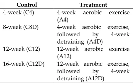
The control and treatment group was each divided into four sub-groups. (Table 1).
IMMUNOHISTOCHEMISTRY
Cardiac ventricular tissue specimens were made on paraffin
blocks, then stained
immunohistochemical with Starr Trek HRP-DAB (Horseradish peroxidase Diaminobenzidine). The primary antibody was an anti-caspase 3 antibody from Abcam (ab4051), with 1: 150 dilution. The primary antibody was incubated overnight at 40C.
PARAMETERS ASSESSMENT
The positive cells expressing caspase-3 were cells showing brown cytoplasm. Slide was photographed using a binocular light microscope (Olympus BX51) with a 400x magnification, and then we took 5 visual fields randomly for each preparation. Photo preparations that have been obtained are then processed with ImageJ program to determine the amount of protein expression of caspase 3. Evaluation is based on the percentage of imunoreactivity expression of caspase-3 by dividing the number of positive cells with total number of cells. Ethical approval was obtained from the Research Ethics Committee of Faculty of Medicine Universitas Indonesia.
RESULTS
Data were analyzed using One-Way ANOVA test that were continued with Post Hoc:LSD. We analyzed more than two sample groups. Post-aerobic exercise group (A4 & A12), will be compared with control group (C),
post-aerobic exercise group (A4&A12) will be compared with detraining group (A4D&A12D), post 4-week aerobic exercise group (A4) will be compared with post12-week aerobic exercise (A12). The difference between groups is considered significant if p < 0.05. Table 2 will show the ANOVA test.
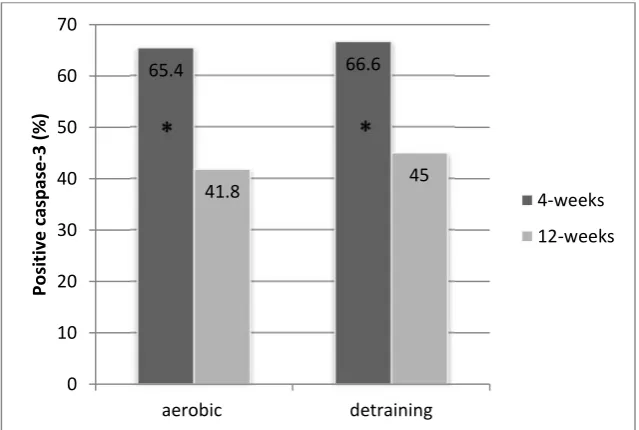
Figure 1 showed a representative immunohistochemistry image from each condition. The percentage of caspase-3 expression in rat left ventricle cardiomyocyte between the control group (C) and aerobic exercise treatment (A) are shown in figure 2. Expression of caspase-3 4 weeks and 12 weeks after physical exercise treatment groups higher than controls (A4: 65.38% 2.54 vs C4: 6.4% 1.78; A12: 41.8% 3.21 vs C12: 5.7% 0.88). Caspase-3 expresion in the detraining groups were also higher than controls (A4D 66.6%±1.89 vs C4D 8.6%±3.60; A12D 45.1%±1,50 vs C12D 7.4%±2.06) Caspase-3 expression in 4-week aerobic exercise group was significantly higher than the 12-week group (A4: 65.4% 2.54 vs A12: 41.8% 3.21, p<0.001). Meanwhile, caspase-3 expression in the 4-week detraining group was significantly higher compared to 12-week detraining group (A4D: 66.6% 1.89 vs A12D: 41.8% 3.21, p<0.001). Caspase-3 expression was not different between aerobic exercise group and detraining group in 4-week nor 12-week group. (A4: 65.4% 2.54 vs A4D 66.6% 1.89, p=0.484; A12: 41.8% 3.21 vs A12D: 45.1% 1.50, p=0.063). (Figure 3).
DISCUSSIONS
caspase-3 indicates an increase in left ventricular cardiomyocyte apoptosis. This finding is different with study conducted by Siu et al. (Siu et al. 2004). They showed that apoptotic index was lower in rat getting physical exercise compared with control (Siu et al. 2004). These results were probably due to the aerobic exercise protocol used in our study was too strenuous for the rats. They used a treadmill five times a week for 8 weeks and gradually increasing speed and duration of treadmill. In the first four weeks, the speed and duration of exercise was gradually increased from 10m/min for 10 min to 28m/min for 55 minutes at the end of the first four weeks. In the last 4 weeks, the rats were given warm-up 4exercises prior to exercise. The speed was 20m/min for 5 minutes, followed by the training session at a speed of 28m/min for 55 minutes.
They classified this exercise protocol as aerobic exercise with moderate endurance level (Siu et al. 2004). Our aerobic exercise protocol was too heavy for the rats in this study. The protocol for our study was 1 week for training adaptation. The treadmill speed was gradually increased every day. In the treatment sessions, aerobic exercise was given for 4 weeks and 12 weeks at a speed of 20m/min for 20 minutes 5 times a week. A 90-second rest breaks every 5 minutes. Physical exercise that was too strenuous can cause ischemia-reperfusion injury. In addition, mechanical factors that cause excessive strain on the muscles which may also be a factor that triggers cardiomyocyte apoptosis (Dyspersyn et al. 2001). Peterson et al proved that apoptosis of heart muscles in obese rats decreased after given physical exercise (Peterson et al. 2008). The rats were
trained on a treadmill for 9 weeks. The speed and duration gradually increased. During the first 3 weeks the rats ran 10m/min for 15 minutes, then gradually increased to 20m/min for 55 min/day (Peterson et al. 2008). The expression of caspase-3 in detraining group was higher than in the 4 and 12 weeks aerobic group, eventhough the differents was not statistically significant. Although this result did not prove that detraining may incease apoptosis, but further study should be made to confirm if detraining really increases apoptosis.
Aerobic exercise is expected to increase antioxidant capacity of the body that is expected to counteract the damaging effects of free radicals (Gomez et al. 2007, Berzosa et al. 2011, Power SK et al. 2002). Minimizing free radicals is the expected to prevent cardiomyocyte apoptosis. Aerobic exercise is also expected to increase stress protective proteins (stress-sensitive protective proteins), including nuclear factor kappa B (NF-kB), insulin-like growth factor (IGF-1), and heat shock protein (HSP90 and HSP70). They are able to reduce the incidence of apoptosis in the cell (Siu PM et al. 2004, Simon HU et al. 2000, Ji LL et al.2004, Gjoyaag TF et al. 206).
exercise group was higher than in the 12-week aerobic exercise group. This showed that physical exercise should be done regularly and continuously (chronic), not just occasionally (acute). In animals given chronic physical exercise small oxidative damage was found when compared to untrained animals, this was probably due to the adaptive response to long-term physical exercise. This adaptive response is the result of cumulative effects of the physical exercise stimulus given repeatedly for a prolonged time (chronic).(Gomez et al. 2007).
This study also showed (Figure 3) the average percentage of caspase-3 expression of 12-week detraining group lower than the 4-week detraining group. It showed that aerobic exercise if done for a prolonged time will provide better adaptation, although detraining may return cardiomyocyte apoptosis to previous levels. Therefore, the capacity of the endogenous antioxidant defense mechanisms in the body can be effective if physical exercise is carried out for a prolonged length of time and not just occasionally. In conclusion, the aerobic exercise protocol used in this study, was not found to decrease left
ventricular cardiomyocyte apoptosis. Detraining did not increase left ventricular cardiomyocyte apoptosis.
LIMITATION
Since this study was part of a larger study, the sample and protocol used was predetermined.
ADVANTAGE
Studies on apoptosis of cardiomyocytes are still not many, especially on apoptosis caused by aerobic exercise. This study can be a future reference for aerobic exercise studies, especially on the dose of aerobic exercise to be used.
ACKNOWLEDGMENT
The authors are grateful to Faculty of Medicine and Health Science, Syarif Hidayatullah State Islamic University, Department of Physiology and Department of Anatomic Pathology, Faculty of Medicine, Universitas Indonesia which have supported this research both material and non-material.
Table 1. Subdivision of control and treatment group
Control Treatment
4-week (C4) 4-week aerobic exercise (A4)
8-week (C8D) 4-week aerobic exercise, followed by 4-week detraining (A4D)
12-week (C12) 12-week aerobic exercise (A12)
16-week (C12D) 12-week aerobic exercise, followed by 4-week detraining (A12D)
Table 2. ANOVA Test
ANOVA
The Percentage of Capase-3 Expression in Rat Left Ventricle Cardiomyocyte
Sum of
Squares df
Mean
Square F Sig.
Between
Groups 20251.702 7 2893.100 529.677 .000 Within
Multiple Comparisons
Dependent Variable The percentage of caspase-3 expression in rat left ventricle cardiomyocyte
LSD
(I) Group (J) Kelompok penelitian Mean Difference (I-J) Std. Error Sig. 95% Confidence Interval Lower Bound Upper Bound aerobic 4 weeks aerobic 12 weeks 23.56179* 1.65258 .000 20.1510 26.9725
aerobic detrain 4 weeks -1.17508 1.65258 .484 -4.5858 2.2357 aerobic detrain 12 weeks 20.33345* 1.65258 .000 16.9227 23.7442 control 4 weeks 58.97370* 1.65258 .000 55.5629 62.3844 control 12 weeks 59.66061* 1.65258 .000 56.2499 63.0714 control 8 weeks 56.73174* 1.65258 .000 53.3210 60.1425 control 16 weeks 58.02365* 1.65258 .000 54.6129 61.4344 aerobic 12 weeks aerobic 4 weeks -23.56179* 1.65258 .000 -26.9725 -20.1510 aerobic detrain 4 weeks -24.73687* 1.65258 .000 -28.1476 -21.3261 aerobic detrain 12 weeks -3.22834 1.65258 .063 -6.6391 .1824 control 4 weeks 35.41190* 1.65258 .000 32.0012 38.8227 control 12 weeks 36.09882* 1.65258 .000 32.6881 39.5096 control 8 weeks 33.16995* 1.65258 .000 29.7592 36.5807 control 16 weeks 34.46186* 1.65258 .000 31.0511 37.8726 aerobic detrain 4
weeks
aerobic 4 weeks 1.17508 1.65258 .484 -2.2357 4.5858 aerobic 12 weeks 24.73687* 1.65258 .000 21.3261 28.1476 aerobic detrain 12 weeks 21.50853* 1.65258 .000 18.0978 24.9193 control 4 weeks 60.14877* 1.65258 .000 56.7380 63.5595 control 12 weeks 60.83569* 1.65258 .000 57.4249 64.2464 control 8 weeks 57.90681* 1.65258 .000 54.4961 61.3176 control 16 weeks 59.19873* 1.65258 .000 55.7880 62.6095 aerobic detrain 12
weeks
aerobic 4 weeks -20.33345* 1.65258 .000 -23.7442 -16.9227 aerobic 12 weeks 3.22834 1.65258 .063 -.1824 6.6391 aerobic detrain 4 weeks -21.50853* 1.65258 .000 -24.9193 -18.0978 control 4 weeks 38.64025* 1.65258 .000 35.2295 42.0510 control 12 weeks 39.32716* 1.65258 .000 35.9164 42.7379 control 8 weeks 36.39829* 1.65258 .000 32.9875 39.8090 control 16 weeks 37.69020* 1.65258 .000 34.2795 41.1009 control 4 weeks aerobic 4 weeks -58.97370* 1.65258 .000 -62.3844 -55.5629 aerobic 12 weeks -35.41190* 1.65258 .000 -38.8227 -32.0012 aerobic detrain 4 weeks -60.14877* 1.65258 .000 -63.5595 -56.7380 aerobic detrain 12 weeks -38.64025* 1.65258 .000 -42.0510 -35.2295 control 12 weeks .68691 1.65258 .681 -2.7238 4.0977 control 8 weeks -2.24196 1.65258 .188 -5.6527 1.1688 control 16 weeks -.95004 1.65258 .571 -4.3608 2.4607 control 12 weeks aerobic 4 weeks -59.66061* 1.65258 .000 -63.0714 -56.2499
aerobic 12 weeks -36.09882* 1.65258 .000 -39.5096 -32.6881 aerobic detrain 4 weeks -60.83569* 1.65258 .000 -64.2464 -57.4249 aerobic detrain 12 weeks -39.32716* 1.65258 .000 -42.7379 -35.9164 control 4 weeks -.68691 1.65258 .681 -4.0977 2.7238 control 8 weeks -2.92887 1.65258 .089 -6.3396 .4819 control 16 weeks -1.63696 1.65258 .332 -5.0477 1.7738 control 8 weeks aerobic 4 weeks -56.73174* 1.65258 .000 -60.1425 -53.3210
aerobic 12 weeks -33.16995* 1.65258 .000 -36.5807 -29.7592 aerobic detrain 4 weeks -57.90681* 1.65258 .000 -61.3176 -54.4961 aerobic detrain 12 weeks -36.39829* 1.65258 .000 -39.8090 -32.9875 control 4 weeks 2.24196 1.65258 .188 -1.1688 5.6527 control 12 weeks 2.92887 1.65258 .089 -.4819 6.3396 control 16 weeks 1.29191 1.65258 .442 -2.1188 4.7027 control 16 weeks aerobic 4 weeks -58.02365* 1.65258 .000 -61.4344 -54.6129
(A) (B)
(C) (D)
(E) (F)
Figure 1.Immunohistochemistry Image. The red arrow shows cell with
caspase-3 (brown in the cytoplasm of rat cardiac left ventricular
cardiomyocytes)(A)negative control.(B)positive control.(C)4-week aerobic exercise (A4).(D)12-week aerobic exercise (A12).(E)4-week aerobic exercise, followed by 4-week detraining (A4D).(F)12-week aerobic
* p < 0.05 between groups
Figure 2. Comparison of caspase-3 expression percentage on treatment group exercise to control
* p < 0.05 between groups
Figure 3. Comparison of the percentage of caspase- 3 expression beetwen 4-week aerobic group and
12-week aerobic group to detraining group.
6.4 8.6 5.7 7.4
65.4 66.6
41.8 45
0 10 20 30 40 50 60 70
A4 A4D A12 A12D
P
o
si
ti
v
e
c
a
sp
a
se
-3
(
%
)
control
treatment
*
*
*
65.4 66.6
41.8 45
0 10 20 30 40 50 60 70
aerobic detraining
P
o
si
ti
v
e
c
a
sp
a
se
-3
(
%
)
4-weeks
REFERENCES
Berzosa C et al. 2011. Acute exercise increase plasma total antioxidant status and antioxidant enzyme activities in untrained men. Journal of Biomeicine and Biotechnology. 2011.
Dewi N Sari, Sutjahjo Endardjo, Dewi IS Santoso 2013. Blood lactate level in Wistar rats after four and twelve week intermittent aerobic training. Medical Journal of Indonesia.2013;22(3).
Dyspersyn GGD, Borgers M 2001. Apoptosis in the heart:about programmed cell death and survival. News Physiol Sci.2001;16volume:41-47. Elmore S 2007. Apoptosis: A review of programmed cell death. toxicol pathol. 2007;35:495-516.
Gomez Cabrera MC, Domenech E, Vina J 2007. Moderate exercise is an antioxidant:upregulating of antioxidant genes by training. Elsevier.2007;44(2008):126-131.
Giam CK, Teh KC 1993. Ilmu kedoteran olahraga. Diterjemahkan oleh Satmoko H. Jakarta:Binarupa Aksara;1993. Gjovaag TF, Dahl HA 2006. Effect of
training and detraining on the expression of heat shock proteins in m.triceps brachii of untrained males and females. Eur J Appl Physiol.2006;98:310-322.
Igney FH, Krammer PH 2002. Death and anti death: tumor resistance to apoptosis. Nat Rev Cancer.2002;2:277-88.
Ji LL, Gomez-Cabrera MC, Steinhafel N, Vina J 2004. Acute Exercise activates nuclear factor (NF)-kappaB signaling pathway in rat skeletal muscle.FASEB J.2004;18:1499-1506.
Kwak HB 2013. Effect of aging and exercise training on apoptosis in the heart. JER. 2013;9:212-19.
Liu Y, Lormes W, Wang L, Reissnecker S, Steinacker JM 2004. Different skeletal muscle hsp70 responses to
high-intensity strength training and low-intensity endurance training. Eur J Appl Physio.2004;91:330-335.
Manchado FB, Gobatto CA, Contarteze RV, Papoti M, De Mello MAR 2005. Maximal lactate steady state in running rats. JEPonline. 2005;8(4):29-35.
Mooren FC, Volker K, editor 2005. Molecular and celullar exercise physiology. USA:Human Kinetics;2005.
Mujik I, Padilla S 2000. Detraining: loss of training-induced physiological and performance adaptations Part I.Sport Medicine. 2000;30(2):79-87.
Peterson JM, Bryner RW, Sindler A, Frisbee JC, Alway SE 2008. Mitochondrial apoptotic signaling is elevated in cardiac but not skeletal muscle in the obese Zucker rat and is reduced with aerobic exercise. J Appl Physiol. 2008;105:1934-1943.
Phaneuf S, Leeuwenburgh C 2001. Apoptosis and exercise. Med Scie Sports Exerc. 2001;33:393-396.
Powers SK, Lennon SL, Quindry J, Mehta JL 2002. Exercise and cardioprotection. Curr Opin Cardiol 2002;17:495-502. Simon HU, Haj-Yehia A, Levi-Schaffer F
2000. Role of reactive oxygen species (ROS) in apoptosis induction. Apoptosis.2000;5:415-418.
Siu PM, Bryner RW, Martyn JK, Alway SE 2004. Apoptotic adaptations from exercise training in skeletal and cardiac muscles. The FASEB.2004;18:1150-52. The American Heart Association Statistics
Committee and Stroke Statistics Subcommittee 2009. Heart Disease and Stroke Statics 2009 Update: A Report from the American Heart Association Statistics Committee and Stroke Statistics Subcommittee. Circulation 2009; 119:e21 – e181.