CrossMark
Abstract
Objective:This study was an observational analytic using cross-sectional study, where all data are observed once at the time. In this study, the amount of sample reviewed were 30 samples, which consisted of 4–9 years old children. Panoramic radiographs were collected based on target population, which fulfilled sample criteria from reconciled patient of Dental Hospital Department of Dental Radiology Hasanuddin University.
Material and Methods:The tooth eruption is estimated according to Demirjian’s method by assessing growth and development process of tooth using panoramic radiography. The difference between
chronological age and dental age is determined using Demirjian’s method based on radiology analysis of panoramic radiography. Results: The estimating the score of dental age using Demirjian method. After that, the dental age and chronological age was analyzed to obtain the mean difference. Based on Wilcoxon test, the mean value was obtained as p:0.011 (p<0.05), this result shows that there is significant difference between chronological age and dental age. Conclusion:As a conclusion the chronological age and dental age can be assessed by reviewing the panoramic radiography using Demirjian’s method.
Keywords:Dental age, Chronological age, Demirjian’s method, Panoramic radiography
Cite this Article:Yunus B, Wardhani Y. 2016. Differences between chronological age and dental age using demirjian’s method based upon a radiology study using panoramic radiography at the dental hospital Hasanuddin university. Journal of Dentomaxillofacial Science 1(2): 96-101.
DOI:10.15562/jdmfs.v1i2.6
In
troduction
The tooth eruption is a physiological process in the form of tooth movement, which starts from the place of tooth development inside the alveolar bone, then the tooth penetrates the gingival until it finally reached the occlusal plane.1,2
The chronological age is determined based on the date, month and year of birth.3
In general, the somatic development is related with the chrono-logical age as in the measurement of somatic matu-rity, such as the bone age, menstruation and the body height. The somatic maturity can be used to estimate the chronological age if there is no other accurate age information available.4,5
This infor-mation is important in medical practices and for dentists to evaluate the development of the patient. The chronological age is frequently not enough in marking the growth stage and somatic maturity of the patient, therefore determining the biological age is needed.6
Dental age, or what they usually call the biological age, is the estimation of age, which is calculated based on the growth and development of the human beings. The dental age gives information that if the growth of a person has reached a certain stage.7
The tooth eruption period has some flaws where its reliability
is still questioned because it is difficult to point out the exact eruption period because the event occurs fast, so the marking is done clinically. Moreover, it is affected by the local factors like systemic disease, as well as eating habits. At the same time, the calcifi-cation stage method is used as a more reliable crite-rion (criteria) to determine the tooth maturation stage. Tooth calcification method gives a very clear description in deciding teeth maturation.7
Hence, the dental age in this research is determine by the method of tooth calcification stage.
In this research, the Demirjian’s method is used to determine the dental age of the patients, which is the process of permanent tooth bud’s calcification. From tooth bud, there is no calcification until the final forming of tooth root.8
This research is done to find the difference between chronological age and dental age by testing the reliability of the Demirjian’s method that analyzed teeth’s calcification stage to acquire the dental age of the patient. Therefore, the distribution of dental age estimation using the Demirjian’s method observed using the panoramic radiography photo needs to be known, which will later be subtracted by the chronological age to find the difference between the chronological age and the dental age.
1Department of Dental Radiology, Faculty of Dentistry, Hasanuddin University, Makassar, Indonesia 2Faculty of Dentistry, Hasanuddin University, Makassar, Indonesia
*Correspondence to: Yulia Wardhani, Faculty of Dentistry, Hasanuddin University, Makassar, Indonesia [email protected]
Received: 12 April 2016 Revised: 15 August 2016 Accepted: 17 August 2016 Available Online: 31 August 2016
Differences between chronological age and dental
age using Demirjian’s method based upon a
radiology study using Dental Hospital Department
of Panoramic Radiography Hasanuddin University.
Material and Methods
The design used in this research is observational analysis with cross-sectional design, done in Dental Hospital Department of Dental Radiology Hasanuddin University in Makassar, Indonesia on 1st April–31st May 2015.
Populations of research are children aged 4–9 years old, who visited Dental Hospital Department of Dental Radiology Hasanuddin University. Research samples are panoramic radiography and the chronological age of the patients, which were obtained from the identity of the patients who were referred to Department of Dental Radiology during April until May 2015. The samples were acquired by consecutive sampling technique, a method of acquiring sample based
on some specific criteria such as the assigned time interval or sample numbers or patients.
The data was analyzed using SPSS 18.0 (SPSS Inc, Chicago, IL, USA) program. The research protocol was approved by the ethical committee, Faculty of Medicine, Hasanuddin University.
Results
The research was done on 47 patients of Dental Hospital and Radiology Section of Oral, the place of the research. From the data gathered from April to May, some data were dropped out due to some exclusion criteria. Therefore, the distribution was done on 30 subjects. Descriptions of chronological age and dental age were acquired by Demirjian’s method, based on radiology study using panoramic radiography. The research results are shown on the distribution table 1.
Table 1 shows the characteristics of samples distribution in a total of 30 people (100%). In this research, the number of females are larger than the number of males with 18 females (60%) and 12 males (40%). The average of chronological age reaches up to almost 7 years old or more than 6 years old, meanwhile, the average of dental age acquired by Demirijan’s method reaches up to more than 7 years old. Based on chronological age cate-gory, the most number of samples was found on 6–7 years old and 8–9 years old category, each was 12 samples in total (40%). Only six samples with the chronological age of 4–5 years old were acquired in this research. In contrast, based on the dental age, the age 6–7 years old category has the most number of samples, with 14 people in total (46.7%), meanwhile age 8–9 years old came second, with Table 1 shows the characteristics of research samples distribution
Sample characteristics Frequency (n) Percentage (%) Mean ± SD
Gender
Male 12 40
Female 18 60
Chronological age (year) 6.91 ± 1.40
4–5 6 20
6–7 12 40
8–9 12 40
Dental age (year) 7.20 ± 1.26
4–5 7 23.3
6–7 14 46.7
8-9 9 30
Total 30 100
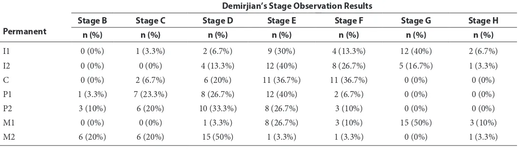
Table 2 Shows Demirjian’s method: observation of stages of permanent teeth sample distribution
Permanent
Demirjian’s Stage Observation Results
Stage B Stage C Stage D Stage E Stage F Stage G Stage H
n (%) n (%) n (%) n (%) n (%) n (%) n (%)
I1 0 (0%) 1 (3.3%) 2 (6.7%) 9 (30%) 4 (13.3%) 12 (40%) 2 (6.7%)
I2 0 (0%) 0 (0%) 4 (13.3%) 12 (40%) 8 (26.7%) 5 (16.7%) 1 (3.3%)
C 0 (0%) 2 (6.7%) 6 (20%) 11 (36.7%) 11 (36.7%) 0 (0%) 0 (0%)
P1 1 (3.3%) 7 (23.3%) 8 (26.7%) 12 (40%) 2 (6.7%) 0 (0%) 0 (0%)
P2 3 (10%) 6 (20%) 10 (33.3%) 8 (26.7%) 3 (10%) 0 (0%) 0 (0%)
M1 0 (0%) 0 (0%) 1 (3.3%) 8 (26.7%) 3 (10%) 15 (50%) 3 (10%)
M2 6 (20%) 6 (20%) 15 (50%) 1 (3.3%) 1 (3.3%) 0 (0%) 1 (3.3%)
people in total (30%). The dental age 4–5 years old category has the lowest number of samples with seven samples in total (23.3%).
Table 2 shows the Demirjian’s method: obser-vation of stages of permanent teeth sample distri-bution. The result showed from 30 I1 teeth that have been examined, about 12 I1 teeth (40%) have reached stage G and only one I1 tooth (3.3%) that is still in the stage C. In addition, only two teeth
have entered in the stage H. Table 2 also shows that there are 12 I2 teeth (40%), which reached stage E and only one I2 tooth (3.3%) that has reached the stage H. In the canine teeth, 11 teeth (36.7%) have reached stage E and F but none of the teeth has reached the stage G or H. Other observations show that 12 (40%) P1 teeth have reached the stage E but still there is one P1 tooth in stage B. About 10 P2 teeth reached stage D, which is 10 P2 teeth (33.3%) and only 3 P2 teeth reach the stage F. In addition, of the 30 M1 teeth observed, three M1 teeth have reached stage H and still one tooth that is at the stage D. In contrast, in the M2 teeth, only one M2 tooth that has reached stage H and six teeth (20%) are at stage B.
Table 3 shows the chronological age and dental
age average distribution in years by gender, chrono-logical age category and dental age category. The dental age reaches 5.38 years old. Meanwhile, in the 6–7 years old category, the dental age is also higher than the chronological age. However, it is inversely proportional as seen in the 8–9 years old chrono-logical age category in which the chronochrono-logical age is higher than the dental age. Based on the dental age category, as seen in the 4–5 years old dental age category, the chronological age reaches 5.14 years old, while the dental age is only 5.25 years old. In the 8–9 years old dental age category, the chrono-logical age and dental age has the same average.
Table 4 shows the chronological age and dental
age category by gender distribution. The study shows, at the 4–5 years old chronological age and dental age, the number of men is more on chrono-logical age category, while the number of women is as much on the dental age category. In the 6–7 years old chronological and dental age category, the number of men is more on the dental age compared to chronological age category but the number of women on both categories is the same. As for the 8–9 years old dental and chronological age, the number of men and women on chronological age category is more than the dental age.
Table 5 shows the overall differences of chrono-logical age and dental age that were obtained from Demirjian method. The results showed that the chronological age only reached 6.91 years, while the dental age obtained from Demirjian method reaches 7.2 years. Table 5 also shows the normality test results to determine the statistical test used in this study. The Shapiro –Wilknormality test results Table 5 Shows chronological age and dental age overall
differences
n (%) Chronological age Dental age
p–––value
Mean ± SD Mean ± SD
30 (100%) 6.910 ± 1.409a 7.200 ± 1.264 0.011*
Note: Normality test; Shapiro–Wilk test: p > 0.05; normal data distribution. Sign Rank test: p < 0.05.
Table 3 Shows chronological age and dental age average (years) distribution by gender, chronological age category and dental age category
Female 6.889 ± 1.490 7.044 ± 1.407
Chronological age
Total 6.910 ± 1.409 7.200 ± 1.264
showed that p>0.05 is only available on the chrono-logical age group. This means only the chronochrono-logical age group data is normally distributed, while the dental age data obtained from Demirjian’s method is not normal. This does not qualify the parametric test that requires the entire data is normally distrib-uted, thus the non-parametric test used in this study is Wilcoxon Signed-Rank test. Based on the Wilcoxon test results, we found the value of p:0.011 (p<0.05), which means that there are differences in chronological age and dental age is significant.
Discussion
This study was conducted to determine the differences in chronological age and dental age and was analyzed from the images and panoramic radio-graphs and measured using Demirjian’s method. The chronological age obtained from age dentition/ tooth eruption seen by date, month and year of birth of the patient. Dental age is obtained by looking at the growth and development of patient’s teeth using eight stages assigned by Demirjian’s method.
Maturity can be determined by the stage of tooth eruption and tooth calcification. Tooth eruption is the process of tooth movement toward occlu-sal plane, starting from the tooth root formation. Time eruption is a clinical maturation index. Tooth eruption time method has its drawbacks including: difficult to determine the exact time of the eruption because of the activities that take place quickly, clinically judged and influenced by local factors, systemic disease and diet so the reliability is ques-tionable. While the dental calcification stage is used as a more reliable criterion to determine the tooth maturation stage, the tooth calcification gives a very clear picture in determining teeth maturation.15
According to Flores et al.16
tooth maturation is expressed as dental age because it is clinically easier to be determined. By determining the maturation of teeth, dental calcification stage is more widely used than tooth eruption.17
This study also used the dental calcification stage observation that is also used by Demirjian et al. on the observation of eight tooth calcification stages defined in the Demirjian’s method. To assess the process of tooth calcification, panoramic radiographs guide can be used to evaluate the teeth on each inspection.
Panoramic radiography is one of the extra-oral radiographs which is often used in dentistry to obtain a complete picture of the whole maxillofacial.10
This study takes an x-ray panoramic radiographs photo to analyze the dental calcification according to the provisions laid down by the eight stages of Demirjian method, that is
figure 5, Stage A: Occlusal point calcification, with-out fusion of other calcification parts, Stage B: The fusion of the mineralization point where the occlu-sal surface contoured the teeth is already seen, Stage C: Calcification of the dental crown has been completed and dentin disposition starts, Stage D: The formation of the crown has been completed, Stage E: The root length of the teeth is shorter than the crown height, Stage F: The root length of the teeth exceeds the crown height, Stage G: The formation of the root has been completed, but the apical foramen is still open, Stage H: Apical fora-men was closed.9,18
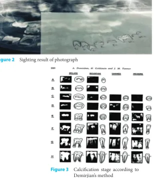
Figure 1 A panoramic photograph result from a 5 years old boy
Figure 2 Sighting result of photograph
Application Stage Demirjian’s Method
1. The panoramic photograph result is shown (digital).
2. A picture sighting shows seven left mandibular teeth starting from central incisor until the second molar.
3. After having the sighting results, the devel-opment stage of roots and the crowns of the seven left mandibular teeth analyzed (except the third molar) were observed through panoramic radiography and classified according to the valuation parameter of calcification stage of teeth by Demirjian’s method.
4. Each calcification stage has a score that has been determined. Scores are differentiated by gender.
5. After the classification and scoring of each tooth is completed, then all the scores of the roots and crowns development stage of central incisor, lateral incisor, canine, first and second premolars, first and second molar that have been determined by Demirjian et al. are added. 6. The addition results of seven teeth scores are then converted into a conversion of teeth maturity table that have been set by Demirjian’s method to determine the patient dental age
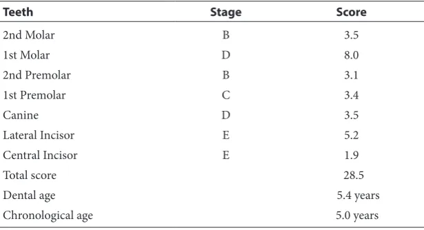
table 6.
Based on the study results, after the sighting and adding the scores and determining the dental age of the entire sample, it was found that the chrono-logical age and the dental age of the man has a difference of 0.491, whereas in women, the chrono-logical age and dental age only differed by 0.155. Based on chronological age categories, seen in the age category of 4–5 years old, the chronological age reaches
4.8 years old but the dental age reaches 5.38 years old. Meanwhile, in the age category of 6–7 years factors: ethnic variations, genetic and environmental factors: such as socioeconomic status, nutrition and lifestyle.
Cheraskin et al.5
found that the chronological age and dental age showed not only a significant relationship between men and women but also that the maturity of the teeth can be used as an indicator to determine the chronological age.
Based on the research results, the normality test results showed that the data is normally distributed in the chronological age group only, while the dental age group data were not normally distributed. This proves that the results of this study were based on Wilcoxon test with a value of p:0.011 (p<0.05), which means that there are significant differences in chronological age and dental age.
Conclusion
The difference of chronological age and dental age in each population were different, the dental age have a big margin than the chronological age, the chronological age have a big margin than the dental age and also the average of the chronological age and dental age were equal. Based on the research results it can be concluded that there are significant differences in chronological age and dental age.
As a suggestion in doing panoramic radiogra-phy techniques, an operator must really master photo techniques and ways to approach pediatric patients who sometimes are not cooperative and lastly a dentist is expected to understand and apply Table 6 Shows teeth maturity to determine patient dental age
Conflict of Interest
The authors report no conflict of interest.
References
1. Indriyanti R, Pertiwi AS, Sasmita IS. Pola erupsi gigi per-manen ditinjau dari usia kronologis pada anak usia 6 sam-pai 12 tahun. Bandung: Laporan Penelitian FKG UNPAD; 2006. p. 1–25.
2. Mokhtar M. Dasar-dasar orthodonti: pertumbuhan dan perkembangan kraniofasial. Medan: Bina Insani Pustaka; 2002;2: 245–224.
3. David S. Perbandingan usia kronologis berdasarkan gam-baran radiografis dari tahapan erupsi gigi molar ketiga rahang bawah dengan metode olze antara pasien laki-laki dan perempuan di RSGM Prof. Soedomo tahun 2008-2013. Yogyakarta: Universitas Gadjah Mada Electronic Theses & Dissertations (ETD); 2013.
4. Rai B, Anand SC. Tooth developments: an accuracy of age estimation of radiographic methods. World J Medical Sci 2006;2: 130–132.
5. Mc-Kenna CJ, James H, Taylor JA, et al. Tooth development standards for South Australia. Aus Dental J 2002;3: 223–227.
6. Rakosi T, Jonas I, Greber TM. Orthodontic diagnosis: color atlas dental medicine thieme 1992;1: 98–107. 7. Uysal T, Sari Z, Ramoglu SI, et al. Relationships between
dental and skeletal maturity in Turkish subjects. Angle Orthod 2004;5: 657–664.
8. Bosmans N, Ann P, Medhat A. et al. The application of Kvaal’s dental age calculation technique on panoramic dental radiographs. Forensic Sci Int 2005.
9. Bagherian A, Sadeghi M. Assessment of dental maturity of children aged 3.5 to 13.5 years using the Demirjian method in Iranian population. J oral Sci 2011;53: 37–42. 10. Behrman, Kliegman, Arvin, et al. Ilmu kesehatan anak.
15th ed. Jakarta: EGC; 1999. p. 83.
11. Hegde RJ, Sood PB. Dental maturity as an indicator of chronological age: radiographic evaluation of dental age in 6 to 13 years children of Belgaum using Demirjian methods. J Indian Soc Pedo Prev Dent 2002;4: 132–137.
12. Tamba S. Waktu erupsi gigi permanen ditinjau dari usia kronologis pada anak usia 6 sampai 12 tahun di SD ST Antonius V Medan. FKG USU; 2010. p. 46–50.
13. Chiego DJ. Oral histology. Available at http://crse.dent. umich.edu. 2006.
14. Kurita LM, Menezes AV, Casanova MS, et al. Dental maturity as an indicator of chronological age: radiograph assessment of dental age in a Brazilian population. J Appl Oral Sci 2007;2: 99–104.
15. Nassar AS. The relationships between cervical vertebral maturation and dental calcification among Malays. Malaysia: Master of Science USM; 2008. p. 1–24. 16. Flores C, Nebbe B, Major PW. Use of skeletal maturation
based on hand-wrist radiographic analysis as a predictor of facial growth: a systemic review. Angle Ortho 2004;74: 118–124.
17. Janson GR. A review of the most commonly used dental age estimation techniques. Odon Forensic J 2001;19: 9–17. 18. Siswanto F, Sjahruddin L. Correlation between mandible length and dental calcification on Deutero-Malay children aged 8–16 years. Fakultas Kedokteran Gigi Trisakti; 2009. p. 198–200.
19. Willems G, Van-Otmen A, Spiessens B, et al. Dental age estimation in Belgian children: Dermijian’s technique revisited. J Forensic Sci 2001;464: 893–895.
20. Yan-jin, Lou Xi. Assessment of dental age of children aged 3.5 to 16.9 years using Demirjian’s method: a meta-analysis based on 26 studies. Plos one 2013;8: 1–7.
21. Whaites E. Essentials of dental radiography and radiology. Third edition. Churchill Livingstone. Edinburgh London. New York: Oxford; 2002.
22. Pasler, Friedrich A. color atlas of Dental Medicine Radiology. Thieme; 2006.