VOL. 6, NO. 1, pp. 10-14, January, 2016
Correlation between Serum Ferritin and Cardiac Troponin I in Major Beta Thalassemia Children
Muhammad Ali Shodikin1*, Renny Suwarniaty2,3, Susanto Nugroho2,3
1Faculty of Medicine, Jember University, Jember, Indonesia
2
Departement of Child Health, Faculty of Medicine, Brawijaya University, Malang, Indonesia 3
dr. Syaiful Anwar Public Hospital, Malang, Indonesia
ABSTRACT
Major beta thalassemia (MBT) is a hereditary disease which synthesies defects in beta chains of haemoglobin, it is causes red blood cell destruction and the symptoms of anemia. Red blood cell destruction, frequent blood transfu -sion and low adherence to routine use of iron chelator lead to iron accumulation in the heart, liver and endocrine organs. Accumulation of iron in the myocard can lead acute myocardial infarction. One of cardiac markers that had been used for the diagnosis of myocardial infarction was cardiac troponin I (cTnI). The aim of this research is find the correlation between serum ferritin levels and cTnI in MBT children. A descriptive analytic research was conducted using a cross sectional design. The subjects were divided into 2 groups, the MBT group and the control group. In both groups, the serum ferritin and cTnI levels ere evaluated. Data were analyzed using ttest and Pear -son correlation test. Eleven children in the MBT group and 11 children in the control group were involved in this study. In the MBT group, the mean of serum ferritin and cTnI levels were 4292.5 µg/L and 0.20 ng/mL respectively. The mean of serum ferritin levels in the MBT group were higher than in the control and statistically signifi -cant (p= 0.0004). The mean of serum ferritin levels in the MBT group were higher than in the control and statistically significant (p= 0.0004). The mean of serum cTnI in the MBT group were higher than in the control, but sta -tistically not significant (p= 0.82). In the MBT group, there was a weak corellation between serum ferritin and cTnI levels (r= 0.34).
Keywords: children, cTnI, ferritin, Major beta thalassemia
Major beta thalassemia (MBT) is a hereditary dis-ease which synthesies defects in beta chains of haemoglobin. MBT causes red blood cell destruction, leads to anemic symptoms and need for regular blood transfusions [1, 2, 3]. Haemolysis and regular blood transfusions can lead to iron overload in the liver, en-docrine organs and myocardium [1, 3] Iron overload can be evaluated easily and verified by measuring serum ferritin levels [1, 4, 5]. High serum ferritin levels increase the risk of myocardial infarction in adults.6,7 Myocardial infark can be diagnosed by the cardial biomarker cardiac troponin I (cTnI) [8]. In Indonesia, the correlation between serum ferritin level and cTnI in MBT children has never been studied.
Study design
A descriptive analytic research was conducted using a cross sectional design, on May 2015, at the Pediatric Departement and Clinical Pathology Departement of Syaiful Anwar General Hospital (SAGH) Malang, In-donesia, and approved by the ethics committee of SAGH. The subjects were divided into 2 groups, the MBT group and the control group.
The patients
Inclusion criteria of MBT subjects were children 1 -18 years old, diagnosed as MBT by haemoglobin elec-trophoresis, needing regular blood transfusion and ap-proved by their parents after informed consent. Inclu-sion criteria for the control groups were children 1 -18 years old, not diagnosed as MBT, never had regular blood transfusion and approved by their parents after informed consent. Respondents were excluded if they had autoimmune haemolytics anemia, iron deficiency
JTLS | J. Trop. Life. Science 10 Volume 6 | Number 1 | January | 2016
MATERIALS AND METHODS INTRODUCTION
*Corresponding author: Muhammad Ali Shodikin
anemia, aplastic anemia, severe infections, malignancy, severe malnutrition and congenital or acquired heart disease.
Assays
In both groups were perform laboratory examina-tion of peripheral blood to checks the serum ferritin and cTnI levels. Serum ferritin levels were measured by immunoturbidimetry assays using FERR3:ACN165, Roche/Hitachi cobas c system analyzer (US). Serum cTnI level were measured by sandwich im-munochroma-tography using AIM TROPONIN I Q Rapid test (Indonesia).
Statistical analyses
The differences in serum ferritin and cTnI levels be-tween the MBT and control groups were analyzed by a t-test. The correlation between serum ferritin and cTnI levels in the MBT group were analyzed by a Pearson correlation test. Confidence interval 95% (α= 0.05) Data were analyzed using SPSS for Windows 16.0.
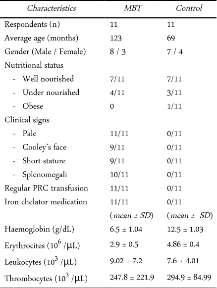
The subjects of this study were 22 children, 11 chil-dren in the MBT group and 11 chilchil-dren in the control group. The mean of age of the MBT and the control groups were 123 months and 69 months respectively. In the MBT group, 8 of 11 were male and 7 of 11 were in good nutritional status. In the control group, 7 of 11 were male and 7 of 11 were had a good nutritional sta-tus. In the MBT group, palor was in all patients, Coo-ley’s face in 9 of 11, spleen enlargement in 10 of 11 and short stature in 9 of 11. Whereas in the control group, there was no palor, Cooley’s face, spleen en-largement or short stature. All subjects in the MBT group were got Packed Red Cell (PRC) transfusions regularly every 1 - 2 months as well as iron chelator medication and no one of control group got PRC transfusions or iron chelator (Table 1).
Complete blood count result showed the average of haemoglobin level before transfusion in the MBT group was 6.5 g/dL that lower than in the control group whose haemoglobin level was 12.5 g/dL. The av-erage of erythrocytes in the MBT group and control groups were 2.9 × 10⁶
/µL and 4.86 × 10⁶
/µL respec-tively. The average of leukocyte in the MBT group and control groups were 9.02 × 10³
/µL and 7.6 × 10³ /µL respectively. The average of thrombocyte in the MBT group and control groups were 247.8 × 10³ - Splenomegali 10/11 0/11 Regular PRC transfusion 11/11 0/11 Iron chelator medication 11/11 0/11
(mean ± SD) (mean ± SD) Haemoglobin (g/dL) 6.5 ± 1.04 12.5 ± 1.03
Erythrocites (106 /μL) 2.9 ± 0.5 4.86 ± 0.4
Leukocytes (103 /μL) 9.02 ± 7.2 7.6 ± 4.01
Thrombocytes (103 /μL) 247.8 ± 221.9 294.9 ± 84.99
The mean of serum ferritin level in the MBTgroups was 4292.5 µg/L higher than it was in the control groups with only 136.2 µg/L. Statistics analyses by t test showed there was a significant difference in serum feritin levels between the MBT and control groups (p value = 0.0004). The mean of cTnI in the MBT group was 0.22 ng/mL, higher than it was in the control group with only 0.20 ng/mL. Statistics analyses by t test indicated difference of cTnI level between the MBT and control groups was not significant (p value = 0.82) (Table 2). In the MBT group, the result of Pearson cor-relation test showed there was a weak corcor-relation be-tween serum ferritin and cTnI levels (r value = 0.34) (Figure 1).
The age average of the children in the MBT group was 10 years 3 months. MBT is usually diagnosed when the children are under 2 years old [9]. In this
re-Table 2. Serum ferritin and cTnI levels
Figure 1. Correlation between serum ferritin and cTnI level in MBT children
search, respondents in the MBT group had got regular PRC transfusion every 1-2 months for many years. The older MBT patiens are more susceptible to complica-tions, such as heart, liver and endocrines disturbance [10].
Eight out of 11 children in the MBT group were male, the male to female ratio being 8:3. Another study in thalassemic children in Surabaya had a male to fe-male ratio of 44 : 17 [11] but in Jakarta this ratio was 37 : 36 [12]. Male and female proportions in MBT pa-tients are influenced by autosomal ressessive inheri-tance and not sex chromosomal linked [2, 9].
Seven children out of 11 in the MBT group had good nutritional status. It was shown they had had proper medical and nutritional care. All the patients (11/11) in the MBT group were pale, caused by ery-throcytes damage and ineffective eryery-throcytes synthesis due to globin chain defect [13,14]. Furthermore, almost all patients in the MBT group had splenomegalli but there were no one in the control group. The spleen was the main organ for erythrocyte degradation [2].
A majority of children (9 / 11) in the MBT group had short stature, whereas, all children in the control group had normal stature. In MBT patients, the short stature was caused by growth hormon disturbance, chronic hypoxia due to chronic anemia and cardiovas-cular disturbance [10]. A majority of the children (9 / 11) in the MBT group suffered from Cooley’s face caused by erythropoitic expansion in the bone marrow leading to the skull and face bone deformities [13].
Complete blood count results showed the average of haemoglobin levels before transfusion in the MBT group were lower than those in the control group. Haemoglobin levels in thalassemic children were less than 7 g/dL when diagnosed at the first time [9]. Ane-mia in the MBT children was caused by erythrocyte
damage and ineffective erythrocyte synthesis due to globin chain defect [13, 14]. The laboratory tests for es-tablishing the diagnoses of MBT were erythrocyte in-dex, peripheral blood smear and haemoglobin elec-trophoresis [15]. The average leukocyte and thrombo-cyte counts in both groups were within the normal limits.
The average serum ferritin level in the MBT group was 4292.5 µg/L, which was not only higer than that in the control group but also higher than normal limits (20 – 200 µg/L). Statistics analyses by t test showed significant differences in serum ferritin levels between the MBT and control groups (p value = 0.0004). A study in China also found high serum ferritin levels in MBT patients, 2754 µg/L [16]. Another study on Egyp-tian children with MBT also found high serum ferritin levels, 4510 µg/L [17]. Serum ferritin levels of more than 1000 µg/L. was indicate iron overload [18].
Iron overload in MBT patients is caused by ery-throcyte damage, frequent PRC transfusions and in-creased iron absorbtion [19]. Continual iron overload will lead to iron deposition in the liver, heart and en-docrine organs [1, 3]. Non transferin-bound forms of iron (NTBI) in the cytoplasmic can induce the conver-tion of Fe2+
to Fe3+
and lead increase free radicals such as reactive oxygen species [20]. Free radicals will in-duce lipid peroxidation in the cell organels such as lysosom, mitochondria and cytoplasmic membranes. Lipid peroxidation can disturb the cell function and cause cell necrosis including miocytes in the myocar-dial layer [20]. Iron chelator medication will not only reduced the iron deposition and labile iron but also re-duced free radicals [4].
The average cTnI levels in the MBT group was 0.22 ng/mL which was higher than those in the control group at 0.20 ng/mL, but the differences in cTnI levels between both groups were not statistically significant (p= 0.82). Based on equipment standard were used in this study, the serum cTnI levels were normal if less than 0.8 ng/mL. This result showed the serum cTnI levels in both groups were within normal limits. The normal serum cTnI levels indicated there was no my-ocardial injury in either groups. The mymy-ocardial injury leads to myocyte damage and induces the leakage of cTnI into the systemic circulation. Increasing cTnI lev-els can be detected although in the intial myocardial injuries even if echocardiography can not detect it [19]. The correlation between the serum ferritin and cTnI levels in the MBT group was weak positive corre-lation (r value=0.34). It was mean increasing of serum feritin level will increase the serum cTnI level. Serum
cTnI level higher than normal had 100% sensitivity and 96.3% specificity for diagnosing the myocardial in-farction [21]. cTnI serum levels was higher than mal indicate mycordial injury [22]. Higher than nor-mal cTnI levels increase the mortality rates [21].
Antioxydant enzymes such as catalase, superoxide dismutase and glutation give cell protection from ox-idative stress [23, 24]. The effects of free radicals and oxidative stress on MBT patient can be neutralized by antioxidant additives such as Vitamin E and N-asetyl-cystein [25].
The results of this study showed that serum cTnI levels in the MBT group was within normal limits al-though serum ferritin level was high. This might been cused by regular iron chelator medication; the good nutritional status of the majority of MBT children and the consumption of sufficient antioxidant such as Vita-min A, VitaVita-min C and VitaVita-min E.
The average serum ferritin levels in the MBT group were higher than those in the control group and the statistical difference was significant. The average serum cTnI levels in the MBT group were higher than in the control group, but this was not statistically significant. There was a weak positive correlation between serum ferritin and cTnI levels in the MBT group.
-1. Gallanello R, Origa R (2010) Beta-thalassemia. Orphanet Journal of Rare Disease 5:11.
2. Nienhuis AW, Nathan DG (2012) Pathophysiology and clinical manifestations of the β-thalassemias. Cold Spring Harb Perspect Med. 2:a011726.
3. Talluri SB, Datta V, Guttula SGB ( 2013) An overview on thalassemia. International Research Journal for Inventions in Pharmaceutical Science 1:1-12.
4. Flemming RE, Ponka P (2012) Iron overload in human disease. N Engl J Med. 366:348-59.
5. Faruqi A, Ahmad SI, Ahmed ST (2014) Early detection of cardiac iron overload in thalassemia major patients. Journal of Rawalpindi Medical College 2014;18:166-9. 6. Silvia WD, Biswas S, Uthappa S, Shetty (2003) Ferritin, a
potent threat for acute myocardial infarction? JAPI (1999) Troponin I, myoglobulin, and mass concentration of creatine kinase-MB in acute myocardial infarction. Q J med 92:711-8.
9. Jha R, Jha S (2014) Beta thalassemia-a review. Journal of pathology of Nepal 4: 663-671.
10. Grow K, Abrol P, Vashist M, Yadav R, Sharma S (2013) Associated complication in beta thalassemia patients. IOSR Journal of Pharmacy 3: 22-25.
11. Suwarniaty R, Ontoseno T, Permono B, Sastroasmoro S (2007) Pengaruh kadar feritin serum terhadap fungsi ventrikel kiri pada thalassemia mayor yang mendapat transfusi multipel. Sari Pediatri 9:178-84.
12. Rahayu H (2012) Faktor – faktor yang mempengaruhi performa sekolah pada anak dengan thalasemia yang menjalani transfusi di RSUPN dr. Cipto Mangunkusmo. Tesis di Fakultas Ilmu Keperawatan, Program Studi Magister Keperawatan, Universitas Indonesia Jakarta. 13. Oliveri NF (1999) The β-thalassemias. The New England
Journal of Medicine 341:99-109.
14. Cao A, Galanello R (2010) Beta-thalassemia. Genetics in medicine 12:61-76.
15. Grow K, Vashit M, Abrol P, Sharma S, Yadav R (2014) Beta thalassemia in India: current status and the challenges ahead. International Journal of Pharmacy and Pharmaceutical Science 6:28-33.
16. Au WY, Li CF, Fang JP, Chen GF, Sun X, Li CG, et al. (2014) Assessment of iron overload in very young children with limited thalassemia care in South China. Hemoglobin 38:119-26.
17. El Beshlawy A, El Tagui M, Hamdy M, El Ghamrawy M, Azim KA, Slaem D et al. (2014) Low prevalence of cardiac siderosis in heavily iron loaded Egyptian thalassemia major patients. Ann Hematol. 93:375-9.
18. Pudjiadi AH, Hegar B, Handryastuti S, Idris NS, Gandaputra EP, Harmoniati ED et al. (2011) Talasemia. dalam pedoman pelayanan medis IDAI. Badan penerbit IDAI Jakarta 2011.
19. Shahramian I, Razzaghian M, Ramazani AA, Ahmadi GA, Noori NM, Rezae AR (2013) The correlation between troponin and ferritin serum levels in the patients with major beta-thalassemia. Int Cardiovasc Res J. 7:51-55. 20. Oudit GY, Moe G (2007) Iron-overload cardiomyopathy
associated with iron-overload conditions: incidence, pathophysiology, and treatment. Cardiology Round 12: 3. 21. Peela JR, Jarari AM, Hai A, Rawal AK, Kolla AD,
Sreekumar S, et al. (2010) Cardiac biomarkers: the troponins and CKMB. Ibnosina Journal of Medicine and Biomedical Science 2:190-7.
22. Hirsch R, Landt Y, Porter S, Canter CE, Jaffe AS, ACKNOWLEDGMENT
Ladenson JH, et al. (1999) Cardiac troponin I in pediatrics: normal values and potential use in the assessment of cardiac injury. J Pediatr. 130:872-7. 23. Shazia Q, Mohammad ZH, Rahman T, Shekhar HU
(2012) Correlation of oxidative stress with serum trace element levels and antioxidant enzyme status in beta thalassemia major patients: a review of the literature. Anemia ID270923:1-7.
24. Choudhary M, Vyas RK (2015) Relation of oxidative stress with serum antoxidant enzymes level in thalasemic subjects. International Journal for Pharmaceutical Research Scholars 4:93-8.
25. Rachmilewitz EA, Stern OW, Adamsky K, Amariglio N, Rechavi G, Breda L, et al. (2005) Role of iron in inducing oxidative stress in Thalasemia. Ann N Y Acad Sci. 1054:118-23.