Jurnal Kedokteran Hewan September 2017, 11(3):91-93 P-ISSN : 1978-225X; E-ISSN : 2502-5600 DOI: https://doi.org/10.21157/j.ked.hewan.v11i3.8687
91
CORRELATION BETWEEN PROGESTERONE HORMONE
CONCENTRATION AND NUMBER OF FETUSES OF
ETAWAH CROSSBRED GOAT IN DIFFERENT
GESTATION PERIOD
Syafruddin1, Khairul Rizal², Rosmaidar3, Juli Melia4*, Roslizawaty1, Zuhrawati1, Erdiansyah Rahmi5, and Mahdi Abrar6
1
Laboratory of Clinical Faculty of Veterinary Medicine Syiah Kuala University, BandaAceh, Indonesia 2
Veterinarian Study Program Faculty of Veterinary Medicine Syiah Kuala University, Banda Aceh, Indonesia 3Laboratory of Pharmacology Faculty of Veterinary Medicine Syiah Kuala University, Banda Aceh, Indonesia
4
Laboratory of Reproduction Faculty of Veterinary Medicine Syiah Kuala University, Banda Aceh, Indonesia 5Laboratory of Histology Faculty of Veterinary Medicine Syiah Kuala University, Banda Aceh, Indonesia 6
Laboratory of Microbiology Faculty of Veterinary Medicine Syiah Kuala University, Banda Aceh, Indonesia Correspondingauthor: [email protected]
ABSTRACT
This study aimed to investigate the correlation between progesterone hormone concentration and number of fetus as well as the difference in progesterone hormone concentration from different gestation periods in Etawah crossbred (EC). Five EC goats used in this study were injected with 2 mL of 5.5 mg prostaglandin F2α (PGF2α, Capriglandin) intramuscularly, with an interval of 10 days. Goats that showed symptoms of estrus were mated naturally with male goats. Blood samples were taken on the 7th, 14th, 21st, and 75th days of gestation. Progesterone hormone
concentration was determined using enzyme linked immunosorbent assay (ELISA). The number of fetus was determined at 35th gestational day using ultrasonography (USG). Four out of 5 goats were found to have 1 fetus and 1 goat had 2 fetuses. The progesterone concentration on the 7th, 14th, 21st, and 75th days of gestation in goats with single fetus were 5.21, 7.78, 11.97, and 18.78 ng/mL, respectively, while in goat with two fetuses were 8.44, 14.53, 16.81, and 22.73 ng/mL, respectively. The correlation (r) between progesterone hormone concentration and number of fetus on the 7th, 14th, 21st, and 75th days of gestation were 0.442, 0.854, 0.592, and 0.757, respectively. It is concluded that progesterone
concentrations are highly correlated to the number of fetus in each gestation period in EC goats.
____________________________________________________________________________________________________________________ Key words: EC goats, ELISA, progesterone, number of fetuses
ABSTRAK
Penelitian ini bertujuan mengetahui hubungan konsentrasi hormon progesteron dengan jumlah fetus dan mengetahui perbedaan konsentrasi hormon progesteron pada periode kebuntingan yang berbeda. Penelitian ini menggunakan 5 ekor kambing peranakan Etawah (PE). Seluruh kambing diinjeksi dengan 5,5 mg prostaglandin F2α (PGF2α, Capriglandin) secara intramuskulus, 2 kali injeksi dengan interval 10 hari. Kambing yang menunjukkan gejala estrus dikawinkan secara alami dengan pejantan. Sampel darah diambil pada hari kebuntingan ke-7, 14, 21, dan 75. Pengukuran konsentrasi progesteron dilakukan dengan metode enzyme linked immunosorbent assay (ELISA). Penentuan jumlah fetus dilakukan pada usia kebuntingan 35 hari menggunakan ultrasonografi (USG). Dari 5 ekor kambing yang digunakan, diketahui 4 ekor mempunyai anak tunggal dan 1 ekor mempunyai anak dua. Hasil penelitian menunjukkan bahwa konsentrasi hormon progesteron pada hari kebuntingan ke-7, 14, 21, dan 75 masing-masing adalah 5,21; 7,78; 11,97; 18,78 ng/ml pada kambing dengan jumlah fetus satu dan 8,44; 14,53; 16,81 dan 22,73 ng/ml pada kambing dengan jumlah fetus dua. Hubungan (r) konsentrasi hormon progesteron dengan jumlah fetus kambing peranakan Etawah pada hari ke-7, 14, 21, dan 75 kebuntingan masing-masing adalah 0,442; 0,854; 0,592; dan 0,757. Disimpulkan bahwa konsentrasi hormon progesteron mempunyai korelasi sangat kuat dengan jumlah anak pada kambing PE.
____________________________________________________________________________________________________________________
Kata kunci: kambing PE, ELISA, progesteron, jumlah fetus
INTRODUCTION
Gestation is an important way for female mammals to preserve the offspring of a species. Gestation is started from the fusion of spermatozoa and ovum into a new cell called zygote. Gestational period is the span of time extending from fertilization or conception until parturition. The duration of gestation in goats is 148-154 (Hafez, 2000). According to Feradis (2010), the normal duration of gestation among lambs and goats is about 149 days. Goats have some advantageous traits such as fast breeding; they often give birth to more than 1 (2-4) goat and in tropical regions goats could give birth 3 times in 2 years (Sindoeredjo, 1996).
To maintain a normal gestation, hormones are required in an appropriate proportion. The formation of placenta and fetal endocrine glands create a hormonal interaction between the mother and the fetus. The
hormones essential to maintain gestation are ovarian progesterone and estrogen, as well as gonadotropin and prolactin secreted by adenohypophysis (Feradis, 2010).
Progesterone is one of the important reproduction-related hormones secreted by cells inside luteal corpus luteum (Hafez, 2000). Progesterone is important to prepare uterine environment for implantation and the increase in its concentration during gestation is important to maintain gestation (Dunlap and Stomshak, 2004). According to Feradis (2010), progesterone is important for blastocyst survival before implantation and to maintain gestation by creating an endometrial environment suitable for embryo survival and growth, and also to slow spontaneous uterine motility and reduce myometrium sensitivity towards oxytocin.
Syafruddin et al.
92
the ovaries (Tjiptosumirat, 2009). Siregar (2002) reported that among lambs, progesterone concentration correlate to the number of corpus luteum during corpus luteum formation, while during gestation it is correlated to the number of fetus. Chauhan and Waziri (1991) reported that for fetal number estimation, serum progesterone concentration was significantly higher among lambs with 2 and 3 fetuses compared to single fetus with the values of 19.2, 29.9, and 9.2 ng/mL respectively. Manalu et al. (1996) reported that progesterone hormone concentration during the last two months of gestation among goats with 2 fetuses was higher than goats with single fetus, 11.11 and 5.79 ng/mL, respectively. Therefore, a study is needed to investigate the correlation between progesterone hormone concentration and number of fetuses of Etawah crossbred (EC) goats in different periods of gestation.
MATERIALS AND METHODS
The samples used in this study were 5 female EC goats that have met clinical healthy criteria, had a history of gestation, gave birth to 1 and 2 kids from the last gestation and aged 2-4 years old. All female goats were injected twice with 5.5 mg prostaglandin F2α (PGF2α, Capriglandin) intramuscularly, with an interval of 10 days. Estrus observation was done directly and assisted by male goats every day after the second injection. Estrus observation was done three times a day visually and assisted by male goats at 08.00, 12.00, and 16.00 (GMT +7). The symptoms observed were swelling and redness of the vulva, excessive urination, behavioral change, discharge of transparent liquid from vulva, and standing heat. Goats showing estrus symptoms were then mated naturally.
Goat blood samples were taken on the 7th, 14th, 21st, and 75th days of gestation. Blood used for hormonal assay was taken from jugular vein using 5 mL disposable syringe. The blood was then inserted into vacuum container and then placed inside ice thermos. The blood was transported to Reproduction Laboratory and centrifuged for 15 minutes at 2500 rpm for serum collection. The serum was collected using micropipette and inserted into microtube. Progesterone concentration was measured using enzyme-linked immunosorbent assay (ELISA) method.
The number of fetus was determined using ultrasonography (USG)in which the fetus, uterus, and placenta appeared white (hyperechogenic/hyperechoic) or gray (isoechogenic/hypoechoic), while the amnion and uterine lumen appeared as black (hypoechogenic/ anechoic). The number of fetus was determined at the 35th day of gestation age based on the observation of embryonic sac, amniotic fluid, fetus, and fetal heart.
Data Analysis
The correlation between progesterone hormone concentration and the number of goat fetus was analyzed using simple regression and correlation.
RESULT AND DISCUSSION
The image of USG showed that four of the five EC goats had single fetus while 1 EC goat had 2 fetuses. The progesterone hormone concentration of the EC goats is shown in Figure 1.
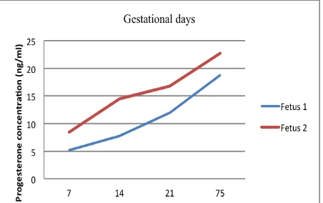
Figure 1. Progesterone hormone concentration (ng/mL) at different gestation days
Figure 1 showed that progesterone hormone concentration on the 7th, 14th, 21st, and 75th days of gestation in goat with single fetus were 5.21, 7.78, 11.97, and 18.78 ng/mL, respectively, while in goat with 2 fetuses were 8.44, 14.53, 16.81, and 22.73 ng/mL in goat, respectively. Figure 1 also showed that progesterone hormone concentration of EC goats with single fetus was lower compared to those with two fetuses. Budiarsana and Sutama (2001) also reported a similar result, whereby EC goats with single fetus had a lower progesterone hormone concentration compared to EC goats with multiple fetus.
The average progesterone concentration on the 7th day of gestation in EC goats with 2 fetuses was twofold higher compared to single fetus (8.44 and 5.21 ng/mL, respectively). This is because the progesterone concentration during formation of corpus luteum is correlated to the number of corpus luteum in the ovaries, as reported previously by Tjiptosumirat (2009) and Manalu and Sumaryadi (1995). Moreover, Jarell
and Dziuk (1991) observed that after gestation, the
number of fetus and the number of corpus luteum significantly influence progesterone level. In goat, Selvaraju et al. (2007) found that the increasing of corpus luteum number from 0, 1, 2, 3, and >3 resulted in the increasing of progesterone concentration from 0.0±0.0, 3.21+0.13, 4.21±0.36, and 5.17±1.15 ng/mL, respectively.
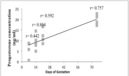
The result of simple regression and correlation analysis showed that the correlation (r) between progesterone hormone concentration and the number of fetus on the 7th, 14th, 21st, and 75th days of gestation were 0.442 (medium correlation), 0.854 (very strong correlation), 0.592 (medium correlation), and 0.757 (strong correlation), respectively (Figure 2). In line with
this study, Adriani et al. (2007) reported that the
Syafruddin et al.
93 Determinant coefficient (R2) showed that 72.9% of
the variation in the number of fetus can be explained by the variation in progesterone hormone concentration while the remainder is influenced by other variables. Based on regression coefficient, the regression equation obtained is Y= 0.214+ 0.108 X. The value of 0.108 means that every 1 ng/mL increases in progesterone concentration in EC goats would increase the number of fetus by 0.108. Hence, the higher the progesterone concentration, the higher the number of goat fetus produced.
Figure 2. Scatter plot of the correlation between progesterone hormone concentration and the number of Etawah crossbred goat fetus on the 7th, 14th, 21st, and 75th days of gestation
The correlation was only medium on the 7th day of gestation because corpus luteum had just started to secrete progesterone hormone. Arinbawa et al. (2012) stated that the increase in progesterone level is correlated to the development of corpus luteum during estrus cycle. Corpus luteum started to function on the 1st day after estrus, indicating that luteinization had started after ovulation, during progesterone hormone is produced. Llewelyn et al. (1995) also stated that the increase in progesterone level is accounted by the production of progesterone by corpus luteum at the initial of gestation which then continues to increase during luteal phase.
Progesterone level continues to increase until the 14th and 21st day of gestation. On the 14th day, progesterone hormone concentration in goats with single fetus vs 2 fetuses was 7.78 vs 14.53 ng/mL while on the 21st day was 11,97 vs 16,81 ng/mL. Budiarsana and Sutama (2001) reported that the increase in progesterone level occurred in the first 2-3 weeks after mating. Jarell and Dziuk (1991) also reported a similar result wherein the increase in progesterone level has occurred since the beginning of gestation until the 13th day of gestation, at which point the level starts to decrease. This decrease is caused by the shrinkage of corpus luteum at day 12-14; hence if the ovum was not fertilized, the production of progesterone would eventually decrease (Hafez, 2000).
The average progesterone hormone concentration on the 75th day of gestation in EC goat with two fetuses was higher compared to single fetus (18,78 vs 22,73 ng/mL) which was in line with previous observation by Adriani et al. (2007). The average of progesterone
hormone concentration in EC goats during 21 weeks of gestation in this study was 8.56±3.13 for single fetus and 10.73±3.50 for two fetuses. All the above observations indicated that progesterone hormone concentration was correlated with the number of fetus. Similarly, Siregar (2002) also proved that progesterone concentration in mid-gestation is correlated to the number of fetus.
CONCLUSION
Based on the result, it is concluded that progesterone concentration is highly correlated to the number of fetus in each gestation period in EC goats.
ACKNOWLEDGEMENTS
The authors would like to thank the Ministry of Research, Technology, and Higher Education who have funded this study through Penelitian Hibah Bersaing year 2016.
REFERENCES
Adriani, S. Adi, Toha, Wasmen, and M.I. Ketut. 2007. Pertumbuhan prenatal dalam kandungan kambing melalui superovulasi.
J. Biossci. 14(2):44-48.
Arinbawa, I.W.P., I. Gusti, and G.O.P. Tjok. 2012. Gambaran hormon progesteron sapi bali selama satu siklus estrus.
Indonesia Med. Vet. 1(3):330-336.
Budiarsana, I.G.M. and I-K. Sutama. 2001. Fertilitas kambing Peranakan Etawah pada perkawinan alami dan inseminasi buatan. Prosiding Seminar Nasional Teknologi Peternakan dan Veteriner. Puslitbang Peternakan, Bogor:85-92.
Chauhan, F.S. and M.A. Waziri. 1991. Evaluation of rectal-abdominal palpation technique and hormonal diagnosis of pregnancy in small ruminant. Indian J. Anim. Reprod.12:63-67. Dunlap, K.A. and F. Stomshak. 2004. Nongenomic inhibition of
oxytocin binding by progesterone in the ovine uterus. Bio. Reprod. 79:65-69.
Feradis. 2010. Bioteknologi Reproduksi pada Ternak. Alfabeta. Bandung.
Hafez, E.S.E. 2000. Reproduction in Farm Animal. 6th ed. Lea and
Febiger, Philadelphia.
Jarell, V.L. and P.J. Dziuk. 1991. Effect of number of corpora lutea and fetuses on concentrations of progesterone in blood of goats.
J. Anim. Sci. 69:770-773.
Llewalyn, C.A., J.S. Ogaa and M.J. Obwolo. 1995. Influence of season and housing on ovarian activity of indigenous goats in Zimbabwe. Trop. Anim. Health and Prod. 27(3):175-185. Manalu, W. and M.Y. Sumaryadi., 1995. Hubungan antara
konsentrasi progeteron dan estradiol dalam serum induk selama kebuntingan dengan massa fetus pada akhir kebuntingan.
Prosiding Seminar Nasional Sains dan Teknologi Peternakan. BPT. Ciawi. Bogor:57-62.
Manalu, W., M.Y. Sumaryadi, and N. Kusumorini. 1996. Effect of fetal number on concentration of circulation maternal serum progesterone and estradiol of does during late pregnancy. Small Rumin. Res. 23:117-124.
Selvaraju, M., D. Kathiresan, and T.G. Devanathan. 2007. Serum progesterone profile during oestrus and early pregnancyin Malabari goats. Tamilnadu J. Vet. Anim. Sci. 3(1):47-48. Sindoredjo, S. 1996. Pedoman Pemeliharaan Kambing Perah.
Balai Pustaka, Jakarta.
Siregar, T.N. 2002. Pengukuran profil progesteron sebagai suatu metode diagnosis kebuntingan dini dan kelahiran kembar pada domba lokal. Med. Ked. Hewan. 18(2):73-77.