See discussions, stats, and author profiles for this publication at: https://www.researchgate.net/publication/289494676
Expression and Purification of PhoR Sensor-Domain Histidine Kinase of
Mycobacterium tuberculosis in Eschericia coli
Article · June 2015 DOI: 10.5454/mi.9.2.1
CITATIONS
0
READS
141
7 authors, including:
Some of the authors of this publication are also working on these related projects:
Development of graphene-based materials from 'green' sources of carbonView project
Overexpression of Middle Hepatitis B Surface AntigenView project Ernawati Giri Rachman
Bandung Institute of Technology
27PUBLICATIONS 118CITATIONS
SEE PROFILE
Fenryco Pratama
Bandung Institute of Technology
4PUBLICATIONS 0CITATIONS
SEE PROFILE
Oktira Roka Aji
Ahmad Dahlan University
4PUBLICATIONS 0CITATIONS
SEE PROFILE
Arum Patriati
Gadjah Mada University
11PUBLICATIONS 22CITATIONS
SEE PROFILE
All content following this page was uploaded by Ernawati Giri Rachman on 22 March 2016.
PhoP/PhoR histidine kinase two-component-system in Mycobacterium tuberculosis has been studied as potential target for new antitubercular drugs.
The system is not found in mammals and the M.
tuberculosis PhoP/PhoR amino acid sequence shows
little homology with other prokaryotic proteins (Suwanto 2012). These findings implied that PhoP/PhoR have low non-targetting potential, either on humans or other organisms, a required characteristic
Vol.9, No.2, June 2015, p 51-57 DOI: 10.5454/mi.9.2.1
Expression and Purification of PhoR Sensor-Domain Histidine Kinase of
Mycobacterium tuberculosis
in
Escherichia coli
AND
1Department of Biotechnology, School of Life Sciences and Technology, Institut Teknologi Bandung,
Jalan Ganesha 10, Bandung, West Java 40132, Indonesia;
2Department of Chemistry, Faculty of Mathematics and Life Sciences, Institut Teknologi Bandung,
Jalan Ganesha 10, Bandung, West Java 40132, Indonesia;
3Center for Science and Technology of Advanced Materials, National Nuclear Energy Agency (BATAN),
Gedung 43 Kawasan PUSPIPTEK Serpong, Serpong, Banten 15314, Indonesia
Globally, tuberculosis (TB) remains a leading cause of death. The emergence of multidrug-resistant strains (MDR-TB) and extensively drug-resistant strains (XDR-TB) has fuelled the discovery for novel drugs and drug targets for its successful and better treatment. One of the potential candidates for drug target is PhoR sensory
protein histidine kinase, a part of the Two Component System (TCS) PhoP/PhoR in Mycobacterium tuberculosis
(Mtb). This protein system was known for its role on regulating hundred of Mtb virulence factors, from genes for cell wall and lipid synthesis to genes for adaptation in human leukocyte and hypoxia response. Previous studies have successfully characterized, isolated, and cloned theputative sensory domain of PhoR protein gene into
pRSET vector expression system. In this study, Escherichia coli was transformed with pRSET-SensPhoR and
o
cultivated at 37 C under IPTG induction to express PhoR sensor-domain protein. Most of the proteins were overexpressed in the form of inclusion bodies. Subsequent protein purification in Ni-NTA system under refolding condition on urea gradient was performed to isolate PhoR sensor-domain protein in soluble form. Arginine was supplemented in purified protein solution to prevent aggregation during long term storage. While highly purified protein was acquired, small angle X-ray scattering (SAXS) analysis was conducted to obtain 3-dimensional (3D) protein structures in solution.
Key words: multi-drug resistance, rational drug design, tuberculosis, two-component signal transduction system of histidine kinase
Secara global, tuberkulosis (TB) masih merupakan salah satu penyakit infeksi penyebab kematian tertinggi.
Semakin maraknya kemunculan kasus TB akibat infeksi oleh strain multidrug-resistant (MDR-TB) dan
extensively drug-resistant (XDR-TB) telah mendorong dilakukannya pengembangan obat dan target obat baru
untuk pengobatan yang lebih efektif dan aman. Salah satu kandidat target obat baru yang cukup menjanjikan adalah protein sensor PhoR histidine kinase, anggota dari protein pensinyalan sistem dua komponen (TCS)
PhoP/PhoR di Mycobacterium tuberculosis (Mtb). Sistem PhoP-PhoR dikenal berperan dalam regulasi ratusan
faktor virulensi dari Mtb, mulai dari gen untuk sintesis dinding sel dan lipid hingga gen untuk adaptasi di leukosit manusia dan respon hipoksia. Penelitian sebelumnya telah berhasil mengkarakterisasi, mengisolasi dan mengklon bagian DNA yang merupakan domain sensor dari protein PhoR kedalam sistem vektor ekspresi
pRSET. Pada penelitian ini, Escherichia coli ditransformasi dengan vektor pRSET-SensPhoR dan dikultivasi
o
pada temperatur 37 C dibawah induksi IPTG untuk mengekspresikan domain sensor protein PhoR. Sebagian besar protein diekspresikan dalam bentuk badan inklusi. Purifikasi protein menggunakan sistem Ni-NTA dalam kondisi refolding di gradien urea dilakukan untuk mengisolasi domain sensor protein PhoR dalam bentuk terlarut. Larutan protein disuplementasikan dengan arginin untuk mencegah pembentukan aggregat protein hasil refolding dan purifikasi selama proses penyimpanan jangka panjang. Ketika domain sensor protein PhoR dengan
kemurnian yang tinggi telah diperoleh, analisis small angle X-ray scattering(SAXS) dilakukan untuk
mempelajari struktur 3-dimensi protein tersebut di dalam larutan.
Kata kunci: desain obat secara rasional, kebal aneka obat, sistem transduksi sinyal dua komponen histidine
kinase, tuberkulosis
1 + 1+ 1
ERNAWATI ARIFIN GIRI-RACHMAN * , FENRYCO PRATAMA , OKTIRA ROKA AJI , ARUM
3 2 3 1
PATRIATI , IHSANAWATI , EDY GIRI RACHMAN PUTRA ,
MAELITA RAMDANI MOEIS
*Corresponding author; Phone: +62-81809418805, Fax:
+62-22-2534107; Email:
+ These authors contributed equally in this work.[email protected]
for new drug target. Previous studies also demonstrated that interrupting PhoP/PhoR two-component-system drastically decreased M. tuberculosis virulence in vivo
(Walters et al. 2006, Gonzalo-Asensio et al. 2008). PhoP/PhoR two-component-system also induces the expression of 114 virulence-related genes (Pathak et al.
2011), including those involved in lipoarabinomanan (a cell wall component) metabolism and M.
tuberculosis resistance inside macrophage (Walters et
al. 2006, Gonzalo-Asensio et al. 2008). Inhibition of PhoR sensor-domain will block the first step in the signal transduction pathway and shut down the expression of the virulence-related genes. Since many genes are affected by the inhibition, anti-tubercular drug which attack the PhoR sensor-domain should be more effective in treating M. tuberculosis infection and have shorter duration of treatment than existing drugs.
Previous studies by Suwanto et al (2012) have successfully characterized, isolated, and cloned PhoR sensor-domain coding sequence from M. tuberculosis
strain H37Rv into pRSET expression vector. In this study, we over expressed recombinant PhoR sensor-domain protein in E. coli BL21 (DE3) and conducted subsequent purification process to isolate the protein. Protein in pure condition is required to be proceed for structural study. The resulting PhoR sensor-domain 3D structure could be used for further characterization of protein function (Shumilin et al. 2012). Moreover, structural information will provide basis for in silico screening of new anti-tubercular drug using rational drug design, primarily from natural chemical products found in Indonesia. Instead of determining the 3D structure of protein crystal, solution scattering, a new promising method for studying conformational changes of protein in physiological conditions has been established (Svergun et al. 2003, Jacqueset al. 2010) and was proposed in this work. Overall 3D structure of protein in low-resolution can be provided from the small-angle scattering (X-ray, SAXS or neutron, SANS) of protein solution as an important complementary method of protein crystallography due to the limitation in preparing the protein single crystal. An isotropic scattering function I(q) which is proportional to the averaged of over all structure orientations from a single protein molecule has to be attained in order to reconstruct the protein structure from solution. This constraint is only achieved from a monodisperse system as non-interacting of identical particles (scatterers) in dilute solutions.
MATERIALS AND METHODS
Cloning Summary and Overexpression of Recombinant PhoR Sensor-Domain Protein.
Previous study by Suwanto et al (2012) has amplified DNA segment encoding sensor-domain of PhoR protein (GenBank, NC_000962.3, NP_215272.1) from
Mycobacterium tuberculosis H37Rv genome using
PCR technique with forward (5'-GAATTCGTCACCT CGATGTTGCAGCA-3') and reverse (5'-CCATGGTT AGACGTCGGCCAGATCAATG-3') primers. The amplified DNA then was cloned primarily into pGEM-T easy vector before transfered into pRSEpGEM-T expresion vector. The expressed protein will be observed around 17 kDa according to in silico predicted product
pRSET-SensPhoR vector was transformed into E. coli BL21(DE3) using heat shock method (Sambrook and Russel, 2001). A single transformant colony was
-1
grown in LB medium containing 0.1 mg mL
o
ampicillin at 37 C and agitated at 200 rpm until the optical density at 600 nm reached 0.4-0.8. The overexpression was induced with 1 mM IPTG for 4-6 h.
o
Cells were harvested by centrifugation at 2000 g, 4 C, for 30 min.
Protein Purification. The cell pellet from overexpression cultures was resuspended in lysis buffer containing 20 mM Tris-HCl pH 8.0, 1 mM PMSF, disrupted by sonication, and then centrifuged at
o
12,000 g, 4 C, for 10 min to separate soluble proteins (supernatant) from insoluble proteins and cell debris (pellet). The pellet was washed twice with Triton X-100 -Urea (2% triton X-X-100, 2M urea, 0.5 M NaCl, 20 mM Tris-HCl pH 8.0) and once with Tris-HCl 20 mM pH 8.0 followed by sonication and centrifugation at
o
12,000 g, 4 C, for 10 min. The pellet containing inclusion bodies was dissolved in binding buffer containing 8 M urea, 0.5 M NaCl, 5 mM imidazole, 2.5 mM β-mercaptoethanol, and 20 mM Tris-HCl pH 8.0 stirred for 30-60 min, and then centrifuged at 12000 g,
o
4 C, for 15 min. The resulting supernatant was filtered using millipore membrane of 0.22 μm. The filtrate was prepared for purification.
Protein purification was carried out using nickel Ni-NTA-agarose system on 50 mL of Econo column (Biorad) in the refolding state at flow rate of 0.5-1 mL
-1 o
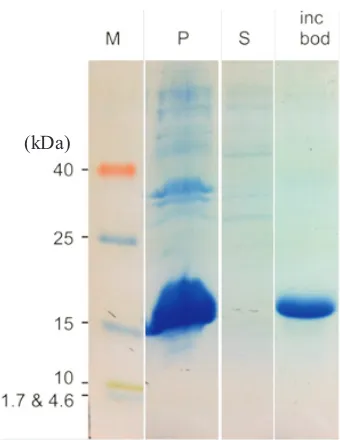
min , 25 C. The column was equilibrated with solution containing 0,5 M NaCl, 5 mM imidazole, 8 M urea, 2.5 mM β-mercaptoethanol,20 mM Tris-HCl pH 8.0 (GE Life Sciences). The protein was refolded using a urea linear gradient of 8-0 M and then eluted with 0.5 M NaCl, 250 mM imidazole, 2.5 mM β-mercaptoethanol, 20 mM Tris-HCl pH 8 added with 200 mM arginine to prevent precipitation formation during storage. Results from protein overexpression and purification were profiled by SDS-PAGE (Fig 1). To confirm the identity of recombinant PhoR sensor-domain protein, slices of acrylamide gel containing a desired band were sent to Center for Mass Spectrometry & Proteomics University of Minnesota for LC-MS analysis.
Small Angle X-ray Scattering (SAXS) for Determining Protein Structure in Solution. The purified protein was dialyzed using servapor membrane (molecular weight cut off /MWCO = 12-14 kDa, Serva) with protein sample:dialysis buffer volume ratio of 1:22 to dispose imidazole and β-merchaptoetanol and to reduce the NaCl concentration in the purified sample to 200 mM. The sample was concentrated using Nanosep Ultrafiltrator (MWCO/Molecular Weight Cut-off = 10 kDa, Pall). After filtration by millipore membrane of 0,22 µm, acquired protein in final concentration of 1.5 – 5 mg
-1
mL then was subjected for SAXS of protein in solution. Sample equilibration was conducted prior to data collection by dialysing protein sample in its solution buffer. Both of equilibrated protein sample and the background solution from its dialysed buffer were exposed to X-ray beam for 10 h and collected at
o
every hour, at 4 C. The collected scattering data of protein samples was subtracted by the background solution data in obtaining corrected scattering data of proteins. The corrected 2-dimensional scattering data was then radially averaged to obtain the scattering
Overexpression of Recombinant PhoR Sensor-Domain Protein. Since pRSET containing T7 promoter, IPTG induction was conducted to increase the expression of the recombinant protein in E. coli
BL21 (DE3). SDS PAGE result showed that recombinant PhoR sensor-domain protein was overexpressed and found dominantly in pellet fraction
of cell lysate to supernatant fraction (Fig 1). ubsequent process to isolate and characterize inclusion bodies protein from pellet fraction confirmed that the estimated molecular weight of 17 kDa protein existed in this form (Fig 1).
Protein Purification. PhoR sensor-domain protein was constructed in pRSET vector which contains hexahistidine as fusion tag. This tag facilitates rapid purification of recombinant protein by immobilized metal ion affinity chromatography (IMAC). Purification was performed alongside the refolding process and highly purified protein was obtained from the single Ni-NTA column running (Fig 2A). Post-purification observation showed that the purified protein was unstable and aggregated after
o
storage at -80 C (Fig 2B). Arginine in final concentration of 0.2 M should be added into the protein solution immediately after elution to prevent further aggregation.
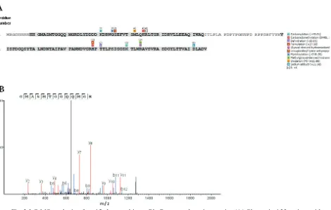
LC-MS analysis was conducted to determine the peptide sequences of ~17 kDa band of purified protein. Several chemical modifications on the protein residues (Fig 3) were performed prior to trypsin digestion to increase peptide fragments generation and readibility. All spectra of detected fragments were searched againts sequence reference of recombinant PhoR sensor-domain using peptide de novo sequence software PEAK 7.5 (Ma et al. 2003). The result then confirmed the identity of recombinant PhoR sensor domain protein with 79% sequence coverage (Fig 3).
Small Angle X-ray Scattering for Determining Protein Structure in Solution. The scattering intensity I(q) vs. momentum transfer q of recombinant PhoR sensor-domain protein in two different
-1 -1
concentrations, (A) 3.9 mg mL (B) 1.56 mg mL was showed (Fig 4). The scattering intensity still increased in low q-range indicating that this protein has a naturally propensity to aggregate under the buffer conditions used in the preparation whereas the protein solution was diluted two times. This aggregated form was also confirmed using dynamic light scattering (DLS) measurements which dominant peak referred to particle with size 11 nm and molecular weight 1052 kDa, significantly different with 3 nm and 17 kDa of PhoR sensor-domain actual features.
DISCUSSION
Fig 1 SDS PAGE of PhoR sensor-domain recombinant protein expressed in E. coli. Lane M shows molecular mass marker (in kDa). Lane P contains pellet-cell lysate; Lane S contains supernatant-cell lysate; lane Incbod the inclusion bodies. Sample was run on 20% acrylamide and visualized using Coomassie Brilliant Blue staining.
54 GIRI-RACHMAN ETAL. Microbiol Indones
University of Oklahoma. The software had developed on basis of logistic regression model with interaction of parameters that accurately predicted 96% insoluble protein from large set of data base (Diaz et al. 2010). Results from the analysis estimated that PhoR sensor-domain protein was expressed as insoluble protein inside E. coli. The intrinsic factor of protein might affect the inclusion bodies formation of PhoR sensor domain in E.coli rather than overexpression factor since significant amount of protein was acquired in insoluble fraction (Fig 1) (Diaz et al. 2010). On
different approach, The higher numbers of β-sheet structure than α helices in a PDC fold-like
characteristic prediction (Geneious) of PhoR sensor domain (Suwanto 2012) was considered to be responsible for the insolubility character of this protein (Vilasi et al. 2006).
Protein purification and refolding was performed to obtain PhoR sensor-domain protein in pure and native form. Refolding protein is an elaborate process and achieving correct folded protein is sometimes tricky. In other cases, expressing the sensor domain of histidine kinase in particular vector that contain soluble promoting tag such as GST and MBP is preferable (Cheung et al. 2012; Tan et al. 2014). Nevertheless, inclusion bodies production has several advantages for
Fig 2 SDS-PAGE of purified recombinant PhoR sensor-domain protein purificationin 20% acrylamide (A). Lane M shows molecular mass marker (in kDa); Lane E the elution fraction. The gel was stained with Coomassie Brilliant Blue. Precipitation of purified protein was observed in the sample without additives (B).
(kDa)
kDa
kDa
kDa
isolating and purifying particular protein such as high number of protein production, resistance from cellular protease, and shorter purifying step. The point that is stated later proven well in this experiment while recombinant PhoR sensor-domain protein could be purified merely in single column process using Ni-NTA agarose with high level of protein purity acquired. The heterogenity of protein sample might be decreased significantly during isolation of inclusion bodies step including removal of common co-purified contaminant
of E.coli protein in nickle column (Bolanos-Garcia and
Thomas 2006), simplifying the subsequent process of protein purification to separate protein target from trace amount of other proteins. Importantly, the lack of cysteine in primary sequence of PhoR sensor-domain protein trimming the elaborate process of protein refolding by finding out the suitable condition for arrangement of correct disulfide bridge. Nevertheless, limitation was found in futher analysis of PhoR sensor-domain recovery after refolding process due to the
Intensity (%)
Fig 3 LC-MS analysis of purified recombinant PhoR sensor-domain protein. (A) Shown is 155 amino acid sequences of expressed PhoR sensor-domain. The peptide sequence coverages by spectral analysis are bold and highlighted (79%). Colorized boxes indicate the chemical modifications on particular residues of recombinant PhoR sensor-domain. List of chemical modifications is displayed at the right side. (B) The
MS/MS spectrum of peptide GMASMTGGQQMGR (m/z 656,16), which is the fragment of
recombinant protein sensor-domain (amino acid residues 11-23) digested by trypsin, is shown as a representative spectra.
Fig 4 PhoR sensor-domain was analyzed using SAXS, in two different concentrations, (A) 3.9 mg and (B)
-1
1.56 mg mL . Analyzes was at 4 C.
56 GIRI-RACHMAN ETAL. Microbiol Indones
protein absence in enzymatic activity and lack of information about the protein ligand.
Post-purification observation showed that the purified and refolded protein was precipitated after
o
storage in -80 C. Supplementation with additive such as arginine significantly reduced progression of protein aggregates. Arginine might effectively prevent protein aggregation by several ways. Ho et al. (2003) showed that addition of arginine in the refolding solution of Lysozimes could shift the viral coefficient value into positive and accordingly increased repulsive interactions between proteins. In another mechanism, Tsumoto et al. (2004) proposed that interaction of arginine with exposed aromatic amino acid in protein could increased its solubility that led to repress protein aggregation.It was later strongly indicated by Das et al.
(2007) experiment that cluster of arginine in aqueous solution displaying methyl groups that might interact with hydrophobic surface in protein and then prevented the aggregation.
Purified protein was confirmed accurate to recombinant PhoR sensor domain since mass spectral analysis shown high peptide coverage score (≥50%) and two and more unique peptides were identified(60 unique peptides) (Link and LaBaer 2009).
Crystallization trial demands on high concentration of protein target. Several attempts to produce final solution of PhoR sensor-domain protein at ≥ 10 mg
-1
mL led to sample aggregation (data not shown). Alternative method for analysing protein structure in lower concentration, such SAXS, was preferable since it was able to give useful signal at concentration of
-1 structural models derived in obtaining useful biostructural information from small-angle scattering data if the samples aggregated. It should be monodisperse, identical particles, and the data are measured to low enough q to reliably characterize their largest dimensions as the plot will have a discernible flat region in the lowest q regime (independenton q) – dashed lines. Moreover, it confirmed that subaggregate particles was still continually developed in the sample even after protein filtration and buffer supplementation with arginine. The highly sensitive technique was required to detect protein homogeneity preceding to SAXS analysis or other related methods for protein structural study. Nonetheless, from those initial SAXS experimental works, it can be concluded that the
sample preparation protocols for solution scattering experiment, experimental setup, data reduction and analysis are well established for protein solution.
In conclusion, inclusion bodies of PhoR sensor-domain protein was produced in E.coli by expressing pRSET-Sensor PhoR system under cultivation
o
temperature of 37 C and IPTG induction. While purification and refolding process dominantly isolated the protein in soluble form, protein homogeneity still an issue before proceeding the protein onto structural study. Some improvements in protein production are required for providing a pure monodisperse protein which has a high stability.We propose to optimize this protein expression on alternative condition, host or vector system to obtain the soluble protein at the first time and to prevent elaborate process of protein refolding and storage optimization.
ACKNOWLEDGMENT
This work was supported by the General Directorate of Higher Education (DIKTI), Indonesian Ministry of Education and Culture through 2012 DIKTI decentralization research grant and 2014 ITB Research and Innovation Grant to E.A.G.R. The authors acknowledge Robert Knott, Anna Sokolova and Agata Rekas of Australian Nuclear Science and Technology Organisation (ANSTO) for a benefit discussion and for providing the beam time. The SAXS experiment was conducted under the BATAN-ANSTO Joint Research Project on the Biophysical Study of Virus Like Particles for Targeted Drug and Vaccine Development, (PI : E. G. R. Putra).
REFERENCES
Ma B, Zhang K, Hendrie C, Liang C, Li M, Doherty-Kirby A, Lajoie G. 2003. PEAKS: Powerful software for peptide De Novo sequencing by tandem mass
spectometry. Rapid Commun Mass Spectrom.17(20):
2337-2342. doi: 10.1002/rcm.1196.
Bolanos-Garcia VM, Davies OR. 2006. Structural analysis
and classification of native proteins from E. coli
commonly co-purified by immobilised metal affinity chromatography. Biochim Biophys Acta. 1760(9): 1304–1313. doi: 10.1016/j.bbagen.2006.03.027. Cheung J, Le-Khac M, Hendrickson W. 2009. Crystal
structure of a histidine kinase sensor domain with similarity to periplasmic binding proteins. Proteins 77(1):235–141. doi: 10.1002/prot.22485.
Pasha S, Mann A, Ganguli M, Verma AK, Bhat R, Chandrayan SK, Ahmed S, Sharma S, Kaur P, Singh TP, Srinivasan A. 2007. Inhibition of protein aggregation: supramolecular assemblies of arginine hold the key. PloS One 2(11): e1176. doi: 10.1371/journal.pone.0001176. Diaz A, Tomba E, Lennarson R, Richard R, Bagajewicz MJ,
Harrison RG. 2010. Prediction of protein solubility in
Escherichia coli using logistic regression. Biotechnol
Bioeng 105(2):374–383. doi: 10.1002/bit.22537. Gasteiger E, Hoogland C, Gattiker A, Duvaud S, Wilkins MR,
Appel RD, Bairoch A. 2005. Protein identification and analysis tools on the ExPASy server. (In)
Langi GEP, Moeis MR, Giri-Rachman EA, Ihsanawati. 2012. Molecular cloning and cold shock induced overexpression of the DNA encoding PhoR
sensor-domain from Mycobacterium tuberculosis as a target
molecule for novel anti-tubercular drugs. Proceedings of International Conference on Mathematics and Natural Sciences 2013, Bandung, Indonesia, 8-9
November 2013. Faculty of Mathematics and Natural
Sciences, Institut Teknologi Bandung. doi: 10.1063/ 1.4868830.
Link AJ, LaBaer J. 2009. Proteomics: A Cold Spring Harbor Laboratory Course Manual. New York: Cold Spring Harbor Laboratory Press.
John M. Walker (ed): The Proteomics Protocols Handbook. Humana Press. pp 571-607. doi: 10.1385/1-59259-890-0:571. Gonzalo-Asensio J, Mostowy S, Harders-Westerveen J,
Huygen K, Hernández-Pando R, Thole J, Behr M, Gicquel B, Martín C. 2008. PhoP: a missing piece in the
intricate puzzle of Mycobacterium tuberculosis
virulence. PloS One 3(10):e3496. doi: 10.1371/journal. pone.0003496.
Ho JGS, Middleberg APJ, Ramage P, Kocher HP. 2003. The likelihood of aggregation during protein renaturation can be assessed using the second viral coefficient. Prot Sci. 12(312):708-716. doi: 10.1110/ps.0233703. Jacques DA, Trewhella J. 2010. Small-angle scattering for
structural biology-expanding the frontier while avoiding the pitfalls. Prot Sci. 19: 642-657. doi: 10.1002/pro.351 Laemmli UK. 1970. Cleavage of structural proteins during
the assembly of the head of bacteriophage T4. Nature 227(5259): 680-685. doi: 10.1038/227680a0.
Pathak A, Goyal R, Sinha A, Sarkar D. 2010. Domain structure of virulence-associated response regulator
PhoP of Mycobacterium tuberculosis: role of the linker
region in regulator-promoter interaction(s). J Biol Chem. 285(45): 34309–34318. doi: 10.1074/jbc.M110.135822. Sambrook J, Russell DW. 2001. Molecular cloning: a
rd
laboratory manual. 3 edition. New York: Cold Spring Harbor Laboratory Press.
Shumilin I, Cymborowski M, Chertihin O, Jha KN, Herr JC, Lesley SA, Joachimiak A, Minor W.2012. Identification of unknown protein function using metabolite cocktail screening. Structure. 20(10): 1715–1725. doi: 10.1016/j.str.2012.07.016.
Skou S, Gillilan RE, Ando N. 2014. Synchrotron-based small-angle X-ray scattering of proteins in solution. Nat Protoc. 9(7): 1727–1739. doi:10.1038/nprot.2014.116. Suwanto AS, Ihsanawati, Giri-Rachman EA. 2012.
Molecular cloning of PhoR sensor domain from
Mycobacterium tuberculosis for structure-based
discovery of novel anti-tubercular. J Life Sc. 6(3):268. Svergun DI, Koch MHJ. 2003. Small-angle scattering
studies of biological macromolecules in solution. Rep. Prog Phys. 66: 1735–1782. doi: 10.1088/0034-4885. Tan K, Chhor G, Binkowski TA, Jedrzejczak RP,
Makowska-Grzyska M, Joachimiak A. 2014. Sensor domain of
histidine kinase KinB of Pseudomonas: a
helix-swapped dimer. J Biol Chem. 289(18): 12232–12244. doi: 10.1074/jbc.M113.514836.
Tsumoto K, Umetsu M, Kumagai I, Ejima D, Philo JS, Arakawa T. 2004. Role of arginine in protein refolding, solubilization, and purification. Biotechnol prog. 20(5):1301–1308. doi: 10.1021/bp0498793.
Vilasi S, Dosi R, Iannuzzi C, Malmo C, Parente A, Irace G, Sirangelo I. 2006. Kinetics of amyloid aggregation of mammal apomyoglobins and correlation with their amino acid sequences. FEBS Lett. 580(6): 1681–1684. doi:10.1016/j.febslet.2006.02.018.
Walters SB, Dubnau E, Kolesnikova I, Laval F, Daffe M,
Smith I. 2006. The Mycobacterium tuberculosis PhoPR
two-component system regulates genes essential for virulence and complex lipid biosynthesis. Mol Microbiol. 60(2): 312–330. doi:10.1111/j.1365-2958.2006.05102.x.