Citation: Hossain MK, Paul S, Hossain MM, Islam MR and Alam MGS. Bovine Mastitis and Its Therapeutic Austin J Vet Sci & Anim Husb - Volume 4 Issue 1 - 2017
Austin Journal of Veterinary Science &
Animal Husbandry
Abstract
Mastitis is one of the most costly devastating diseases in the dairy industry. Bovine mastitis can be classiied as clinical or subclinical based on clinical features of the disease. Subclinical mastitis occurrence is more prominent compared to clinical form and its prevalence is more than 20 to 50% of cows in given herds. Both forms cause substantial inancial losses due to reduced milk yield, degraded milk quality, drug costs and veterinary expenses, early culling of cows and increased labour costs for the farmers. The chief causal organisms of mastitis are cell-walled bacteria, even though mycoplasma, algae and yeast have also been identiied to cause mastitis. Numerous inluencing factors like physiological, pathological, environmental or genetic factors may contribute to the occurrence of mastitis in dairy cattle. Presently, different types of antibiotics are prescribed for the treatment of mastitis. However, therapeutic success rate of mastitis is generally poor because of pathological changes in the udder parenchyma, mastitogenic bacterial and related factors, indiscriminate use of antibiotics and poor animal husbandry practices. These evidences focus the necessity for discovering completely newer strategies for treatment of mastitis. This review will highlight the key etiological agents and factors affecting the manifestation of mastitis, and the recent advancement of therapeutic strategies including application of antibiotic-based therapies by doing antibiotic sensitivity test as a prospective approach to prevent and control mastitis in dairy herds.
Keywords: Mastitis; Causal agents; Factors; Antibiotics; Sensitivity test
medicine and increase labor cost for the farmers [12-15]. Bacterial contaminated milk from mastitis infected cows can impede with food manufacturing process or can cause food poisoning; may render unsuited for human consumption, and in some circumstances may have the possibility to transmit disease to humans [16].
Medical therapy involving antibiotics is a key tool in the scheme of mastitis treatment and control. For successful antibacterial mastitis therapy, the active medicine must achieve and conserve concentrations above the minimum inhibitory concentration (MIC) at the center of infection for long enough to breakdown the production and toxin-producing phase of the causal pathogen [17-18]. his may be prohibited by several factors that include pathological changes in the udder parenchyma, mastitogenic bacterial and related factors, poor selection and indiscriminate use of antibiotics, and is also inluenced by the route of administration [16]. Enormous economic losses by mastitis are also experienced due to unmarketable milk or milk-products adulterated with antibiotic residues originating from treatment in the developing nations along with usage of antibiotics as growth promoters predominantly in dairy feedlots in the advanced world [20-21]. he long time use of antibiotics in the treatment of mastitis has directed further problem of occurrence of antibiotic resistant strains, therefore there is continual worry about treatment failure and the resistant strains entering the food chain [22-24]. Treatment failures also lead to longer periods of infectivity, which increase the numbers of infected cows moving in the farm and thus expose the whole herd to the risk of contracting a resistant strain of infection. Continual attention has given by the researchers to discover
Ab b re via tio n s
MIC: Minimum Inhibitory Concentration; SCC: Somatic Cell Count; CMT: California Mastitis Test; kPa: Kilo Pascal; NAGase: N-acetyl-β-D-glucosaminidase; LDH: Lactate Dehydrogenase Activity; EC: Electric Conductivity; MBC: Minimum Bactericidal Concentration, IL: Interleukins; TNF-α: Tumor Necrosis Factor alpha
In tro d u ctio n
Mastitis is one of the important devastating diseases in cattle striking the greatest challenge to the dairy industry [1,2]. Mastitis remains the most economically destructive and zoonotic imminent disease for consumers irrespective of many years of research worldwide with diferent levels of economic losses identiied by diferent countries [2-6]. Milk production is reduced considerably in the afected animals and estimated loss of milk yield may range from 100 to 500 kg per cow per lactation [7]. he predictable loss causing clinical mastitis in cows is nearly 700kg in irst lactation and 1,200kg in the second lactation [8]. In United States, expenses related to mastitis on dairy farms are around U$ 200 per cow per year and an estimated annual loss of 2 billion dollars for dairy industry [9]. Australian dairy industry is losing more than $130 million every year because of poor udder health causing reduced milk production that is mainly associated with mastitis [10]. In India, annual economic loss sufered by dairy industry owing to udder infections is estimated to be more than $120 million [2,11]. Economic consequences of mastitis, clinical or sub-clinical, include reduced milk yield, poorer quality milk, increased culling rate, increased cost of veterinary services and
Review Article
Bovin e Mastitis an d Its Therapeutic Strategy Doin g
An tibiotic Sen sitivity Test
H o s s a in MK1*, P a u l S2, H o s s a in MM3, Is la m MR3
a n d Ala m MGS4
1Departm en t of Surgery an d Theriogen ology, Sylhet
Agricultural Un iversity, Ban gladesh
2Departm en t of Public H ealth an d Epidem iology, Sylhet
Agricultural Un iversity, Ban gladesh
3Departm en t of Medicin e, Sylhet Agricultural Un iversity,
Ban gladesh
4Departm en t of Surgery an d Obstetrics, Ban gladesh
Agricultural Un iversity, Ban gladesh
*Co rre s p o n d in g a u th o r: H ossain MK. Departm en t of Surgery an d Theriogen ology, Faculty of Veterin ary an d An im al Scien ce, Sylhet Agricultural Un iversity, Tilagor, Sylhet-310 0 Ban gladesh
the proper antibiotic to treat and control mastitis doing antibiotic sensitivity test. herefore, this review will summarize the key etiological agents and factors afecting the manifestation of mastitis, and the recent status of therapeutic strategy including antibiotic-based therapies by doing antibiotic sensitivity test as a prospective approach to prevent and control mastitis in dairy herds.
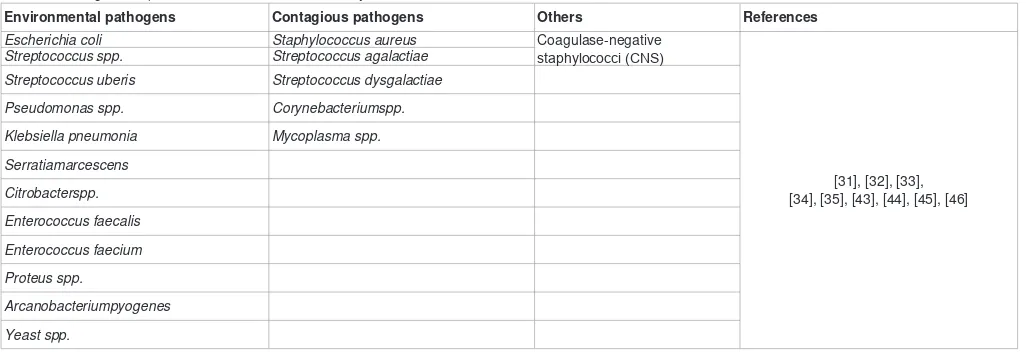
Ma s titis P a th o ge n s
Huge quantities of microorganisms have been known to cause bovine mastitis. he major causal microorganisms are bacteria, but mycoplasma, fungi and algae have also been reported to cause mastitis problems in some herds or regions [25-28]. It is very much interesting, about 137 species and subspecies of possible pathogens can be accompanying with infection of the mammary gland [29]. he proportionate importance of the pathogens has varied between countries, regions and farms, mostly owing to diferences in management and housing systems [30]. Conversely, mastitis in dairy herds is commonly of two types, these are environmental mastitis and contagious mastitis (Table 1).
he primary source of environmental mastitis pathogens is the habitat of the cow like soil, plant material, manure, bedding, and contaminated water or potential pathogens usually in the digestive tract [31,32]. Interaction to environmental pathogens ensues primarily between milking. hese microorganisms usually proliferate nearly 1,000,000 or more cells per gram of bedding. his issue increases the possibility of mammary glands infection leading to clinical mastitis [33,34]. here is a progressive association among the number of coliforms existing in the bedding material and the bacterial load on the teat ends along with the incidence rates of clinical mastitis [35]. Primary environmental pathogens include two types of bacteria: coliform bacteria and species of streptococci other than Strep. Agalactiae. Coliforms bacteria chiely Escherichia coli, Enterobacter aerogenes, Klebsiella pneumonia and Serratia marcescens, and a Streptococcus sp, Streptococcus Uberis are the chief organisms found as the of source environmental mastitis [31,36,37]. Coliform bacteria mostly cause clinical form of mastitis. Environmental mastitis has formerly founded less than 10% of total mastitis cases, but there has been an increase in the occurrence of environmental mastitis more recently [38-40] mostly related with S. Uberis infection. his
pathogen is most frequently linked with chronic mastitis, which does not respond to antibiotic treatment [31].
Contagious mastitis is commenced by bacterial pathogens that grow well on the udder skin and lesions of teat. hey cannot persist for long times in the environment and are usually spread from one cow to another at milking time through the hands of milkers, the milking machine, milk-contaminated fomites or the sponge [41,42]. he pathogens typically interrelated with contagious mastitis are Staphylococcus aureus and Streptococcus agalactiae [43-45]. hough Streptococcus dysgalactiaeis considered as an environmental pathogen, there is indication of its spread from cow to cow as a contagious pathogen triggering mastitis [43]. hese bacteria are common causes of both clinical and sub clinical mastitis. Mycoplasma species are also another source of contagious mastitis. Mycoplasma mastitis may be accessed to the herd by newly purchased animals and can be transmit during milking. Mycoplasma bovis is the chief species occasionally leading to severe complications like abrupt onset, decrease in milk yield, quick transmission and lack of response to treatment [41,46]. Conversely, the most predictable pathogen in the majority of clinical and subclinical mastitis cases in most countries is Staphylococcus Aureus [47-51]. hese bacteria are of enormous importance as they are responsible for more than 25% of intra-mammary infections and undesirably distressing the quality of milk in enormous number of clinical cases [52,53]. hey are also supposed to be the most emerging pathogens leading bovine mastitis as they are commonly isolated as the topmost bacterial pathogens [51,54].
Ris k Fa cto rs o f B o vin e Ma s titis
here are plenty of predisposing factors that can inluence emergence of mastitis at individual and herd level in dairy cattle. he factors may be physiological, genetic, pathological or environmental [55] which are given below:
Age of cows has signiicant afects in occurring mastitis. It has been shown that manifestation of mastitis in infected quarters increases with advancement of age in cows [41,56-59], the highest occurrence are being observed in cows of more than 7 years of age [60]. his may be due to more dilated teat canals in older age, permanent udder tissue damage resulting from the primary infection or due to an increased cellular response to intra mammary infection
Environmental pathogens Contagious pathogens Others References
Escherichia coli Staphylococcus aureus Coagulase-negative
staphylococci (CNS)
[31], [32], [33], [34], [35], [43], [44], [45], [46]
Streptococcus spp. Streptococcus agalactiae
Streptococcus uberis Streptococcus dysgalactiae
Pseudomonas spp. Corynebacteriumspp.
Klebsiella pneumonia Mycoplasma spp.
Serratiamarcescens
Citrobacterspp.
Enterococcus faecalis
Enterococcus faecium
Proteus spp.
Arcanobacteriumpyogenes
Yeast spp.
[61,62]. Another reason may be efective innate host defense mechanism that makes the younger animals less susceptible to infection [63]. Moreover, cow’s parity has signiicant inluence of on prevalence of mastitis in farms. Cows in parity number more than 3 have considerably higher mastitis prevalence than those of parity 2-3 and primiparous one [64]. Primiparous cows have stronger defense mechanism than multiparous cows that make them less susceptible to mastitis.
Several genetic traits may also have substantial efect upon susceptibility to mastitis in bovine. hese genetic traits are natural resistance of cows, comparative distance between teats, teat shape and conformation, position of udders and milk yield and fat content of milk. High yielding dairy cows with high fat contents are reported to be more vulnerable to mastitis [65-67]. Udder conformation and shape of the teat are signiicant genetic traits that may also afect susceptibility to mastitis. Cows with extended teats are more susceptible to mastitis infection compared to cows with reversed teat ends [68,69]. An additional important inluencing factor for mastitis is super numerous teats, which may be responsible for extra reservoirs for potential pathogens leading to manifestation of mastitis.
Lactation has a highly signiicant consequence on prevalence of mastitis. In bovine, there is possibility of increased oxidative stress and reduced antioxidant defense mechanisms immediately
ater parturition, early lactation and during the dry period and the incidence of mastitis is reported to be higher during these times [70]. Somatic cell counts (SCC) mainly neutrophils which is the cow’s natural irst line of defense to prepare the onset of the new lactation, seems to be increased immediately ater parturition, which remains high for a few weeks regardless of the presence or absence of infection [71-73]. Recent studies have shown that higher amount SCC count in later stage of lactation in cows [74]; possibly signifying increased subclinical infection leading to a decrease in milk production.
Herd management and milk hygiene are also considered signiicant risk factors for mastitis [64,75]. he occurrence of bovine mastitis is remarkably higher in less clean udder in contrast to clean one. he farms that have followed pre and post teat dipping practice for milking have been less afected by mastitis infection. Several studies have found that let front quarter (LFQ) are more afected by mastitis than the other quarters and may be due to contaminations from the operators let hands without proper washing and disinfection [76,77]. In farms where milking machines are used for milking, it is essential to conserve ideal pressure like 50kPa for most machines, because extra pressure may lead to incomplete milking and tissue damage in the teat [78]. hese may cause increased risk of both contagious and environmental mastitis. herefore, it is better to use those milking machines that can achieve complete milking as well as fewer incidences of teat injuries [79].
he incidence of mastitis is obviously inluenced by the weather and climatic conditions. A greater percentage of mastitis has been observed to arise mostly during summer and rainy months [80-83]. Bacterial propagation as well as the load of pathogens increases as heat and humidity increases in the environment [84]. On the contrary, another study has reported a greater occurrence of coliform mastitis during the cold months when the temperature was recorded to be less than 21°C [85,86] in the environment.
Plan of nutrition is another important factor that inluences clinical manifestation of mastitis in heifers and cows [87]. Vitamin E has been described to boost the immune response of cows by enhancing the phagocytic properties of neutrophils ater parturition [88,89]. Vitamin E combined with selenium acts as an anti-oxidant substance by preventing oxidative stress [90,91]. Numerous investigations have proved that neutrophils of selenium fed cows are more active at killing mastitis triggering microorganisms than those not complemented with selenium [92-94]. Beta-carotene and Vitamin A have also been recognized to be active in inhibiting the manifestation of mastitis, most possibly owing to their antioxidant and immune-enhancing properties to inluence mucosal surface reliability of the mammary gland [95,96]. Dairy feed supplemented with Zinc and copper contribute in mammary gland health by promoting cellular repair, wound healing and reduction in SCC [95,97,98] assisted by rises in metallothionein synthesis with antioxidant potential.
Ma s titis D ia gn o s tic Ap p ro a ch e s
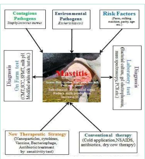
Udder health performance observation is diicult without any reliable and reasonable diagnostic techniques. Hence, there is a continuous necessity for development of advanced diagnostic methods which are readily available, cost efective and more accurate. he key diagnostic tools that are used in detecting mastitis are somatic cell Figure 1: Graphical abstract of bovine mastitis.
Schematic presentation of causes of bovine mastitis and therapeutic strategies to prevent and control of the disease. Environmental and contagious
pathogens and various risk factors causes mastitis occurrence in dairy herds.
Clinical cause’s pain and swelling of udder and subclinical mastitis reduce
the milk production and increase SCC. Mastitis can be diagnosed on farm and by laboratory examination. Various conventional treatment procedure including antibiotics, NSAIDs and new advanced therapeutic strategies like nanoparticles, vaccines, antibiotic susceptibility testing speciic antibiotics are
count (SCC) and bacteriological culturing of milk [33,99]. Presently, techniques such as California Mastitis Test (CMT), modiied white side test, somatic cell count (SCC), trypsin inhibition test, milk pH, N-acetyl-β-D-glucosaminidase (NAGase), quantiication of lactate dehydrogenase activity (LDH), electric conductivity (EC) are less frequently practiced [100]. Diagnosis of mastitis begins with visual distress of the udder and of the milk via fore stripping [101]. If there is any palpable change in quarter or any visible defect in the milk, the afected quarter is deined as clinical mastitis. It is essential not to overlook clinical signs. A rapid, easy and cheap on farm mastitis diagnostic tool is California Mastitis Test. his test can be more readily used by farmers and veterinarians to diagnose and treat the early stages of mastitis and thus devising the ability to stop the magnitude of disease in dairy herds. Increasing the body temperature is one of the signiicant sign associated with mastitis. Assessment of the intensities of inlammation-linked enzymes might also be used for the recognition of mastitis as these display proper association with SCCs. Additional enzymatic tests comprise the revealing of an esterase secreted by somatic cells using an enzymatic assay on a dipstick is another method of diagnosis. Another assay called bioluminescence- determination, built on assessment of the ATP concentrations in somatic cells or the response of somatic cell DNA by luorescent staining, may also be able for trust worthy determination of higher SCC levels and consequently the apparent occurrence of mastitis [102]. Determination of pathogens initiating mastitis is signiicant for control of disease and epidemiological studies. Bacterial culture can be performed at herd, along with cow and quarter level, each with its individual deinite goal. Probable identiication of the species is done interpreting cultural, biochemical and serological examination. he usage of molecular diagnostic tools in pathogen inding has advanced over the last years. PCR-based techniques have been deined for a number of mastitis pathogens [103]. Newly, multiplex PCR devices have been developed to detect quite a lot of pathogens at the same time [104]. Moreover, real-time PCR analyses are being advanced for identiication and quantiication of mastitis pathogens in milk. Two-dimensional gel electrophoresis and mass spectroscopy of relevant proteomics techniques have also directed for identiication of several new proteins involved in mastitis expressed by invading pathogens. his signal can be useful not only to discover new therapeutic goals but also to search new diagnostic biomarkers. he efective application of these novel biomarker devices are still remains a challenge for clinical mastitis diagnosis. Recent progresses in micro luidics and so-called ‘Biochips’ have the capability to modernize diagnostic procedure in early diagnosis of mastitis and to discover and develop speciic therapeutic strategy [105] (Figure 1).
P rin cip le s o f B o vin e Ma s titis Th e ra p y
herapeutic success of mastitis depends mainly on accurate diagnosis, severity of udder pathology, drug selection, and relevance of route of administration, supportive treatment, and elimination of predisposing factors [106]. Actually, there is no standard treatment regime yet developed for mastitis; however, it is encouraged to treat clinical forms of mastitis according to the severity of the udder inlammation. Once cows are infected with mastitis, the aim is to diminish the length of infection by treatment of infected cows or culling of cows from the farm. Efective and appropriate treatment of mastitis used all over the season called lactation therapy or dry cow
therapy at the end of the season is vital [107].
Before starting antibiotic treatment it should be ensure that the gland is milked out as completely as possible. his will eliminate millions of bacteria, milk clots and debris that can delay the magnitude of antibiotic treatment [106,108]. Some bacteria can also release toxins along with mastitis in infected cows that can make them very sick. In that case, milking out will also remove these toxins. It is recommended milking cows more frequently to remove the infective bacteria and toxins as much as possible. Oxytocin inluences contraction of muscle cells that can squeeze milk from the glands and into the udder. he drug helps relief the cow milk out completely if the animal is being exposed out in between regular milking [108].
Bovine mastitis is generally treated by intra-mammary infusion of drugs [109,110]. his route is drug of choice in subclinical, mild or moderately severe form of mastitis, and is practiced as an aid to parenteral administration in severe cases. Some clinicians choose only intra-mammary administration even in severe disease [111]. It is noteworthy, intra-mammary drugs should be dispensed throughout the udder and be quickly immersed into the general blood circulation for efective functions. In severe case, spreading of drugs through the udder may be decreased by inlammation or blockage of milk ducts by debris. In that case, parenteral administration may over whelm these complications, even though it is common to administer agents simultaneously by the intramammary route [112]. Acutely inlamed udders should be milked out repeatedly, with the support of oxytocin if needed. Combined systemic and intramammary treatment with well-suited antibiotics, supplemented with supportive therapy like cold application and anti-inlammatory drug is recommended in per-acute or per-acute clinical mastitis cases with systemic clinical signs [110].
It is commonly desired to treat infectious mastitis consistent with the antimicrobial drug sensitivity outline of the pathogens and clinical experiences [111]. he fundamental rule in selecting the drug is to select as narrow a spectrum as possible against a speciic pathogen and shorten side-efects. Initially, broad-spectrum antibiotics must be given on the basis of pharmacokinetic properties and formulation of the drugs [109,113,114]. Response to treatment of disease rises with persistence of the antibiotic in the udder. herefore, the concentration of the drug must exceed the minimum inhibitory concentration (MIC) value for the pathogen, if possible also the minimum bactericidal concentration (MBC) value in the udder [106]. In case of bacteriostatic drugs, high concentrations in the target organ should be maintained.
no distribution to the gastrointestinal tract and similar clearance from organs, tissues and blood [111,114,120]. Immediately ater diagnosis, acute mastitis therapy should begin using a remarkably bio available antibiotic, with required supportive drugs [107,121,122]. Treatment duration will depend on disease anamnesis, clinical signs, and microbiological diagnosis. It is not worthy that prolonged treatment means a long milk withdrawal period, which makes the treatment expensive, so treatment duration should not be so long. Chronic mastitis that cannot be cured throughout lactation, culling is the apparent solution. Dairy cows that have had clinical mastitis throughout lactation should be prescribed drying of therapy.
Speciic Therapy for Various Forms of
Ma s titis
Per acute mastitis
Suitable parenteral antibiotic with identical or synergistic action are usually preferred for 3–5 days, depending on clinical cure and special care must be taken to prevent shock. he intravenous route must be chosen to accomplish highest parenchymal difusion [109,110]. While the response to antibiotic and other supportive therapy is unsatisfactory, extra measures may be necessary to avoid death or to treat a severely afected quarter. When the infected quarter is severely damaged or gangrenous, intra-mammary infusion is unproductive, and if it is economically reasonable teat amputation may support drainage. In severe cases tying of the mammary veins has also been encouraged to decrease the uptake of toxins into the circulation [123].
Acute clinical mastitis
In acute mastitis, recurrent failure of intramammary antimicrobial therapy is due to poor or irregular dissemination of the drug in the acutely swollen udder parenchyma. Parenteral and local antibiotic therapy may be chosen applying either the same antibiotic, or well-suited antibiotics with identical or synergistic action for 3–5 days, depending on clinical medication. However, intravenous route is the best to achieve extreme parenchymal difusion [109,110]. Primarily, intramammary antibiotic preparation should be infused in double dose into the exaggerated quarters and a single infusion is suggested into each of the healthy quarters to inhibit new intramammary infection. Ater the preliminary infusion, intramammary antibiotic preparations should be infused 12-hourly and continued for 3–5 days as recommended by the manufacturer. For enhanced bioavailability of the drug, a great volume should be injected intramuscularly at two sites [114,123-125]. Mastitis caused by Gram-positive bacteria is potentially best treated using macrolide antibiotics and gentamicin, or polymyxin B for Gram-negative infections. Penicillin G is the drug of choice for mastitis infection caused by Clostridium perfringens and Bacillus cereus organism [124,126]. Mastitis caused by penicillin-sensitive coagulase-negative staphylococci, S. agalactia and S. dysgalactiaeis usually treated by penicillin for 2–3 days [126]. he organism S. Uberis may be more challenging and treatment should be continued for 3–5 days. he intracellular bacteria Staphylococcus aureus has been identiied as therapy-resistant and treatment for 5 days has made better results [126].
In case of mild clinical mastitis, bacteriocidal drugs can be used as intramammary therapy to act only against Gram-positive bacteria. Application frequency of intramammary preparations depends on
the pharmacokinetics of the selected drug. Procaine penicillin that are absorbed slowly is suicient to apply once daily; drugs for example cephalexin, erythromycin that are quickly absorbed must be given twice a day [122]. Frequent intramammary administration of slow-release formulations is more dynamic than using fast-slow-released osage forms [111]. here are very limited antimicrobial drugs appropriate for treating coliform mastitis. Some drugs for example tetracycline, novobiocin-penicillin, chloramphenicol and streptomycin with high concentrations have been found to suppress phagocytosis in the udder. his consequence is boosted if corticosteroids are integrated with the drugs.
Sub-acute clinical mastitis
It is most predominant form of clinical mastitis in dairy herds where the duration of treatment is based on the causal organism and clinical progress [127,128]. Intra-mammary drugs are rapidly absorbed and usually administered and thus can usually be prescribed for 3 days at 12 hours intervals supplemented by repeated hand stripping to eliminate secretion and debris [111]. Ater the disappearance of clinical signs [128] treatment should be continued for at least another 24 hours to eliminate the bacterial contamination. In high-yielding dairy cows, 2 syringes of intra-mammary antibiotic preparation may be infused initially into the afected quarter. he introduction of cefoperazone for intra-mammary administration with single-dose treatment is a signiicant advance to treat sub-acute form of mastitis [129].
Chronic mastitis
In chronic form of mastitis, parenteral and intramammary antibiotic treatment may be used for 3–5 days in combination with anti-inlammatory products, but the prospects remains disappointing. Infusion of a 5% or 10% dextrose solution in combined with antibiotics for 2-3 days at 12 hours intervals has been suggested to which the microbes are sensitive into the afected quarter [123,130] but the prognosis also remains poor. herefore, it is usually necessary to cull the cow or to destroy the afected quarter by means of infusing concentrated ether to eliminate an important potential source of bacterial infection for healthy quarters [131,132].
Subclinical mastitis
mastitis when the inluencing factors are very limited [134,135,136]. In chronic infections, proven by earlier clinical incidents and persistent high SCC, may not respond to dry-cow therapy and should be culled from the dairy herd [128].
Dry cow therapy
Dry cow therapy with antibiotics has been recommended as one of the choices to control intra-mammary infections and inhibit progress of mastitis [137,138]. he cow is at the maximum danger of obtaining new intra-mammary infections with both contagious and environmental pathogens during the dry period [139,140]. It has been shown that about 61% of new infections are achieved during this period [140]. herapy with antibiotics during dry period is beneicial as there is no need to remove milk from treated quarters. Antibiotics are applied near the end of lactation [141] and may persist in the udder in high concentrations to destroy pathogenic bacteria for 20-70 days. he applied antibiotic has enhanced penetration capacity because of long time contact and curing intramammary infections unless resistance to novel antibiotics is developed by the entering pathogen [142]. Dry cow therapy has been described to remove nearly 100% of mastitis caused by S. agalactiae [139,143]. Conversely, dry cow therapy is less efective to inhibit S. aureus mastitis compared to streptococcal mastitis [138]. he treatment success rate of dry cow therapy against S. aureus mastitis is about 50% and vaccination against this pathogen throughout the dry season may improve the antibiotic eiciency [140]. Dry cow treatment for a duration of two weeks exhibited substantial decrease in the number of clinical mastitis cases owing to infection with S. dysgalactiae and S. uberis [144].
N e w Th e ra p e u tic S tra te gie s fo r Ma s titis
Nanotechnology
Wide ranges of antibiotics have been used for past 40 years in control of bovine mastitis. However, antibiotic resistance develops mainly due to their overuse and for other possible causes and creates the cure rate very poor. Nanotechnology has endowed researchers to produce nanosized particles less than 100nm in size, apply them
in a wide range of sectors, mostly in drug delivery system. he nanoparticles may be revealed prospective delivery methods in the treatment of bovine mastitis from when they are used by phagocytes. For example, silver nanoparticles exhibited inhibitory activity against S. aureus isolated from subclinical mastitis [145] and to be perfect for a wonderfully economical antimicrobial solution. Furthermore, nowadays antibiotics are incorporated in most of the nanoparticles delivery system to combat against microorganisms. Amoxicillin incorporated into nanoparticles exhibited biologically dynamic against S. aureus, E. coli, and S. agalactiae [146]. he key challenge seems to be obtaining an abundant antibiotic loading inside these delivery systems.
Mastitis vaccines
Mastitis vaccines have been in use with the expectation of decreasing the occurrence mastitis in dairy farming and upholding the proit of industry. he practice of killed whole cell vaccines to handle infective organism on-farm in dairy cattle is common like other disease vaccine. A lot of studies have been prepared to produce vaccine against mastitis, but very few have claimed acceptable results. It is important to discover a vaccine capable of protective against an extensive range of strains as multiple strains can be existent within a herd and within a single cow [147]. It is deined that only one vaccine will not prevent mastitis afected by the diverse numbers of pathogens and their diferent strategies of pathogenesis. Coliform vaccines can decrease the prevalence, severity and length of infections in both milking cows and heifers [148]. Vaccines against S.aureus can decline the occurrence of new intra-mammary contamination as well as increase the impulsive remedy, and should be practiced in herds where this kind of organism is a main issue [149,150]. Conversely, it should be noticed that there are lots of chance of vaccine failure if the farms are not immunized against similar type organism and it will make extra burden for diary industry.
Bacteriophage therapy for mastitis
Bacteriophage therapy is one of the alternate treatments for
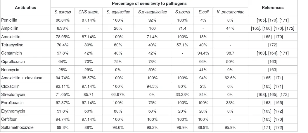
Antibiotics Percentage of sensitivity to pathogens References
S.aureus CNS staph. S. agalactiae S.dysagalactiae S.uberis E.coli K. pneumoniae
Penicillin 86.84% 87.14% 100% 92% 100% 4% 0% [165], [170], [171] Ampicillin 8.33% - 20% 100 71.4 - 44% [165], [166], [170], [172]
Amoxicillin 78.95% 87.14% 100% 71.4% 100% 18% - [165], [170]
Tetracycline 70.4% 80% 60% 40% 57.1% 40% - [172]
Gentamicin 97.8% 42% 40% 42% - 94.4% 98.7 [163], [164], [171]
Ciproloxacin 64% 70% 75% 73% - 66% 50% [163]
Neomycin 28% 29% 0% 50% - 41% 0% [163]
Amoxicillin + clavulanat 94.74% 98.57% 100% 100% 100% 94% 62.6% [165], [171]
Cloxacillin 92.11% 97.14% 100% 94.5% 80% 2% 0% [165], [171]
Streptomycin 71.05% 85.71 66.67% 0% 33.33% 84% 0% [163], [165], [172]
Enroloxacin 97.37% 97.14% 100% 75% 100% 100% 33% [163], [165]
Erythromycin 51.8% 60% 80% 60% 20% 20% 0% [163], [172] Ceftifour 94.74% 97.14% 100% 100% 100% 100% - [165], [170]
Sulfamethoxazole 99.3% 88% 98.6% 96.2% 96.9% 88.9% 95.9% [171], [172]
mastitis which practices pathogen speciic bacteriophages in the cure of a bacterial infection. Bacteriophages are viruses capable to invade and destroy bacteria [151]. Phages are proved to be determinants as new antimicrobial agents for veterinary applications. Certain studies specify that phage therapy could create advantageous efect against E.coli and S.aureus inluenced mastitis infection [148]. Still, further research is necessary to discover the therapeutic prospective of bacteriophages to treat mastitis associated bacterial infections.
Cytokines
In mammary gland, the roles of the various immune system mechanisms against infection are not successful. Conversely, cytokine generation and leukocyte populations carry out signiicant roles in host defense system and pathophysiological devices throughout the bacterial infection [148]. Numerous cytokines like complement component, interleukins (IL) and tumor necrosis factor alpha (TNF-α) have been recognized as key actors during the acute-phase reaction. hey play chemotactic action liable for leukocyte enrollment and motivation and they can also advance the bactericidal action of phagocytes. Numerous experimental works point out that use of cytokines with or without antibiotics signiicantly improved the cure rate in S. aureus infected mastitis. Cytokines therapy appears to be a promising method; however, advanced research has to be done to accept therapeutic attention.
Antibiotics therapy doing Antibiotic sensitivity test
Antibiotics have extensively reduced the hazard simulated by mastitis pathogens since their discovery in the 20th century. In the
dairy industry, antibiotic treatment is the key strategy for bovine mastitis treatment. On the contrary, beyond the expenses of its usage, the occurrence of resistance and the non-responsiveness of cattle to antibiotics have developed a serious issue. he random and misuse of antibiotics in bovine mastitis may represent intent diiculties linked with the development of antibiotic resistance and the appearance of resistant bacteria in food chain [153]. Furthermore, bacteria, which are accomplished to exist within the cell and mammary gland, and to form abscesses, are further challenging to combat because of the restriction of their interaction with antibiotics. his is the example of S. aureus where the ratio of remedy by presently permitted antibiotics like pirlimycin is approximately 10–30 % [153]. Bannerman and Wall [154] determined that treatment accessible for the treatment of mastitis formed by S. aureus keep at suboptimal and therefore proven inefective [148,155]. herefore, there is an urgent need to ind efective tool to apply speciic antibiotic which can efectively prevent and control bovine mastitis. Currently, Antibiotic susceptibility testing against microbes is considering as one of the choosing methods to treat bovine mastitis in dairy herds (Table 2).
he practice of these drugs joined with improvements in housing, sanitation, nutrition and the introduction of extensive vaccination programs for key environmental pathogens, has managed to ininite fall in occurrence of mastitis that was previously prevalent and frequently fatal. hese advances are now a days extremely threatened by emergence and spread of new microbes those are resistant to economical and efective irst-line drugs. Beta-lactamase producing bacteria S. aureus and B. fragilis candiminish the activity of penicillin. Various mastitogenic strains of B. fragilis are unafected to ampicillin, amoxicillin and cephalotine [134,156], and certain strains yield
cephalosporinase, which diminishes them resistant to cephalosporin [157]. he microbe P. aeruginosa is not susceptible to ampicillin, cefoxitin, cephalothin, cefamandole oroxacillin [157]. hus mastitis caused antibiotic resistant microbes fail to respond to treatment, ensuing constant illness and more threat of cull or death. Presently, there is also evidence of multiple drug resistance in the common bacterial agents liable for mastitis in bovines. Treatment failure also result increase number of infected cows afecting the farm and thus express the whole herd to the danger of contracting a resistant strain of infection [158,159]. Signiicant concern has been stretched over the last few years regarding the regulation of antibiotic use, due to the worrying rise of bacterial resistance. Antibiotic sensitivity testing is regarded as method of choice to treat the mastitis infected cows in dairy herds.
examined by disk difusion method and found that all of the S. aureus isolates were resistant to cetiofur and all of the S. agalactiae isolates were resistant to streptomycin. he multiple drug resistance amongst the cultures involved in mastitis has also been described by several other investigators [33,148]. Most of the cultures exhibited resistance to the third generation cephalosporins. his excessive advent of resistance to cephalosporin’s is unpleasant owing to its extensive usage against microbes as per adapted by most of the clinicians as the irst line of treatment in recent years. herefore, the probable long-term haphazard usage of these antibiotics has directed to the advent of resistance in mastitogenic bacteria counter to third generation cephalosporin [167,168]. Increase of resistance among bacteria is a matter of noticeable alarm in the ield of veterinary medicine speciically when the organisms render an animal unproductive. It is clinically thoughtful to practice susceptibility outcomes to guide treatment results for some pathogens [169]. hus, constant screening of cultures for antibiotic susceptibility outline will be advantageous for picking a suitable antibiotic and also to recognize the shiting drits of antibiotic resistance for evolving antibiotic usage strategy and for controlling the indiscriminate practice of inluential antibiotics like cephalosporin as primary treatment.
Co n clu s io n s
Bovine mastitis is truly important production disease due to its impact in the quality and quantity of milk production. he dairy industry sufers from substantial inancial losses due to mastitis all over the world. Practice of hygienic methods during milking, milk collection, lactation and dry cow therapy, dietary supplements and culling are likely to reduce but not control the incidence of both clinical and subclinical mastitis. he efects of mastitis on dairy cattle health and milk production highlight an urgent need to develop sensitivity test is thus playing a key role to treat the mastitis case more efectively.
References
1. Albenzio M, Taibi l, Muscio A. and Sevi A. Prevalence and etiology of
subclinical mastitis in intensively managed locks and related changes in the yield and quality of milk. Small Ruminant Res. 2002; 43: 219- 226.
2. Dua K. Incidence, aetiology and estimated loss due to mastitis in India- An update. Ind Dairy man. 2001; 53: 41-48.
3. Singh PJ, Singh PB .A study of economic losses due to mastitis in India. Ind J Dairy Sci. 1994; 47: 265-272.
4. Bramley AJ, CullorJS, Erskine RJ, Fox LK, Harmon RJ, Hogan JS, et al.
Current concepts of bovine mastitis, 4th Ed. 1996. National Mastitis Council,
Madison, WI.
5. Fang WH, Hu SH, Jiang CS, Huang IQ, Liu HR. Bovine mastitis - a
Chinese approach to its prevalence, causal pathogens, losses and control. Proceedings of the 6th International Symposium on Veterinary Epidemiology
and Economics. Ottawa, Canada, 1991.
6. Swinkels JM, Hogeveen H, Zadoks RN. A partial budget model to estimate
economic beneits of locational treatment of subclinical Staphylococcus
aureus mastitis. J Dairy Sci. 2005; 88: 4273-4287
7. NAAS. Mastitis Management in Dairy Animals. Policy Paper No. 61, National
Academy of Agricultural Sciences, New Delhi, India. 2013.
8. Wilson DJ, González RN, Hertl J, Schulte HF, Bennett GJ, Schukken YH, et
al. Effect of clinical mastitis on the lactation curve: A mixed model estimation using daily milk weights. J. Dairy Sci. 2004; 87: 2073-2084.
9. Bogni C, Odierno L, Raspanti C, Giraudo J, Larriestra A, Reinoso E, Lasagno
M, et al. War against mastitis: Current concepts on controlling bovine mastitis pathogens. Science against microbial pathogens: communicating
current research and technological advances. A. Méndez-Vilas (Ed.). 2011.
10. Dairy Australia. Dairy Australia Strategic Plan. 2011; 2011-2015.
11. Tuteja FF, Kapur MP, Sharma A, Vinajaka AK. Studies on bovine subclinical
mastitis: Prevalence and microlora. Indian Vet J. 1993; 70: 787-791.
12. Brightling P, Mein GA, Malmo J, Ryan DP. Countdown down under: farm guidelines for mastitis control. In Countdown Downunder: Dairy Research and Development Corporation. 1998.
13. Seegers H, Fourichon C, Beaudeau F. Production effects related to mastitis
and mastitis economics in dairy cattle herds. Vet Res. 2003; 34: 475–491.
14. Chagunda MGG, Friggens NC, Rasmussen MD, Larsen T: A Model for
Detection of Individual Cow Mastitis Based on an Indicator Measured in Milk. J Dairy Sci. 2006; 89: 2980–2998.
15. Halasa T, Nielen M, De Roos APW, Van Hoorne R, de Jong G, Lam TJGM,
et al. Production loss due to new sub-clinical mastitis in Dutch dairy cows
estimated with a test-day model. J Dairy Sci. 2009; 92: 599–606.
16. Tiwari JG, Babra C, Tiwari HK, Williams V, Sharon DW, Gibson J, et al.
Trends In Therapeutic and Prevention Strategies for Management of Bovine
Mastitis: An Overview. J Vaccin. 2013; 4: 2.
17. Thomson K, Rantala M, Hautala M, Pyörälä S, Kaartinen l. Cross-sectional prospective survey to study indication-based usage of antimicrobials in
animals: Results of use in cattle. BMC Vet. Res. 2008; 4: 15.
18. Kaneene JB, Miller R: Description and evaluation of the inluence of
veterinary presence on the use of antibiotics and sulfonamides in dairy
herds. J. Am. Vet.Med. Assoc. 1992; 201: 68-76.
19. Sharma N, Gautam A, Upadhyay SR, Hussain K, Soodan JS, Gupta SK.
Role of antioxidants in udder health: a review. Indian J. Field Vet. 2006; 2: 73–76.
20. Gogoi J. Public health importance of Staphylococcus aureus isolated from
cases of bovine mastitis and milkers. MVSC thesis. Assam Agricultural University, India. 1997.
21. Zafalon LF, Nader Filho A, Oliveira JV, Resende FD. Subclinical mastitis
caused by Staphylococcus aureus: Cost beneit analysis of antibiotic therapy in lactating cows. Arq Bras Med Vet Zootec. 2007; 59: 577-585.
22. White DG, McDermott PF. Emergence and transfer of antibiotic resistance. J Dairy Sci. 2001; 84: E151-E155.
23. Virdis S, Scarano C, Cossu F, Spanu V, Spanu C, et al. Antibiotic Resistance
in Staphylococcus aureus and Coagulase Negative Staphylococci Isolated from Goats with Subclinical Mastitis. Vet Med Int. 2010; 517060.
24. Collins NA, Moses M, Moneoang SM, Corneleus B .Antibiotic-resistant
Staphylococcus aureus isolated from milk in the Maikeng Area, North West
province, South Africa. South Afr J Sci. 2010; 106: 1-6.
25. Sharif A, Umer M, Muhammad G. Mastitis control in dairy production. J Agril
Soc Sci, 2009; 5: 102-105.
26. Wilson DJ, Gonzalez RN, Das HH. Bovine mastitis pathogens in New York
and Pennsylvania: prevalence and effects on somatic cell count and milk
production. J Dairy Sci. 1997; 80: 2592-2598.
27. Barkema HW, Schukken YH, Lam TJ, Beiboer ML, Wilmink H, et al.
Incidence of clinical mastitis in dairy herds grouped in three categories by
bulk milk somatic cell counts. J Dairy Sci. 1998; 81: 411-419.
28. Fox LK, Kirk JH, Britten A. Mycoplasma mastitis: a review of transmission
and control. J Vet Med B Infect Dis Vet Public Health. 2005; 52: 153-160.
29. Watts JL. Etiological agents of bovine mastitis. Vet Microbiol.1988; 16:
30. Hogeveen H, Pyorala S, Waller KP, Hogan JS, Lam JGMT, Oliver SP, et
al. In Proceedings of the National Mastitis Council 50th Annual Meeting:
Arlington. Canada, 2011.
31. Jones GM. Understanding the basics of mastitis. Virginia Cooperative
Extension, Publication No. 404-233, Virginia State University, USA, 2006;
1-7.
32. Philpot WN, Nickerson SC. Mastitis: Counter Attack. Westfalia Surge LLC.
Illinois. USA. 1999.
33. El-Hamid MIA. Bovine Mastitis: Current Concepts and Future Control Approaches. Austin Clin Microbiol. 2016; 12: 1007.
34. Bradley AJ, Green MJ. Clinical mastitis in dairy cows after ‘blitz’ therapy. Vet
Rec. 1997; 141: 179-180.
35. Hogan JS, Smith KL, Hoblet KH, Todhunter DA, Schoenberger PS, et al.
Bacterial counts in bedding materials used on nine commercial dairies. J Dairy Sci. 1989; 72: 250-258.
36. Boyer PJ. Outbreak of clinical mastitis in dairy cows following ‘blitz’ therapy.
Vet Rec. 1997; 141: 55.
37. Edmondson PW. Clinical mastitis in dairy cows after ‘blitz’ therapy. Vet Rec.
1997; 141: 108.
38. Bradley AJ, Green MJ. Adaptation of Escherichia coli to the bovine mammary gland. J. Clin Microbiol. 2001; 39: 1845-1849.
39. Bradley AJ, Leach KA, Breen JE, Green LE, Green MJ. Survey of the
incidence and aetiology of mastitis on dairy farms in England and Wales. Vet
Rec. 2007; 160: 253-257.
40. Dairy Australia. Environmental mastitis increasing. The Land, 2011.
41. Harmon RJ. Physiology of mastitis and factors affecting somatic cell counts. J Dairy Sci. 1994. 77: 2103-2112.
42. APHIS (Animal and Plant Health Inspection Service) Info sheet. Prevalence
of contagious mastitis pathogens on U.S. dairy operations, 2007. USDA.
2008.
43. Smith KL, Hogan JS. Epidemiology of mastitis. Proceedings of 3rd
International Mastitis Seminar, 1995.Tel Aviv, Israel. 1995; S6: 3-12.
44. Barkema HW, Schukken YH, Zadoks RN. Invited Review: The role of cow,
pathogen, and treatment regimen in the therapeutic success of bovine Staphylococcus aureus mastitis. J Dairy Sci. 2006; 89: 1877-1895.
45. Monecke S, Kuhnert P, Hotzel H, Slickers P, Ehricht R. Microarray based
study on virulence-associated genes and resistance determinants of
Staphylococcus aureus isolates from cattle. Vet Microbiol. 2007; 125:
128-140.
46. BurgtGVD, Main W, Ayling R. Bovine mastitis caused by Mycoplasma bovis.
Vet Rec. 2008; 163: 666.
47. Workineh S, Bayleyegn M, Mekonnen H, Potgieter LN. Prevalence and
aetiology of mastitis in cows from two major Ethiopian dairies. Trop Anim Health Prod. 2002; 34: 19-25.
48. Barrett DJ, Healy AM, Leonard FC, Doherty ML. Prevalence of pathogens
causing subclinical mastitis in 15 dairy herds in the Republic of Ireland. Ir Vet
J. 2005; 58: 333-337.
49. Middleton JR. Staphylococcus aureus antigens and challenges in vaccine
development. Expert Rev Vaccines. 2008; 7: 805-815.
50. Unnerstad HE, Lindberg A, Persson Waller K, Ekman T, Artursson K, et al.
Microbial aetiology of acute clinical mastitis and agent-speciic risk factors. Vet Microbiol. 2009; 137: 90-97.
51. Pereira UP, Oliveira DG, Mesquita LR, Costa GM, Pereira LJ. Eficacy of
Staphylococcus aureus vaccines for bovine mastitis: a systematic review.
Vet Microbiol. 2011; 148: 117-124.
52. Owens WE, Watts JL, Boddie RL, Nickerson SC. Antibiotic treatment of
mastitis: comparison of intramammary and intramammary plus intramuscular therapies. J Dairy Sci.1988; 71: 3143-3147.
53. Haveri M. Staphylococcus aureus in bovine intramammary infection: molecular, clinical and epidemiological characteristics. Academic dissertation, submitted to Department of Production Animal Medicine,
Faculty of Veterinary Medicine, University of Helsinki, Finland. 2008.
54. Pyörälä S, Taponen S. Coagulase-negative staphylococci-emerging mastitis
pathogens. Vet Microbiol. 2009; 134: 3-8.
55. Sordillo LM. Factors affecting mammary gland immunity and mastitis
susceptibility. Liv Prod Sci. 2005; 98: 89-99.
56. Emanuelson U, Persson E. Studies on somatic cell counts in milk from
Swedish dairy cows. I. Non-genetic causes of variation in monthly test-day
results. Acta Agri Scand. 1984; 34: 33-44.
57. Busato A, Trachsel P, Schällibaum M, Blum JW. Udder health and risk
factors for subclinical mastitis in organic dairy farms in Switzerland. Prev Vet
Med. 2000; 44: 205-220.
58. Sharma H, Maiti SK, Sharma KK. Prevalence, etiology and antibiogram of microorganisms associated with sub-clinical mastitis in buffaloes in Drug, Chhattisgarh state. Intl J Dairy Sci. 2007; 2: 145-151.
59. Sharma N, Maiti SK. Incidence, etiology and antibiogram of sub clinical
mastitis in cows in drug, Chhattisgarh. Ind J Vet Res. 2010; 19: 45-54.
60. Schukken YH, Grommers FJ, Van de Geer D, Brand A. Incidence of clinical
mastitis on farms with low somatic cell counts in bulk milk. Vet Rec. 1989;
125: 60-63.
61. Radostits OM, Gay CC, Blood DC, Hinchcliff KW. Mastitis. In: Veterinary
Medicine, A Textbook of the Diseases of Cattle, Sheep, Pigs, Goats and Horses. 9thEdn, W B Saunders Co. Philadelphia, USA. 2000; 603–612.
62. Rahman AM. Bhuiyan MU, Kamal MM, Shamsuddin M. The Bangladesh
Veterinarian. 2009; 26: 54 – 60.
63. Dulin AM, Paape MJ, Nickerson SC. Comparison of phagocytosis and
chemiluminescence by blood and mammary gland neutrophils from
multiparous and nulliparous cows. Am J Vet Res. 1988; 49: 172-177.
64. Sarker SC, Parvin MS, Rahman AK MA and Islam MT. Prevalence and
risk factors of subclinical mastitis in lactating dairy cows in north and south
regions of Bangladesh, Tropical Animal Health and Production. 2013; 45: 1171-1176.
65. Rajala PJ, Gröhn YT. Disease occurrence and risk factor analysis in Finnish
Ayrshire cows. Acta Vet Scand. 1998; 39: 1-13.
66. Grohn YT, Erb HN, McCulloch CE, Saloniemei HS. Epidemiology of
mammary gland disorders in multiparous Finnish Ayrshire cows. Prev Vet
Med. 1990; 8: 241-252
67. Lactation Resource Library. Other Factors Affecting Milk Yield and
Composition. 2009.
68. Rathore AK. Relationships between teat shape, production and mastitis in
Friesian cows. Br Vet J. 1976; 132: 389-392.
69. Seykora AJ, McDaniel BT. Udder and teat morphology related to mastitis
resistance: a review. J Dairy Sci. 1985; 68: 2087-2093.
70. Sharma N, Singh NK, Singh OP, Pandey V, Verma PK. Oxidative stress and
antioxidant status during transition period in dairy cows. Asian-Aust J Anim
Sci. 2011; 24: 479-484
71. Bradley A, Green MJ. Use and interpretation of somatic cell count data in
dairy cows. In Pract. 2005; 27: 310-315.
72. Berry, D.P., Lee, J.M., Macdonald, K.A., Stafford, K., Matthews, L. and
Roche, J.R. Associations among body condition score, body weight, somatic cell count, and clinical mastitis in seasonally calving dairy cattle. J Dairy Sci. 2007; 90: 637-648.
73. Plastridge WN. Bovine mastitis: A review. J Dairy Sci. 1958; 41: 1141-1181.
74. Peeler EJ, Green MJ, Fitzpatrick JL, Morgan KL, Green LE. Risk factors
associated with clinical mastitis in low somatic cell count British dairy herds. J Dairy Sci. 200; 83: 2464-2472.
sub-clinical mastitis in smallholder dairy cows in Tanzania. Tropic Anim Prod. 2004; 36: 581–592.
76. AbdullahiSJ, Jibril A, Mohammed AA, FasinaFO. Sub-clinical mastitis and
associated risk factors on lactating cows in the Savannah Region of Nigeria. BMC Vet Res. 2012; 8: 134
77. Mgedela RH, Ryoba R, Karimuribo ED, Phiri EJ, Loken T, Reksen O et
al. Prevalence of clinical and sub-clinical mastitis and quality of milk on smallholder dairy farms in Tanzania. J South African Vet Assoc. 2009; 80: 163–168.
78. Blood DC, Radostits OM .Veterinary medicine. In: textbook of the diseases
of cattle, sheep, pigs, goats and horses, (7th edn), WB Saunders Co. Philadelphia, USA.1989; 501.
79. Mein GA, Schuring N. Lessons from scrapbooks and scrapheaps of history.
Bulletin-International Federation. 2003.
80. Reneau J. Gear up for warm weather mastitis management now. Dairy Star. 2012
81. Costa EO, Ribeiro AR, Watanabe ET, Melville PA. Infectious bovine mastitis
caused by environmental organisms. Zentralbl Veterinarmed B. 1998; 45:
65-71.
82. Akyuz A, Boyaci S, Cayli A. Determination of critical period for dairy cows
using temperature humidity index. J Anim Vet Adv. 2010; 9: 1824-1827.
83. Ribeiro AR, Garinojr F, Silva JAB, Watanabe ET, Vallue CR, et al.
Seasonality on the occurrence of environmental bovine mastitis. Nopgama.
2001; 4: 19-22.
84. Godden S, Rapnicki P, Stewart S, Fetrow J, Johnson A, et al. Effectiveness
of an internal teat seal in the prevention of new intramammary infections during the dry and early-lactation periods in dairy cows when used with a dry cow intramammary antibiotic. J Dairy Sci. 2003; 86: 3899-3911.
85. Shathele MS. Weather effect on bacterial mastitis in dairy cows. Intl J Dairy Sci. 2009; 4: 57-66.
86. Ranjan R, Gupta MK, Singh KK. Study of bovine mastitis in different climatic
conditions in Jharkhand, India. Vet World. 2011; 4: 205-208.
87. Heinrichs AJ, Costello SS, Jones CM. Control of heifer mastitis by nutrition.
Vet Microbiol. 2009; 134: 172-176.
88. Spears JW, Weiss WP. Role of antioxidants and trace elements in health
and immunity of transition dairy cows. Vet J. 2008; 176: 70-76.
89. Hogan JS, Weiss WP, Todhunter DA, Smith KL, Schoenberger PS. Eficacy
of an Escherichia coli J5 mastitis vaccine in an experimental challenge trial.
J Dairy Sci. 1992; 75: 415-422.
90. Chamberlain AT, Wilkenson JM. Feeding the dairy cow. Chalcombe
Publications. 1996; 90-91.
91. Mustacich D, Powis G. Thioredoxin reductase. Biochem J. 2000; 346: 1-8.
92. Erskine RJ, Eberhart RJ, Grasso PJ, Scholz RW. Induction of Escherichia
coli mastitis in cows fed selenium-deicient or selenium-supplemented diets. Am J Vet Res. 1989; 50: 2093-2100.
93. Grasso PJ, Scholz RW, Erskine RJ, Eberhart RJ. Phagocytosis, bactericidal
activity, and oxidative metabolism of milk neutrophils from dairy cows fed selenium-supplemented and selenium-deicient diets. Am J Vet Res. 1990;
51: 269-274.
94. Underwood EJ, Suttle NF. In: The Mineral Nutrition of Livestock. Underwood
EJ and Suttle NF (eds), CABI Publishing, New York. 1999.
95. Sordillo LM, Shafer-Weaver K, DeRosa D. Immunobiology of the mammary
gland. J Dairy Sci. 1997; 80: 1851-1865.
96. Chew BP. Role of carotenoids in the immune response. J Dairy Sci. 1993; 76: 2804-2811.
97. Prasad AS, Bao B, Beck FW, Kucuk O, Sarkar FH. Antioxidant effect of zinc
in humans. Free Radic Biol Med. 2004; 37: 1182-1190.
98. Bruno DR. Mastitis, Mammary Gland Immunity, and Nutrition. Mid- South
Ruminant Nutrition Conference 19 Arlington, Texas. 2010.
99. Bogni C, Odierno L , Raspanti C, Giraudo J , Larriestra A , Reinoso E, et
al. War against mastitis: Current concepts on controlling bovine mastitis pathogens. Science against microbial pathogens: communicating current
research and technological advances. Formatex. 2011.
100. Joshi S, Gokhale S. Status of mastitis as an emerging disease in improved
and periurbandairy farms in India. Ann New York Acad Sci. 2006; 1081:
74-83.
101. Reneau JK. Somatic cell counts: measures of farm management and milk quality. In: Proceedings of National Mastitis Council Ann. 2001. Mtg. p. 29-37. Reno, NV.
102. Frundzhyan V, Parkhomenko I, Brovko L, Ugarova N. Improved
bioluminescent assay of somatic cell counts in raw milk. J Dairy Res. 2008;
75: 279-83.
103. Zadoks R, Schukken Y. Use of molecular epidemiology in veterinary
practice. Veterinary Clinics of North America: Food Animal Practice. 2006;
22: 229-261.
104. Phuektes P, Browning G, Anderson G, Mansell P. Multiplex polymerase chain reaction as a mastitis screening test for Staphylococcus aureus, Streptococcus agalactiae, Streptococcus dysgalactiaeand Streptococcus
uberisin bulk milk samples. J Dairy Res. 2003; 70: 149-155.
105. Viguier C, Arora S, Gilmartin N, Welbeck K, O’Kennedy R. Mastitis detection: current trends and future perspectives. Trends in Biotech. 2009; 27: 8-10. 106. Du Preez JH. Bovine mastitis therapy and why it fails. Tydskr.S.Afr.vet.Ver.
2000; 71: 201–208.
107. Du Preez JH. Reasons why mastitis therapy is unsuccessful. S. Afr J Dairy
Sci. 1989; 21: 21–29.
108. Barlow J. Mastitis Therapy and Antimicrobial Susceptibility: a Multispecies Review with a Focus on Antibiotic Treatment of Mastitis in Dairy Cattle. J
Mammary Gland BiolNeoplasia. 2011; 16: 383–407.
109. MacDiarmid S C. Drugs used in the antibacterial therapy of mastitis.
Proceedings of a post-graduate short course. 1980, Palmerston North, Massey University, New Zealand. 1980: 103–110.
110. Ziv G. Practical pharmacokinetic aspects of mastitis therapy – 1: parental
treatment. Agri Practice: 1980a; 227–290.
111. Prescott JF, Baggot JD. Antimicrobial therapy in veterinary medicine.
Blackwell Scientiic Publications, Boston. 1988.
112. Du Preez JH, Giesecke WH. Mastitis. In Coetzer JAW, Thomson GR, Tustin RC (eds) Infections diseases of livestock with special reference to southern Africa, Vol. 2. Oxford University Press, Cape Town South Africa. 1994; 1564–1595.
113. Du Preez JH. Speenkanaalinfeksies by melkkoeie: diagnose, voorkoms,
aard, terapie, omvang en betekenis. DVScthesis, University of Pretoria.
1985.
114. Ziv G. Drug selection and use in mastitis: systemic vs local therapy. J Amer Veter Medi Assoc. 1980d; 176: 1109–1115.
115. Rasmussen F. Studies on the mammary excretion and absorption of drugs. Carl F Mortensen, Copenhagen. 1966.
116. Rasmussen F. Excretion of drugs in milk. In Brodie BB, Gillette J (eds)
Handbook of experimental pharmacology. Springer Verlag, New York. 1971; 28: 390–402.
117. Sjoqvist E, Borga O, Orme ML. Fundamentals of clinical pharmacology drug treatment. In Avery G S (ed.) Drug treatment. Adis Press, Sydney. 1976:
1–42.
118. Miller GE, Banerjee NC, Stowe CM. Diffusion of certain weak organic acids and bases across the bovine mammary gland membrane after systemic
administration. J Pharm and Ther. 1967; 157: 245–253.
119. Giesecke WH. Bovine mastitis. Department of Agricultural Technical
Services, Republic of South Africa, Technical Communication No. 1979;
120. Sandholm M, Honkanen-Buzalski T, Kaartinen L, Pyörälä S. The bovine
udder and mastitis. Gummerus Kirjapaino Oy, Helsink. 1995.
121. Wilson CD. Antibiotic therapy in mastitis control. In Bramley A J, Dodd F H, Griffen T K (eds) Mastitis control and herd management. Technical Bulletin
4, National Institute for Research in Dairying, Reading. 1980. 113–127.
122. Pyörälä S. Therapy of clinical mastitis. In Sandholm M, Honkanen-Buzalski
T, Kaartinen L, Pyörälä S (eds) The bovine udder and mastitis. Gummerus Kirjapaino Oy, Jyväskylä. 1995; 201–208.
123. Robinson TC. Therapy for acute and peracute mastitis. In Bramley AJ, Dodd FD, Griffen TR (eds) Mastitis control and herd management. Technical
Bulletin 4, National Institute for Research in Dairying, Reading. 1980; 128–
134.
124. Jackson E, Bramley J. Coliform mastitis. In Practice 5. 1983; 135–146. 125. MacDiarmid SC. Antibacterial drugs used against mastitis in cattle by the
systemic route. N Zeal Veter J. 1978; 26: 290–295
126. Ziv G. Practical pharmacokinetic aspects of mastitis therapy 1: parental
treatment. Agri Practice. 1980; 227–290.
127. DuPreez JH. Planne teen hoë SST in kuddes. Landbouweekblad.
1999;1094: 24–26.
128. Philpot WN, Nickerson SC. Mastitis:counter attack. Babson Bros, Naperville. 1991.
129. Wilson CD, Agger N, Gilbert GA, Thomasson CA, Tolling ST. Field trials with
cefoperazone in the treatment of bovine clinical mastitis. The Veter Recor.
1986; 118: 17.
130. Schalm OW, Carroll EJ, Jain NC. Bovine mastitis. Lea &Febiger, Philadelphia. 1971
131. DuPreez JH. Reasons why mastitis therapy is unsuccessful. S Afr J Dairy
Sci. 1989; 21: 21–29.
132. DuPreez JH. Planne teen hoë SST in kuddes.Landbouweekblad. 1999;
1094: 24–26.
133. Moore GA, Heider LE. Treatment of mastitis. Veterinary Clinics of North
America: Large Animal Practice. 1984; 6: 323–333.
134. DuPreez JH. The role of anaerobic bacteria in bovine mastitis: a review. J S
AfrVeter Ass. 1989;60: 159–168.
135. Giesecke WH, DuPreez JH, Petzer IM. Practical mastitis control in dairy herds. Butterworths, Durban. 1994.
136. Platonow I, Blobel H. Therapeutic failures in chronic staphylococcal mastitis. 1963. 142; 1097.
137. Oliver SP, Sordillo LM. Udder health in the periparturient period. J Dairy Sci.1988; 71: 2584-2606.
138. Ziv G, Storper M, Saran A. Comparative eficacy of three antibiotic products for the treatment and prevention of subclinical mastitis during the dry period.
Vet Q. 1981; 3: 74-79.
139. Oliver SP, Mitchell BA. Susceptibility of bovine mammary gland to infections during the dry period. J Dairy Sci. 1983; 66: 1162-1166.
140. Todhunter DA, Smith KL, Hogan JS, Schoenberger PS. Gram-negative
bacterial infections of the mammary gland in cows. Am J Vet Res. 1991;
52: 184-188.
141. JanosiSz, Huszenicza G. The use of the dry cow therapy in the control of
bovine mastitis. Vet Med-Czech 2001; 46: 55-60.
142. Babra C, Gogoi TJ, Pier G, Thein TH, Sunagar R, et al. The persistence of
bioilm-associated antibiotic resistance of Staphylococcus aureus isolated
from clinical bovine mastitis cases in Australia. Folia Microbiol. 2013; 58: 1. 143. Bramley AJ, Dodd FH. Reviews of the progress of dairy science: mastitis
control--progress and prospects. J Dairy Res. 1984; 51: 481-512.
144. Hassan Z, Daniel RC, O’Boyle D, Frost AJ. Effects of dry cow intramammary
therapy on quarter infections in the dry period. Vet Rec. 1999; 145: 635-639.
145. Dehkordi SH, Hosseinpour F, KahrizangiAE. An in vitro evaluation of antibacterial effect of silver nanoparticles on Staphylococcus aureus isolated
from bovine subclinical mastitis. Afr J Biotechnol. 2011; 10: 10795–10797.
146. Xue FY. Post-antibiotic effect of amoxicillin nanoparticles against main pathogenic bacteria of bovine mastitis in vitro. J Northwest Sci-Tech UnivAgric For. 2009; 37: 1–6.
147. Bannerman DD, Wall RJ. A novel strategy for the prevention of Staphylococcus aureus-induced mastitis in dairy cows. Information Systems
for Biotechnology (isb) news report. 2005; 1–4.
148. Gomes F,Henriques M. Control of Bovine Mastitis: Old and Recent
Therapeutic Approaches. Curr Microbiol. 2016 ; 72: 377–382.
149. Hoedemaker M, Korff B, Edler B, Emmert M, Bleckmann E. Dynamics
of Staphylococcus aureus infections during vaccination with an
autogenousbacterin in dairy cattle. J Vet Med. 2001; 48: 373–383.
150. Kazemi J, Ahmadi M, Saei HD, Adibhesami M. Antibacterial effect of silver nanoparticles along with protein synthesis-inhibiting antibiotics on Staphylococcus aureus isolated from cattle mastitis. Biol J Microorg. 2014;
2: 15–22.
151. Chibani-Chennoui S, Dillmann ML, Marvin-Guy L, Rami-Shojaei S,
Bru¨ssowH. Lactobacillusplantarum bacteriophage LP65: a new member of the SPO1-like genus of the family Myoviridae. J Bacteriol. 2004; 186: 7069–7083.
152. Alluwaimi AM. The cytokines of bovine mammary gland: prospects for
diagnosis and therapy. Res Vet Sci. 2004; 77: 211–222.
153. White DG, McDermott PF. Emergence and transfer of antibiotic resistance.
J Dairy Sci. 2001; 84: E151–E155.
154. Bannerman DD, Wall RJ. A novel strategy for the prevention of Staphylococcus aureus-induced mastitis in dairy cows. Information Systems
for Biotechnology (isb) news report. 2005; 1–4.
155. Atulya M, Mathew AJ, Rao JV, Rao CM. Inluence of milk components
in establishing bioilm mediated bacterial mastitis infections in cattle: a fractional factorial approach. Res Vet Sci. 2014; 96: 25–27.
156. Du Preez JH. Comparison of various criteria for determining the health
status of the bovine udder. J S Afr Veter Asso. 1987; 58: 9–14.
157. Finegold SM, Rosenblatt JE, Sutter VC, Atteberry HR. Anaerobic infections.
In Thomas B A (ed.) Scope monograph on anaerobic infections (3rd edn). The Upjohn Company, Kalamazoo, Michigan. 1976.
158. Lipsitch M, Singer RS, Levin BR. Antibiotics in agriculture: when is it time to
close the barn door? Proc Natl Acad Sci USA. 2002; 99: 5752–5754.
159. Sischo WM. Stakeholder position paper: dairy producer. Prev Vet Med.
2006; 73: 203–208.
160. CLSI. Performance standards for antimicrobial disk and dilution susceptibility
tests for bacteria isolated from animals: approved standard. M31-A3, 3rd ed. Wayne, PA: CLSI; 2008.
161. Saini V, Riekerink RG, McClure JT, Barkema HW. Diagnostic accuracy
assessment of Sensititre and agar disk diffusion for determining antimicrobial resistance proiles of bovine clinical mastitis pathogens. J Clin Microbiol. 2011; 49: 1568–1577.
162. Walker RD. Antimicrobial susceptibility testing methods and interpretation of
results. 4th ed. Antimicrobial Therapy in Veterinary Medicine. Ames, Iowa: Blackwell. 2006.
163. Alekish1 MO, Al-qudah KM, Al-saleh A. Prevalence of antimicrobial
resistance among bacterial pathogens isolated from bovine mastitis in
northern Jordan. Revue Méd. Vét. 2013; 164: 319-326.
164. ChauhanPM, Thumar HK,Bhagat A, Sharma VK, Chauhan HC, Patel MR.
Comparative eficacy of antibiotic sensitivity tests for management of acute clinical Escherichia Coli mastitis in crossbred cow. J. Livestock Sci. 2016;
7: 41-45.
165. Idriss SE, Foltys V, TančinV, Kirchnerová V, TančinovD , Zaujec K. Mastitis pathogens and their resistance against antimicrobial agents in dairy cows in
166. İkiz S, Saran BB, Bingöl EB, Çetin Ö, Kaşikçi G, Özgur NY, et al. Presence and antibiotic susceptibility patterns of contagious mastitis agents
(Staphylococcus aureusand Streptococcus agalactiae) isolated from milks
of dairy cows with subclinical mastitis. 2013; 37: 569-574.
167. Hornish RE &Kotarski SF. Cephalosporins in veterinary medicine—Ceftiofur
use infood animals. Curr top med chem, 2002; 2: 717–731.
168. Nora Mestorino and Jorge O. Errecalde. Pharmacokinetic – Pharmacodynamic Considerations for Bovine Mastitis Treatment. A
Bird’s-Eye View of Veterinary Medicine Edited by Dr. Carlos C. Perez-Marin.www.
intechopen.com. 2012.
169. Waller KP, HårdemarkV, Nyman AK, Duse A. Veterinary treatment strategies
for clinical mastitis in dairy cows in Sweden.Veter Rec. 2016; 178: 240.
170. Cameron M, Saab M, Heider L, McClure JT, Rodriguez-Lecompte JC,
J Sanchez J. Antimicrobial Susceptibility Patterns of Environmental Streptococci Recovered from Bovine Milk Samples in the Maritime Provinces of Canada. Front Vet Sci. 2016; 15: 79.
171. Rocha B,Mendonça D, Niza-Ribeiro J. Trends in Antibacterial Resistance
of Major Bovine Mastitis Pathogens in Portugal. RCPV. 2014; 109: 79-88.
172. Moges N, Asfaw Y, Belihu K, Tadesse A. Antimicrobial susceptibility of mastitis pathogens from small holder dairy herd in and around Gondar,
Ethiopia. J AnimVeter Ad. 2011; 10: 1616-1622.
Citation: Hossain MK, Paul S, Hossain MM, Islam MR and Alam MGS. Bovine Mastitis and Its Therapeutic Strategy Doing Antibiotic Sensitivity Test. Austin J Vet Sci & Anim Husb. 2017; 4(1): 1030.
Austin J Vet Sci & Anim Husb - Volume 4 Issue 1 - 2017 ISSN: 2472-3371 | www.austinpublishinggroup.com